Aorta Drawing
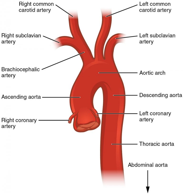
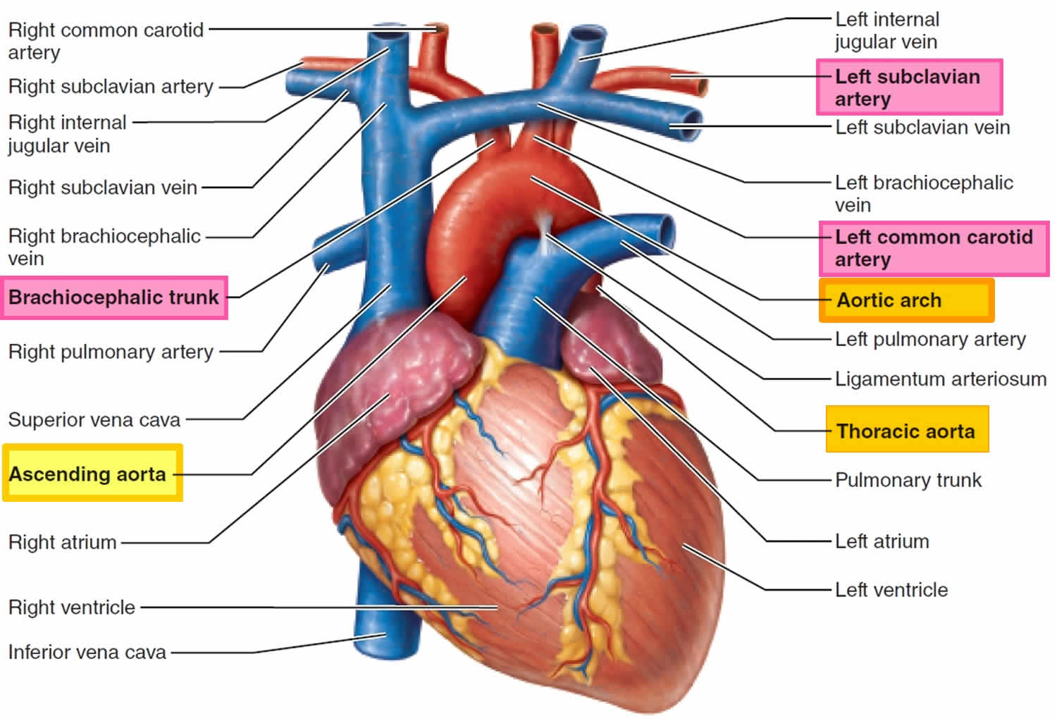
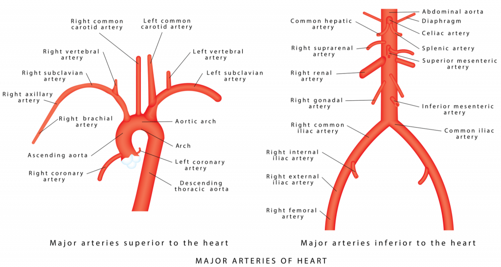
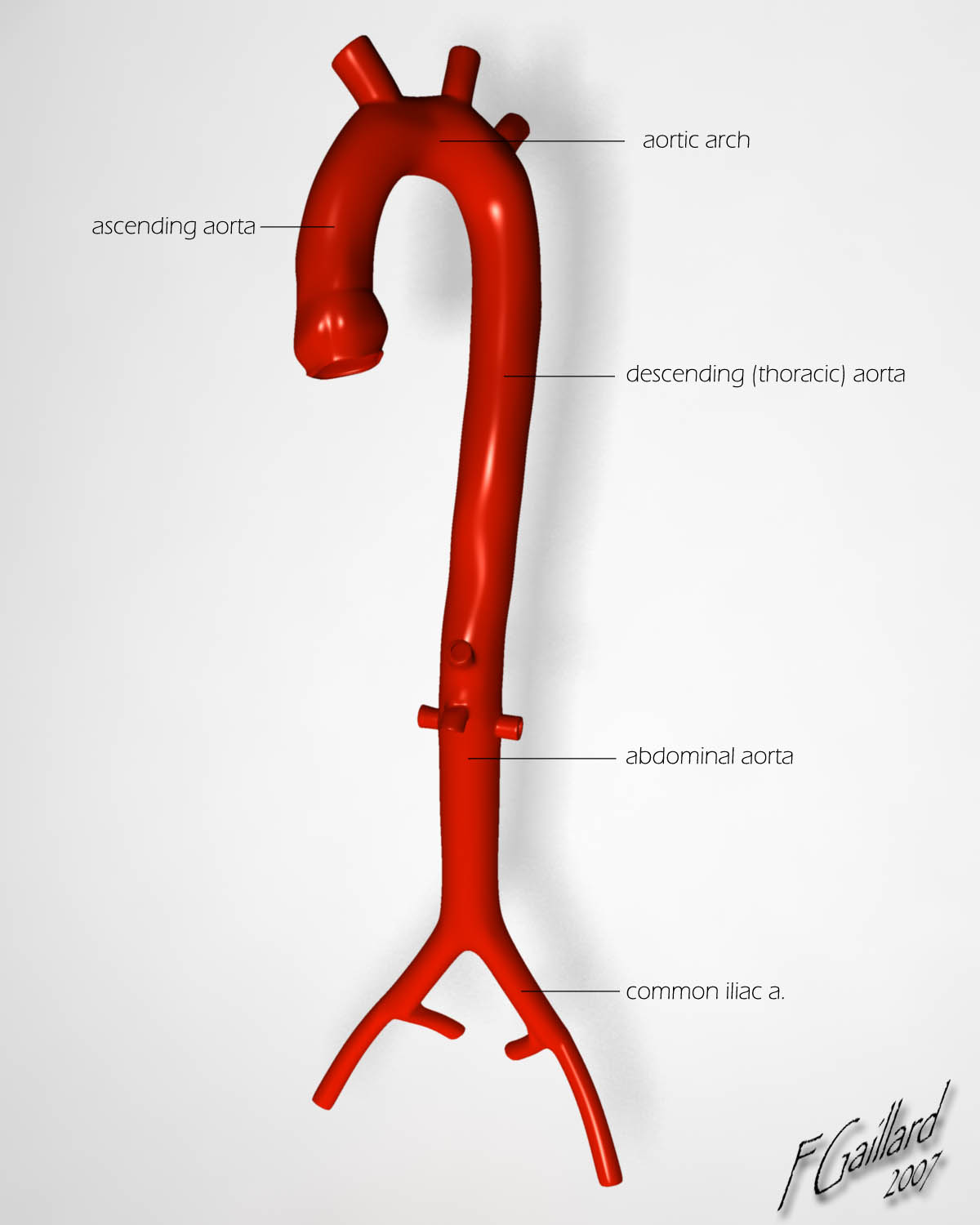
Aorta Drawing - Find an image that displays the entire heart, and click on it to enlarge it. Web major aorta anatomy displaying ascending aorta, brachiocephalic trunk, left common carotid artery, left subclavian artery, aortic isthmus, aortic arch, and descending thoracic aorta. Long, straight segment that runs from your chest (thoracic aorta) to your abdominal area (abdominal aorta). In this video, i'm taking a look at the branches of the thoracic aorta. The thoracic aorta consists of the ascending aorta, aortic arch, and descending aorta. The part that attaches to your heart. It has three semilunar cusps/leaflets: Web the aorta ascends ( ascending aorta ), arches ( aortic arch ), and then descends ( descending aorta) posterior to the heart through the thoracic cavity ( thoracic aorta ), through the diagram and then through the abdominal cavity ( abdominal aorta ). Web the aorta extends from the aortic valve of the left ventricle to the proximal iliac bifurcation at the l4 vertebral level. The ascending aorta, the aortic arch, the thoracic (descending) aorta and the abdominal aorta.
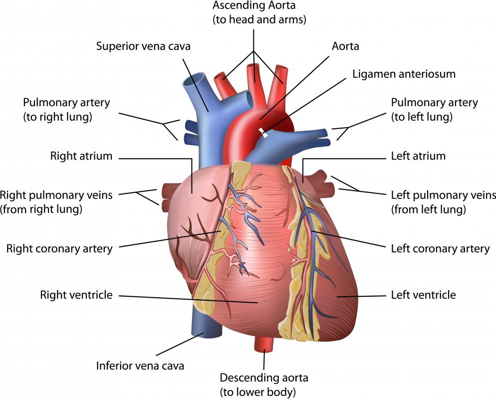
Angle the slightly tampered end of the shape to the left about 120 degrees. Start with the pulmonary veins. It is the largest artery in the human body. Web the aorta ascends ( ascending aorta ), arches ( aortic arch ), and then descends ( descending aorta) posterior to the heart through the thoracic cavity ( thoracic aorta ), through the diagram and then through the abdominal cavity ( abdominal aorta ). Browse 1,200+ aorta drawing stock photos and images available, or start a new search to explore more stock photos and images. Web it includes several distinct parts: Learn the structure, location in the human heart, anatomy, various functions with labelled diagrams. The ascending aorta (where the aorta initially leaves the heart and points toward the head), the arch of the aorta (where the aorta changes direction), and the descending aorta (where the aorta points toward the feet). Explore the heart with the free iheart touc. It bridges the ascending and descending aorta.
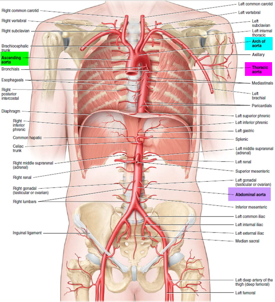
The part that attaches to your heart. The aorta is the largest artery in the body. It has three semilunar cusps/leaflets: To find a good diagram, go to google images, and type in the internal structure of the human heart. Web the aorta ascends ( ascending aorta ), arches ( aortic arch ), and then descends ( descending aorta) posterior to the heart through the thoracic cavity ( thoracic aorta ), through the diagram and then through the abdominal cavity ( abdominal aorta ). The aorta is the largest artery in the human body and arguably one of the most important. Superior and inferior vena cava. It terminates at the level of l4 by bifurcating into the left and right common iliac arteries. In clinical practice, the heart valves can be auscultated, usually by using a stethoscope. Web the aorta can be divided into four sections:
What is an Aorta? (with pictures)
Aorta drawing pictures, images and stock photos. The part that curves upward from your heart. The part that attaches to your heart. Angle the slightly tampered end of the shape to the left about 120 degrees. The aorta is the largest artery in the body.
Aorta Explained Anatomy 101 For patients
Start with the pulmonary veins. It is the largest artery in the human body. In this condition the aorta (the main artery that carries blood from the heart to the body) is narrowed or constricted. Long, straight segment that runs from your chest (thoracic aorta) to your abdominal area (abdominal aorta). It bridges the ascending and descending aorta.

Aorta Drawing Stock Image C020/9510 Science Photo Library
The vessel can be divided into various segments depending on course and location. Web © 2024 google llc. Coa can cause high blood pressure or heart damage. Find an image that displays the entire heart, and click on it to enlarge it. 18k views 2 years ago anatomy.
Aortic Dissection Type A And B Symptoms, Causes, Treatment
Web the aorta ascends ( ascending aorta ), arches ( aortic arch ), and then descends ( descending aorta) posterior to the heart through the thoracic cavity ( thoracic aorta ), through the diagram and then through the abdominal cavity ( abdominal aorta ). Web the aorta extends from the aortic valve of the left ventricle to the proximal iliac.
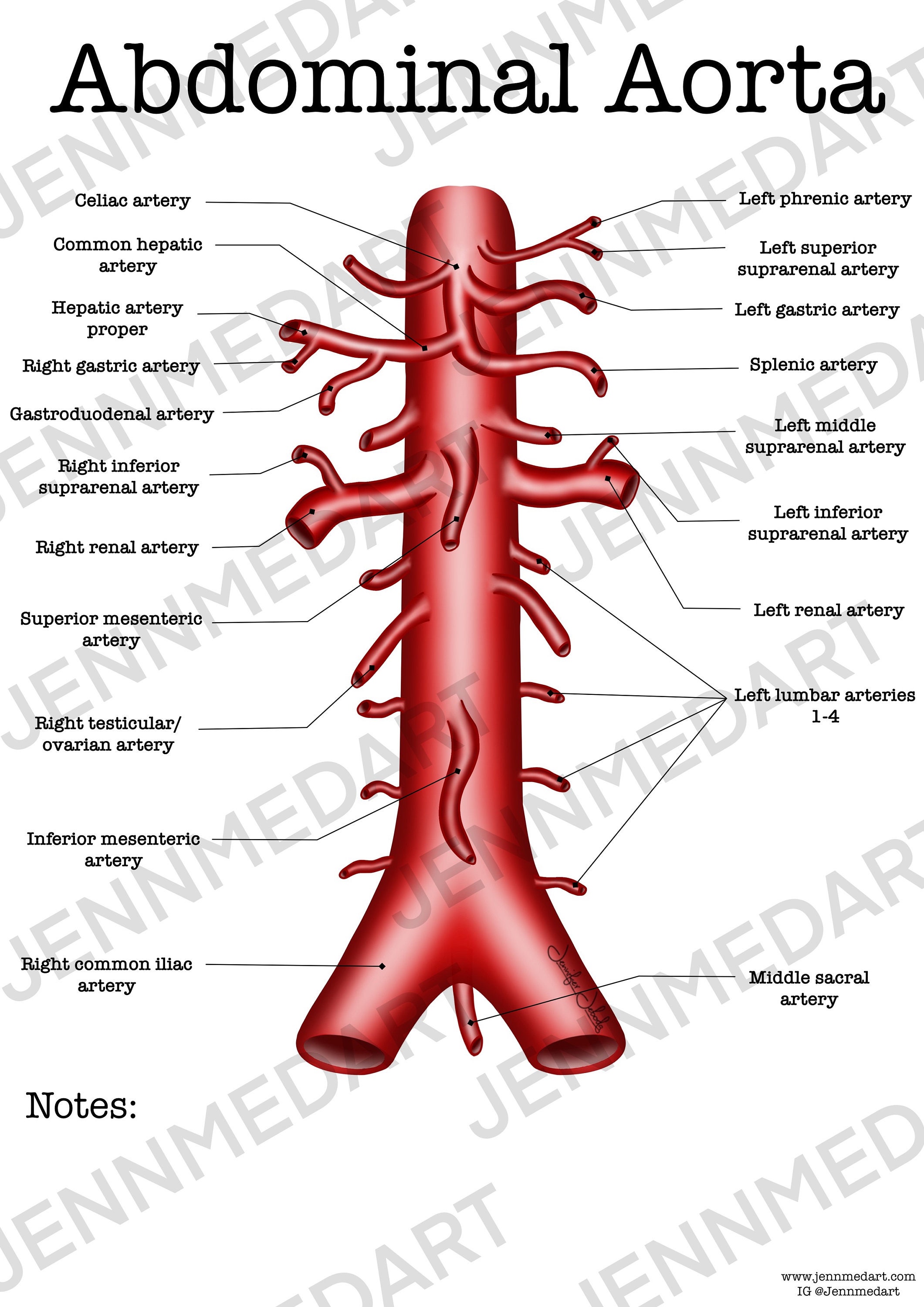
Abdominal Aorta Anatomy Worksheet Single FILLED Digital Download
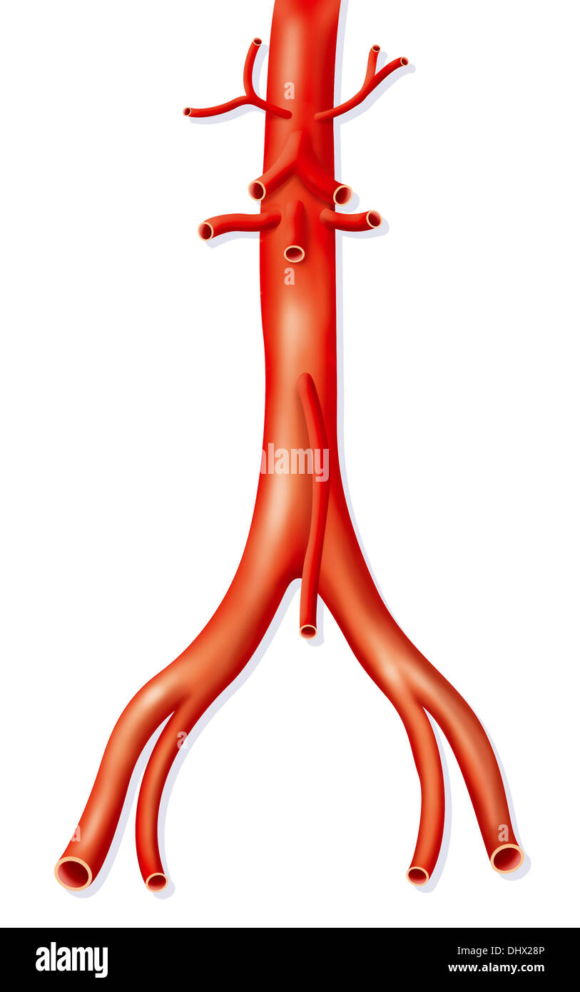
Find an image that displays the entire heart, and click on it to enlarge it. Coa can cause high blood pressure or heart damage. It terminates at the level of l4 by bifurcating into the left and right common iliac arteries. They will be to the lower left of the aorta. Pencil drawing of a human heart in retro style.
Aorta anatomy, function, branches, location & aorta problems
The pulse of life” see an illustration of and learn about the aorta, the largest artery in the body, in the emedicinehealth image collection gallery. Create a curved shape similar to an acorn or apple’s bottom half. The aorta is the largest artery in the body. Explore the heart with the free iheart touc. Web the aorta ascends ( ascending.
Aorta Explained Anatomy 101 For patients
Coarctation of the aorta enlarge image. 18k views 2 years ago anatomy. Explore the heart with the free iheart touc. It bridges the ascending and descending aorta. Superior and inferior vena cava.

AORTA DRAWING Stock Photo Alamy
The aorta classified as a large elastic artery, and more information on its internal structure can be found here. 18k views 2 years ago anatomy. To find a good diagram, go to google images, and type in the internal structure of the human heart. The pulse of life” see an illustration of and learn about the aorta, the largest artery.
Aorta wikidoc
It bridges the ascending and descending aorta. Find an image that displays the entire heart, and click on it to enlarge it. Angle the slightly tampered end of the shape to the left about 120 degrees. It is the largest artery in the human body. The aorta classified as a large elastic artery, and more information on its internal structure.
AORTA DRAWING Stock Photo 62652822 Alamy
Use a pen or pencil to draw the heart's main body. The thoracic aorta consists of the ascending aorta, aortic arch, and descending aorta. It is the largest artery in the body consisting of three parts that each has its special characteristics, most. 104k views 2 years ago cardiology. The part that curves upward from your heart.
The Part Of Your Descending Aorta That’s Contained In Your Chest.
Pencil drawing of a human heart in retro style. Web it includes several distinct parts: Coa can cause high blood pressure or heart damage. To find a good diagram, go to google images, and type in the internal structure of the human heart.
Web The Aorta Is Divided Into Three Parts:
Web the aorta ascends ( ascending aorta ), arches ( aortic arch ), and then descends ( descending aorta) posterior to the heart through the thoracic cavity ( thoracic aorta ), through the diagram and then through the abdominal cavity ( abdominal aorta ). The ascending aorta (where the aorta initially leaves the heart and points toward the head), the arch of the aorta (where the aorta changes direction), and the descending aorta (where the aorta points toward the feet). The aorta is the largest artery in the human body and arguably one of the most important. Draw a tilted and irregular curved shape in the center of your page.
The Aorta Is The Largest Artery In The Body.
18k views 2 years ago anatomy. The ascending aorta, the aortic arch, the thoracic (descending) aorta and the abdominal aorta. Upward curve that occurs shortly after the aorta leaves the heart. They will be to the lower left of the aorta.
The Part That Attaches To Your Heart.
Web the aorta extends from the aortic valve of the left ventricle to the proximal iliac bifurcation at the l4 vertebral level. The aorta is the largest artery in the body. Angle the slightly tampered end of the shape to the left about 120 degrees. The aorta starts at the heart’s left ventricle, arches upwards towards the neck, then curves back downward, extending into the abdomen.