Bacillus Subtilis Drawing
Bacillus Subtilis Drawing - When nutrients become limited, it undergoes a developmental process that results in the formation of a dormant spore. Web the data for rrna input, b. A bacterium for all seasons. Web bacillus subtilis (hereafter b. The spores are usually ellipsoidal, and they help preserve the. It is naturally transformable and has an extremely powerful genetic toolbox. These spores are highly resistant to. Apart from being widely present in nature, it is also a part of the microbial gut flora. Web here, we provide a database of 105 fully annotated genomes of a series of strains with sequential deletion steps of the industrially relevant model bacterium bacillus subtilis starting with the laboratory wild type strain b. Web in this review, we provide a brief summary of b.
To enhance ionization efficiency and avoid interference from coeluting species, several uridine This bacterium can form a tough, protective endospore that allows it to tolerate extreme environmental conditions. It was originally named vibrio subtilis by christian gottfried ehrenberg, and renamed bacillus subtilis by ferdinand cohn in 1872 (subtilis being the latin for fine, thin, slender). Web in this review, we provide a brief summary of b. Apart from being widely present in nature, it is also a part of the microbial gut flora. 1) and is known for its ability to differentiate into metabolically inactive spores (lopez et al. Web bacillus subtilis, the model gram‐positive bacterium: These spores are highly resistant to. Coli/ peak area ratio for each modified nucleosides was normalized to the b. Sizes, shapes, and arrangements of bacteria.
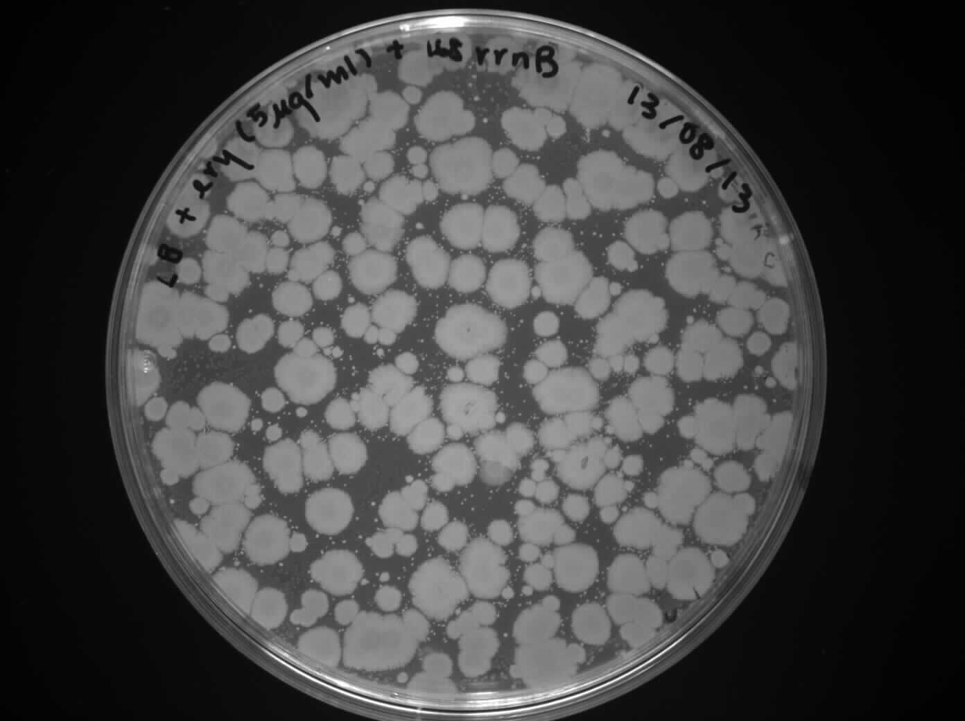
The most important parts of the microscope are labeled. Web bacillus subtilis, the model gram‐positive bacterium: Web interpret results of an endospore stain. Coli/ peak area ratio for each modified nucleosides was normalized to the b. As a library, nlm provides access to scientific literature. Tell how the endospore stain works including the stains involved and how the stains penetrate cells and. It is naturally transformable and has an extremely powerful genetic toolbox. Subtilis 168 and ending with b. Web the data for rrna input, b. Endospores in bacillus subtilis bacteria are mostly formed in the tips of protuberances extending downward from liquid surface pellicles (schaechter 2006).

Bacillus Subtilis ClipArt ETC
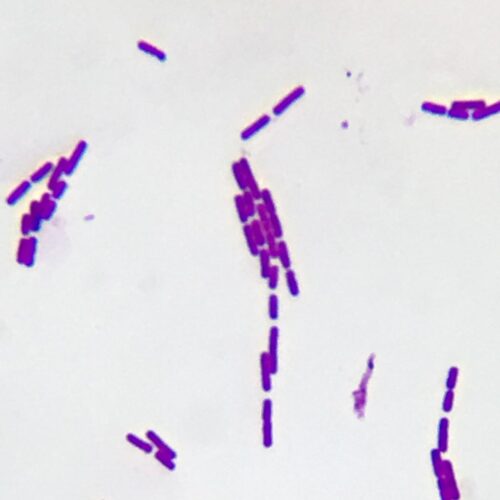
Tell how the endospore stain works including the stains involved and how the stains penetrate cells and. The gram stain, developed in 1884 by the danish bacteriologist hans christian gram (1), differentiates bacteria based on the composition of the cell wall (1, 2, 3, 4). The us food and drug administration (fda) classifies bacillus subtilis as a gras organism. It.
Bacillus subtilis; Natto Bacteria
Bacillus subtilis captured under the u2 biological microscope at 40x. Subtilis sporulation, describe the function of the spore surface layers and discuss the recent progress that has improved our understanding of the structure of the endospore coat and the mechanisms of coat assembly. It was originally named vibrio subtilis by christian gottfried ehrenberg, and renamed bacillus subtilis by ferdinand cohn.

Bacillus Subtilis Arrangement, Characterstics & Shape Lesson
Subtilis 168 and ending with b. The us food and drug administration (fda) classifies bacillus subtilis as a gras organism. The most important parts of the microscope are labeled. Identify when endospores are terminal, subterminal, and central in microscopic images, diagrams, and descriptions. Web in this review, we provide a brief summary of b.

Bacillus subtilis bacteria, illustration Stock Image F020/1916
Web in this review, we provide a brief summary of b. Identify when endospores are terminal, subterminal, and central in microscopic images, diagrams, and descriptions. The spores are usually ellipsoidal, and they help preserve the. To enhance ionization efficiency and avoid interference from coeluting species, several uridine As a library, nlm provides access to scientific literature.
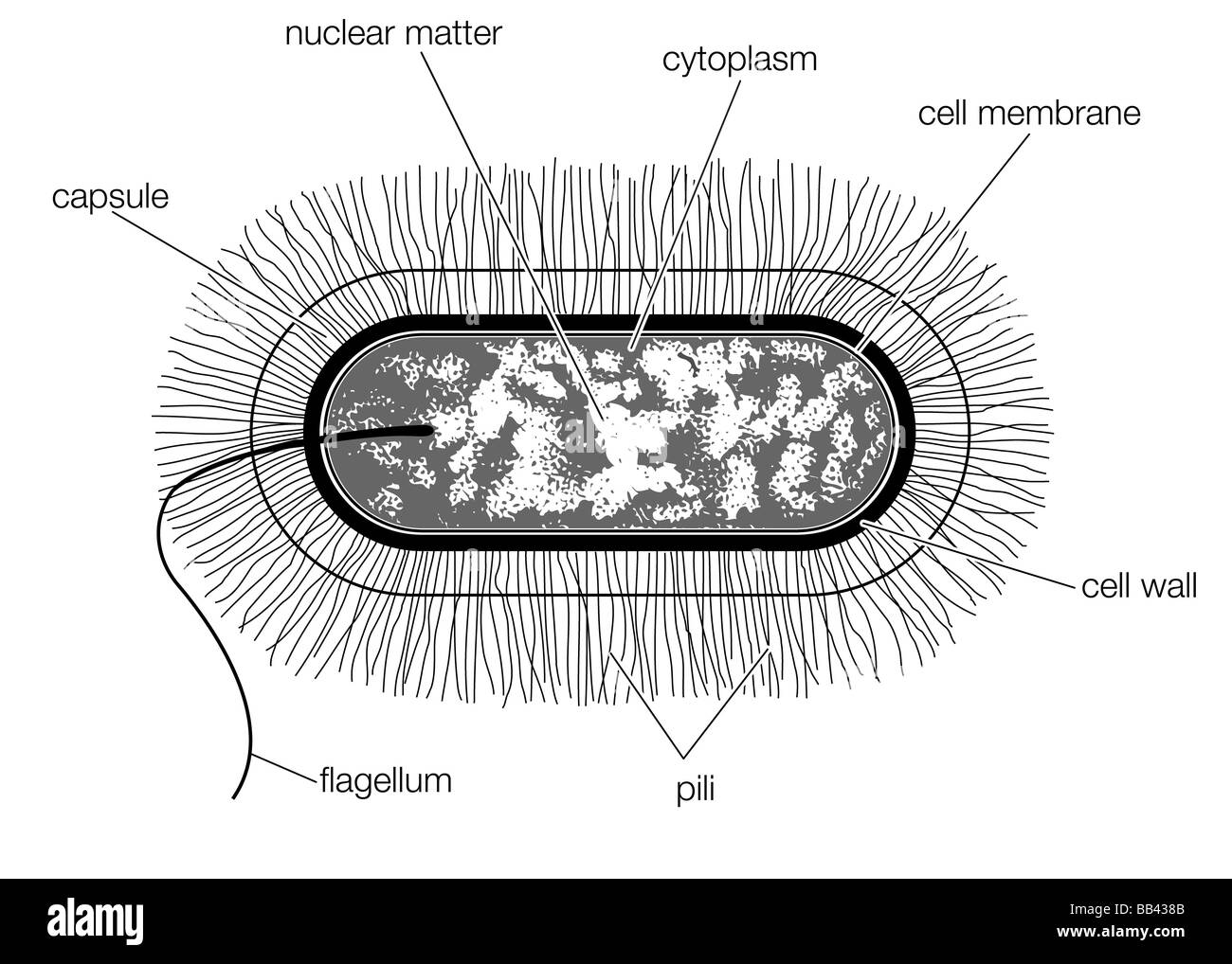
Schematic drawing of the structure of a typical bacterial cell of the
Bacillus subtilis captured under the u2 biological microscope at 40x. Identify when endospores are terminal, subterminal, and central in microscopic images, diagrams, and descriptions. Web the cell wall of bacillus subtilis is a rigid structure on the outside of the cell that forms the first barrier between the bacterium and the environment, and at the same time maintains cell shape.

Bacillus subtilis bacteria, illustration Stock Photo Alamy
List the three basic shapes of bacteria. The us food and drug administration (fda) classifies bacillus subtilis as a gras organism. Apart from being widely present in nature, it is also a part of the microbial gut flora. Web bacillus subtilis, strain 168 remains a case in point, and here we present an updated annotation, based on experimental evidence collected.

Bacillus Subtilis Bacteria Artwork HighRes Vector Graphic Getty Images
Bacillus subtilis captured under the u2 biological microscope at 40x. Web interpret results of an endospore stain. It is fast growing and easy to cultivate. To enhance ionization efficiency and avoid interference from coeluting species, several uridine The most important parts of the microscope are labeled.

Bacillus subtilis bacteria, illustration Stock Image F029/1926
Sizes, shapes, and arrangements of bacteria. Subtilis 168 and ending with b. Web in this review, we provide a brief summary of b. It was originally named vibrio subtilis by christian gottfried ehrenberg, and renamed bacillus subtilis by ferdinand cohn in 1872 (subtilis being the latin for fine, thin, slender). Web bacillus subtilis (hereafter b.
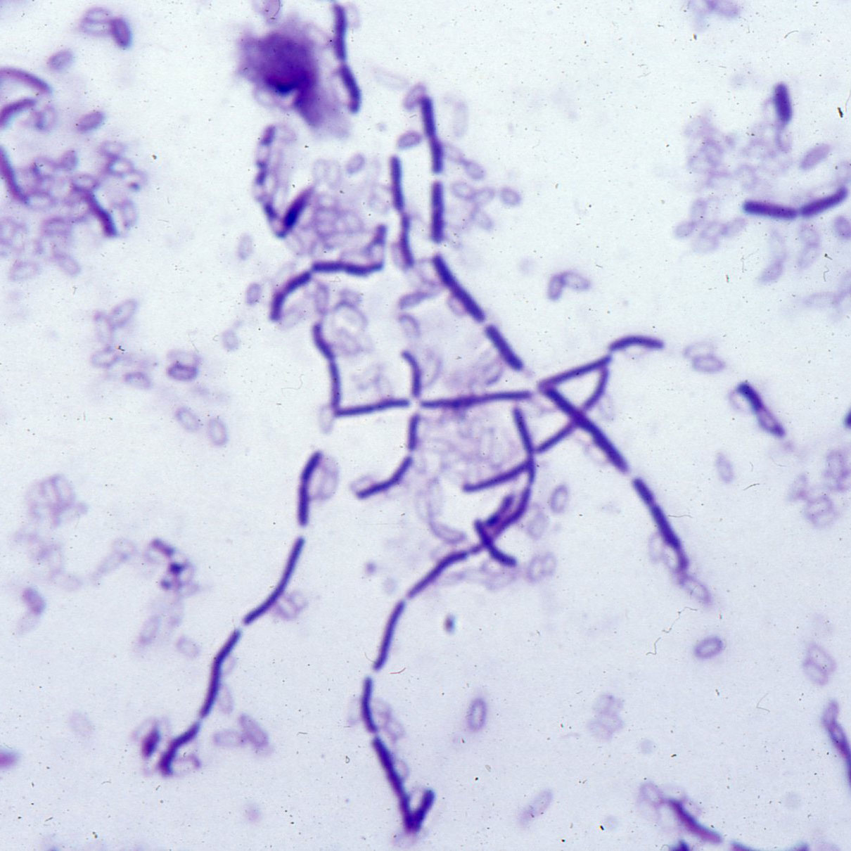
Bacillus subtilis smear with bacilli and spores, bacteria prepared
Coli peak area ratio for the canonical nucleosides (i.e., median value for a, g, u, c) and reported in figure 3. Web bacillus subtilis is a model organism for studying endospore formation in bacteria. The gram stain, developed in 1884 by the danish bacteriologist hans christian gram (1), differentiates bacteria based on the composition of the cell wall (1, 2,.
Morphology Of Bacillus Subtilis
1) and is known for its ability to differentiate into metabolically inactive spores (lopez et al. It is naturally transformable and has an extremely powerful genetic toolbox. The most important parts of the microscope are labeled. Coli peak area ratio for the canonical nucleosides (i.e., median value for a, g, u, c) and reported in figure 3. It was originally.
This Bacterium Can Form A Tough, Protective Endospore That Allows It To Tolerate Extreme Environmental Conditions.
When nutrients become limited, it undergoes a developmental process that results in the formation of a dormant spore. The us food and drug administration (fda) classifies bacillus subtilis as a gras organism. Subtilis 168 and ending with b. It is fast growing and easy to cultivate.
It Was Originally Named Vibrio Subtilis By Christian Gottfried Ehrenberg, And Renamed Bacillus Subtilis By Ferdinand Cohn In 1872 (Subtilis Being The Latin For Fine, Thin, Slender).
To enhance ionization efficiency and avoid interference from coeluting species, several uridine Coli/ peak area ratio for each modified nucleosides was normalized to the b. A bacterium for all seasons. Web the cell wall of bacillus subtilis is a rigid structure on the outside of the cell that forms the first barrier between the bacterium and the environment, and at the same time maintains cell shape and withstands the pressure generated by the cell's turgor.
List And Describe 5 Different Arrangements Of Cocci.
Apart from being widely present in nature, it is also a part of the microbial gut flora. Subtilis pg38, which lacks approximately 40% of the original genome. Web bacillus subtilis is a model organism for studying endospore formation in bacteria. Web here, we provide a database of 105 fully annotated genomes of a series of strains with sequential deletion steps of the industrially relevant model bacterium bacillus subtilis starting with the laboratory wild type strain b.
As A Library, Nlm Provides Access To Scientific Literature.
Bacillus subtilis captured under the u2 biological microscope at 40x. Subtilis sporulation, describe the function of the spore surface layers and discuss the recent progress that has improved our understanding of the structure of the endospore coat and the mechanisms of coat assembly. 1) and is known for its ability to differentiate into metabolically inactive spores (lopez et al. Web interpret results of an endospore stain.