Chromatid Drawing
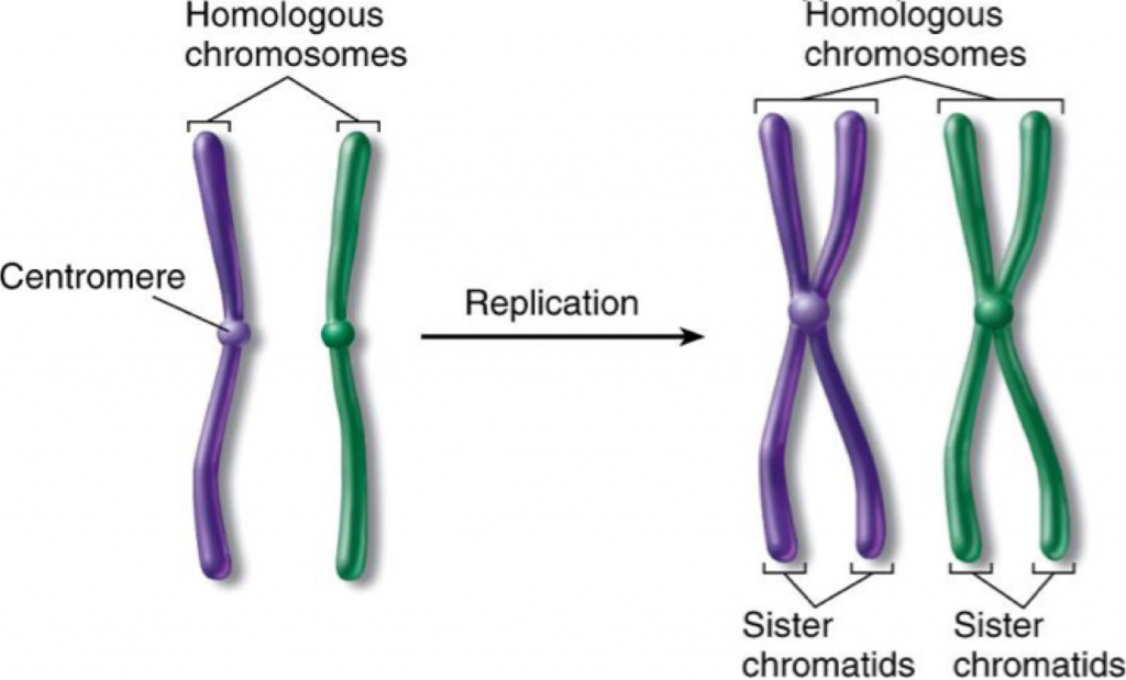
Chromatid Drawing - Web a major reason for chromatid separation is the precipitous degradation of the cohesin molecules joining the sister chromatids by the protease separase (figure 10). Different species have different numbers of chromosomes. Web what is a chromatid? Web as a result, chromatin can be packaged into a much smaller volume than dna alone. Prior to cell division, chromosomes are copied and identical chromosome copies join together at their centromeres. 22 pairs of autosomes and 1 pair of sex chromosomes. During cell division, spindle fibers attach to the centromere and pull each of the sister chromatids to. Long arm is termed q. Joined chromatids are known as sister chromatids. The two copies of the cell’s original chromosome are called “sister chromatids.”.
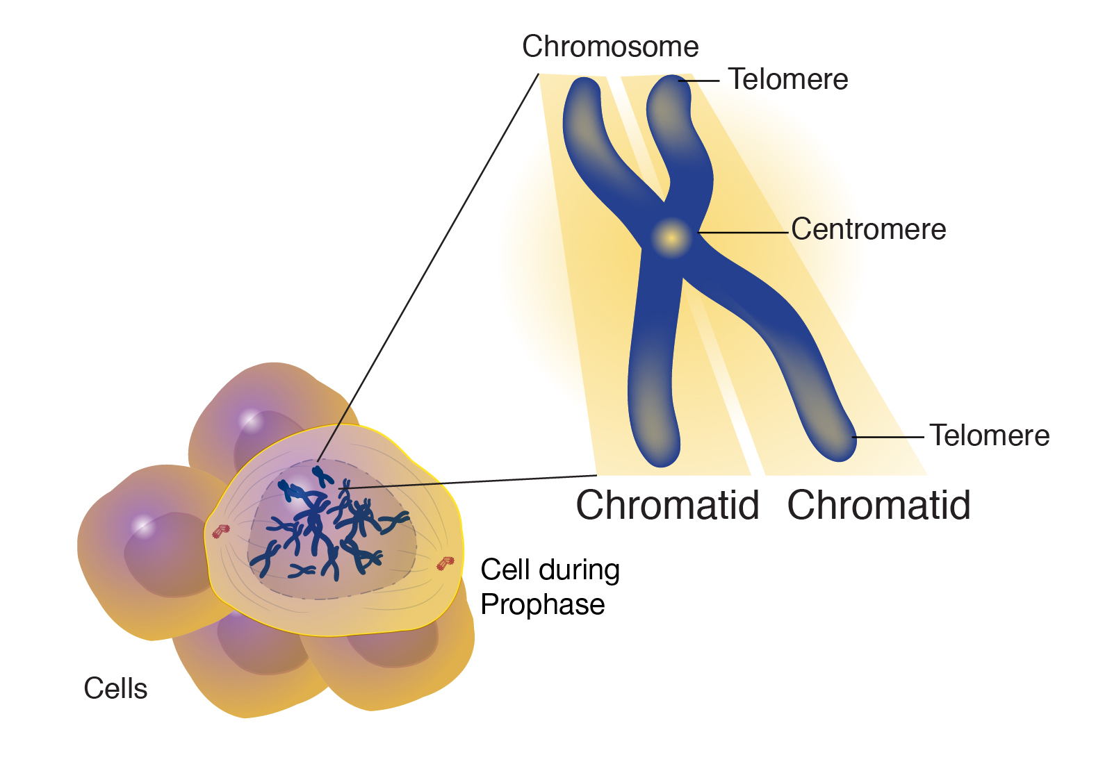
During cell division, spindle fibers attach to the centromere and pull each of the sister chromatids to. For example, humans are diploid (2n) and have 46 chromosomes in their normal body cells. Meanwhile, changes in microtubule length. Prior to cell division, chromosomes are copied and identical chromosome copies join together at their centromeres. Long arm is termed q. Web figure 8.4.3 8.4. Each strand of one of these chromosomes is a chromatid. Web what is a chromatid? Histones are a family of small, positively charged proteins termed h1, h2a, h2b, h3, and h4 (van holde, 1988). A chromatid is one of the two identical halves of a chromosome that has been replicated in preparation for cell division.
Upon separation, every chromatid becomes an independent chromosome. Prior to cell division, chromosomes are copied and identical chromosome copies join together at their centromeres. Web figure 8.4.3 8.4. A chromatid is one of the two identical halves of a chromosome that has been replicated in preparation for cell division. Drawing of chromosomes during mitosis by walther flemming, circa 1880. A chromatid is one half of a replicated chromosome. Web as a result, chromatin can be packaged into a much smaller volume than dna alone. Chromosomes:a threadlike structure of nucleic acids and protein found in the nucleus of most living cells, carrying genetic information in the form of genes. Diagram of replicated and condensed eukaryotic chromosome (sister chromatids). Histones are a family of small, positively charged proteins termed h1, h2a, h2b, h3, and h4 (van holde, 1988).

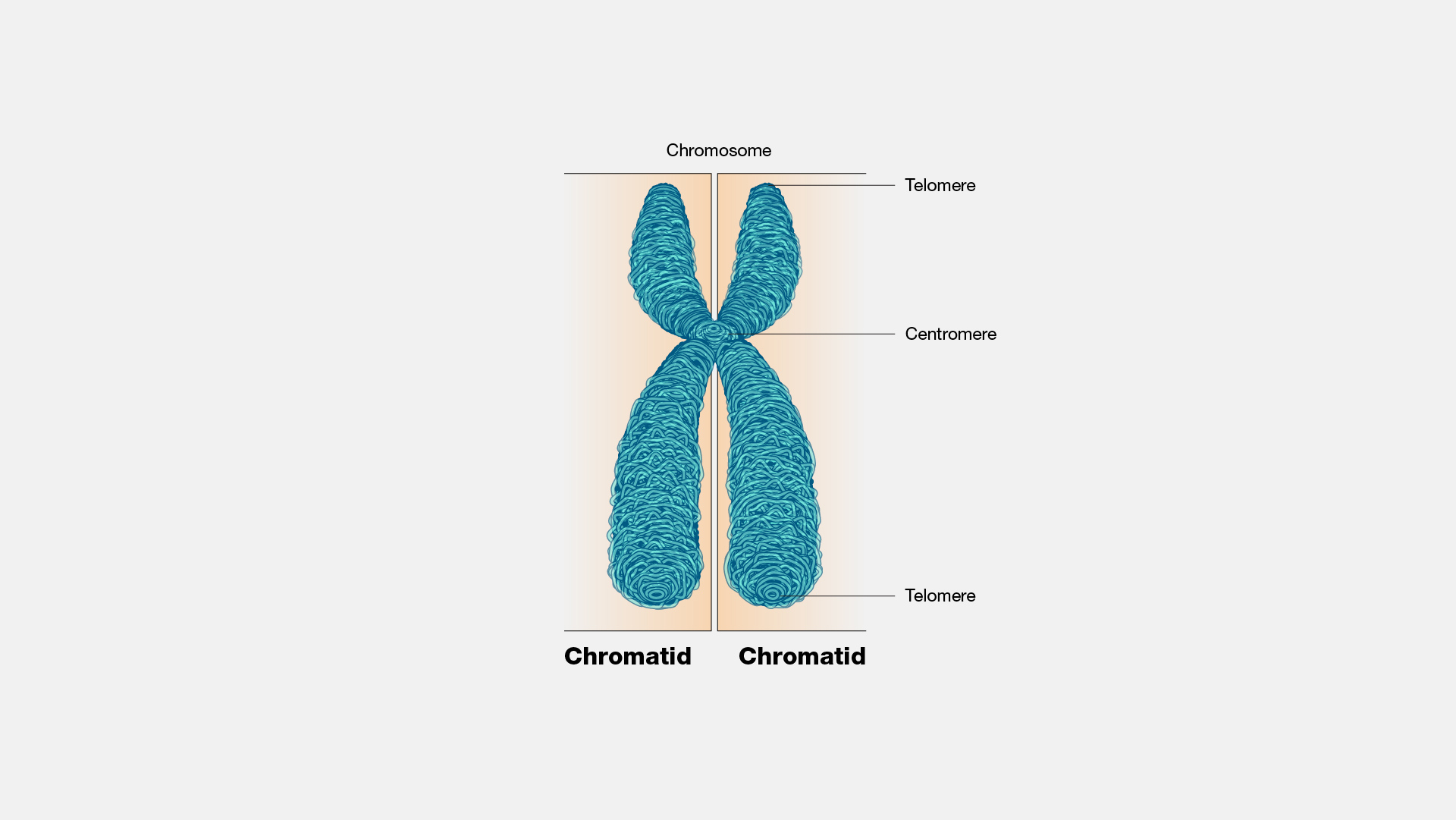
Structure of a chromosome showing two identical chromatids each made up
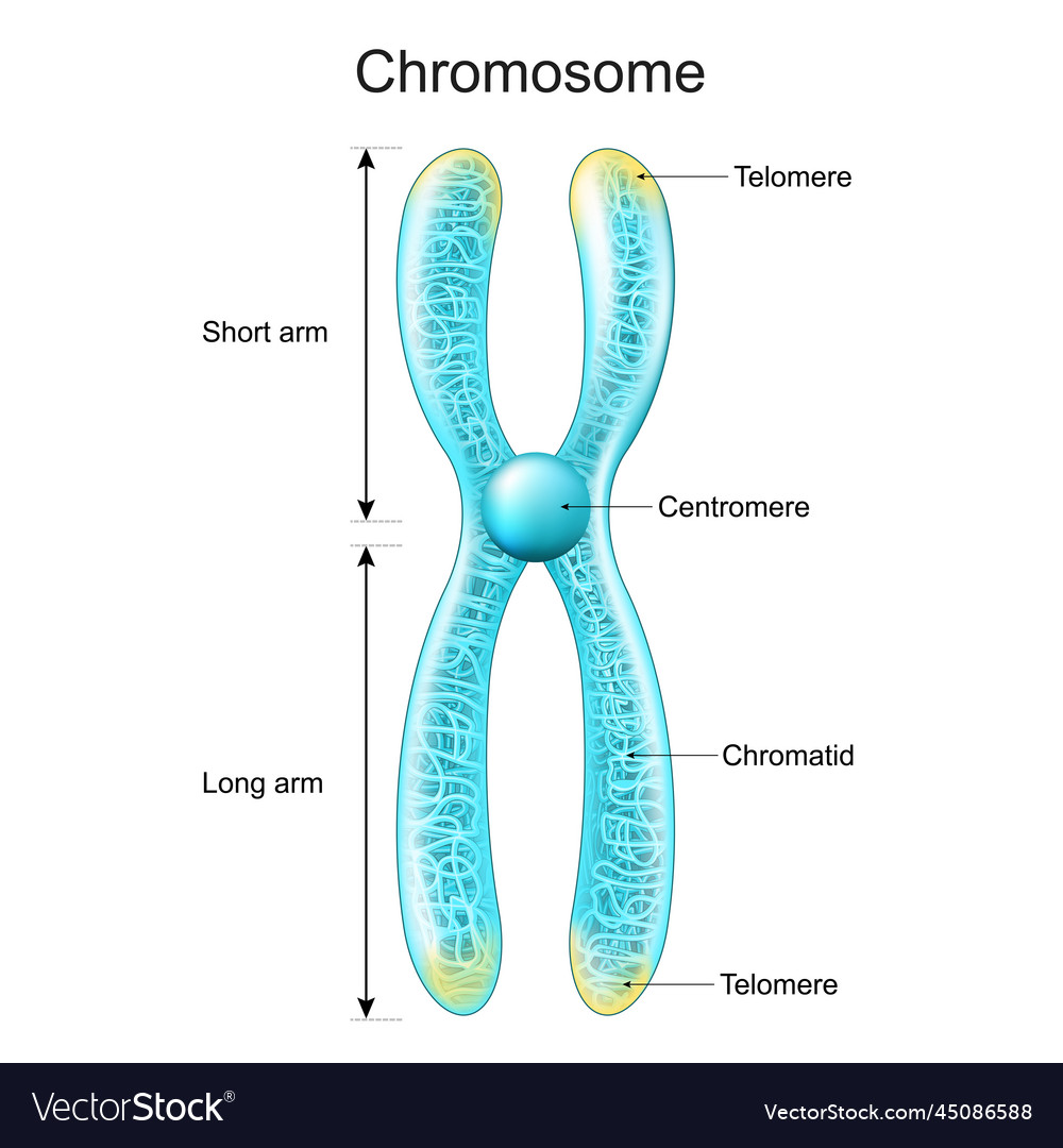
(3) short arm is termed p; Chromatid:each of the two threadlike strands into which a chromosome divides longitudinally during cell division. Prior to cell division, chromosomes are copied and identical chromosome copies join together at their centromeres. Drawing of chromosomes during mitosis by walther flemming, circa 1880. Web as a result, chromatin can be packaged into a much smaller volume.
Chromatid
Meanwhile, changes in microtubule length. Start practicing—and saving your progress—now: Web figure 8.4.3 8.4. Different species have different numbers of chromosomes. The sex cells of a human are haploid (n), containing only one.
Draw the structure of the chromosome and label its parts.
Drawing of chromosomes during mitosis by walther flemming, circa 1880. Upon separation, every chromatid becomes an independent chromosome. Meanwhile, changes in microtubule length. 22 pairs of autosomes and 1 pair of sex chromosomes. Web as a result, chromatin can be packaged into a much smaller volume than dna alone.

At The Beginning Of Cell Division Each Chromosome Consists Of Two
Web as a result, chromatin can be packaged into a much smaller volume than dna alone. Drawing of chromosomes during mitosis by walther flemming, circa 1880. Web a major reason for chromatid separation is the precipitous degradation of the cohesin molecules joining the sister chromatids by the protease separase (figure 10). Joined chromatids are known as sister chromatids. Diagram of.
Sister Chromatids Definition, Formation, Separation, Functions
22 pairs of autosomes and 1 pair of sex chromosomes. A chromatid is one half of a replicated chromosome. Start practicing—and saving your progress—now: Histones are a family of small, positively charged proteins termed h1, h2a, h2b, h3, and h4 (van holde, 1988). Chromosomes:a threadlike structure of nucleic acids and protein found in the nucleus of most living cells, carrying.
Structure of chromosome chromatid centromere Vector Image
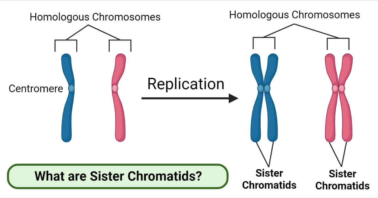
The two copies of the cell’s original chromosome are called “sister chromatids.”. Each strand of one of these chromosomes is a chromatid. Histones are a family of small, positively charged proteins termed h1, h2a, h2b, h3, and h4 (van holde, 1988). Start practicing—and saving your progress—now: Web what is a chromatid?
3.2 Chromosomes The Biology Classroom
The two copies of the cell’s original chromosome are called “sister chromatids.”. Long arm is termed q. Web a major reason for chromatid separation is the precipitous degradation of the cohesin molecules joining the sister chromatids by the protease separase (figure 10). Chromatid:each of the two threadlike strands into which a chromosome divides longitudinally during cell division. The sex cells.
labelled diagram of chromosome RosieAreebah
Web figure 8.4.3 8.4. Web what is a chromatid? For example, humans are diploid (2n) and have 46 chromosomes in their normal body cells. Long arm is termed q. These 46 chromosomes are organized into 23 pairs:

ChromatidStructure, Types, Characteristics, & FAQs
Web courses on khan academy are always 100% free. A chromatid is one half of a replicated chromosome. When a cell is preparing to divide, it makes a new copy of all of its dna, so that the cell now possesses two copies of each chromosome. Joined chromatids are known as sister chromatids. Web figure 8.4.3 8.4.
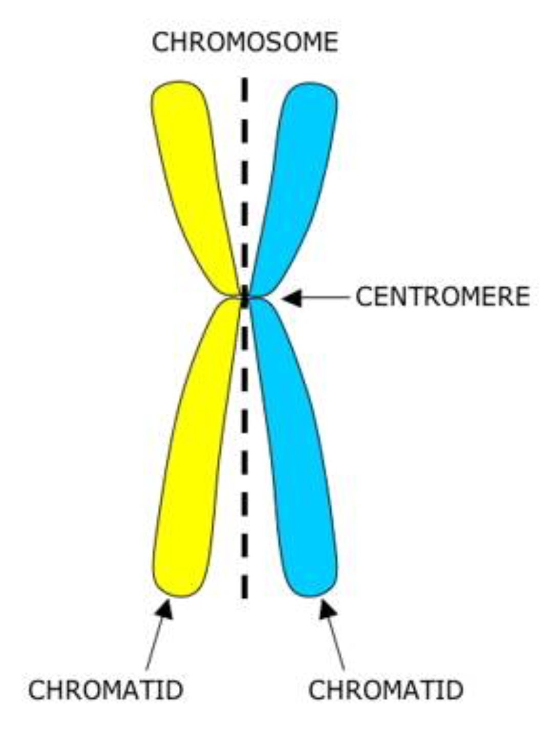
What is the name of the structure that connects the two chromatids
Web what is a chromatid? Web as a result, chromatin can be packaged into a much smaller volume than dna alone. Web a major reason for chromatid separation is the precipitous degradation of the cohesin molecules joining the sister chromatids by the protease separase (figure 10). Histones are a family of small, positively charged proteins termed h1, h2a, h2b, h3,.
Web As A Result, Chromatin Can Be Packaged Into A Much Smaller Volume Than Dna Alone.
These 46 chromosomes are organized into 23 pairs: Start practicing—and saving your progress—now: During anaphase of cell division, the two chromatids will be pulled apart, and chromatid will be. Web what is a chromatid?
22 Pairs Of Autosomes And 1 Pair Of Sex Chromosomes.
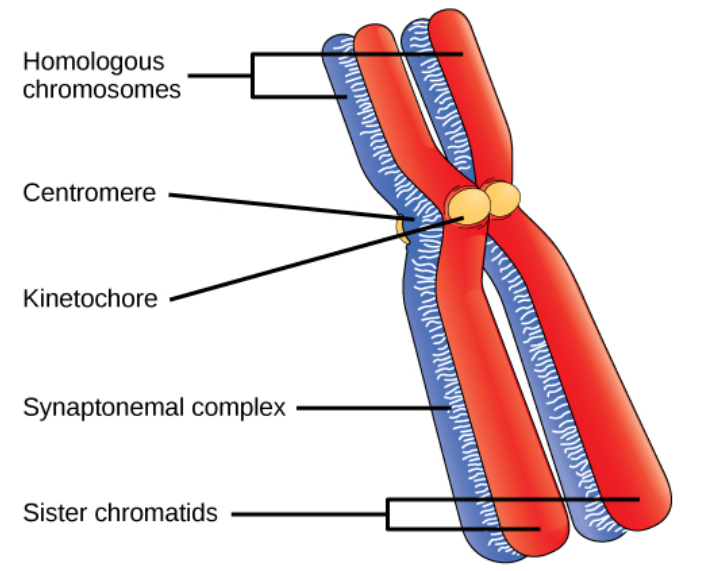
Web figure 8.4.3 8.4. The two “sister” chromatids are joined at a constricted region of the chromosome called the centromere. When a cell is preparing to divide, it makes a new copy of all of its dna, so that the cell now possesses two copies of each chromosome. The sex cells of a human are haploid (n), containing only one.
Chromosomes:a Threadlike Structure Of Nucleic Acids And Protein Found In The Nucleus Of Most Living Cells, Carrying Genetic Information In The Form Of Genes.
For example, humans are diploid (2n) and have 46 chromosomes in their normal body cells. Prior to cell division, chromosomes are copied and identical chromosome copies join together at their centromeres. Drawing of chromosomes during mitosis by walther flemming, circa 1880. Long arm is termed q.
Diagram Of Replicated And Condensed Eukaryotic Chromosome (Sister Chromatids).
Histones are a family of small, positively charged proteins termed h1, h2a, h2b, h3, and h4 (van holde, 1988). Different species have different numbers of chromosomes. Chromatid:each of the two threadlike strands into which a chromosome divides longitudinally during cell division. Each strand of one of these chromosomes is a chromatid.