Chromosomes Drawing
Chromosomes Drawing - These are termed as chromomeres. Web dna structure and function. Web drawing a karyogram online. Redraw the nuclear membrane around the chromosomes and draw a nucleolus inside of each nucleus. The sex cells of a human are haploid (n), containing only one. (3)the visualization results are finally stored into an image file and displayed on the webpage. These instructions are stored inside each of your cells, distributed among 46 long structures called chromosomes. Meiosis involves two divisions, so it’s typically broken down into meiosis i and meiosis ii. Eukaryotic cells, with their much larger genomes, have multiple, linear chromosomes. Chromosome 1 is the largest and is over three times bigger than chromosome 22.
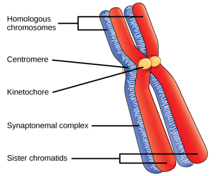
Web the drawing of a genetic map is decomposed into 8 modules inmg2c program as follows: In metaphase i, chromosomes line up in the middle of the cell. Human sperm and eggs, which have only one homologous chromosome from each pair, are said to be haploid ( 1n ). When a sperm and an egg join in fertilization, the two haploid sets of chromosomes. Anaphase i separates homologous pairs, while telophase i forms two new cells with a. Web to put that another way, meiosis in humans is a division process that takes us from a diploid cell—one with two sets of chromosomes—to haploid cells—ones with a single set of chromosomes. Web the 46 chromosomes of a human cell are organized into 23 pairs, and the two members of each pair are said to be homologues of one another (with the slight exception of the x and y chromosomes; Web chromosomes undergo segregation and independent assortment during meiosis. Note that each daughter cell has half the number of chromosomes as. These paired chromosomes are called homologous.
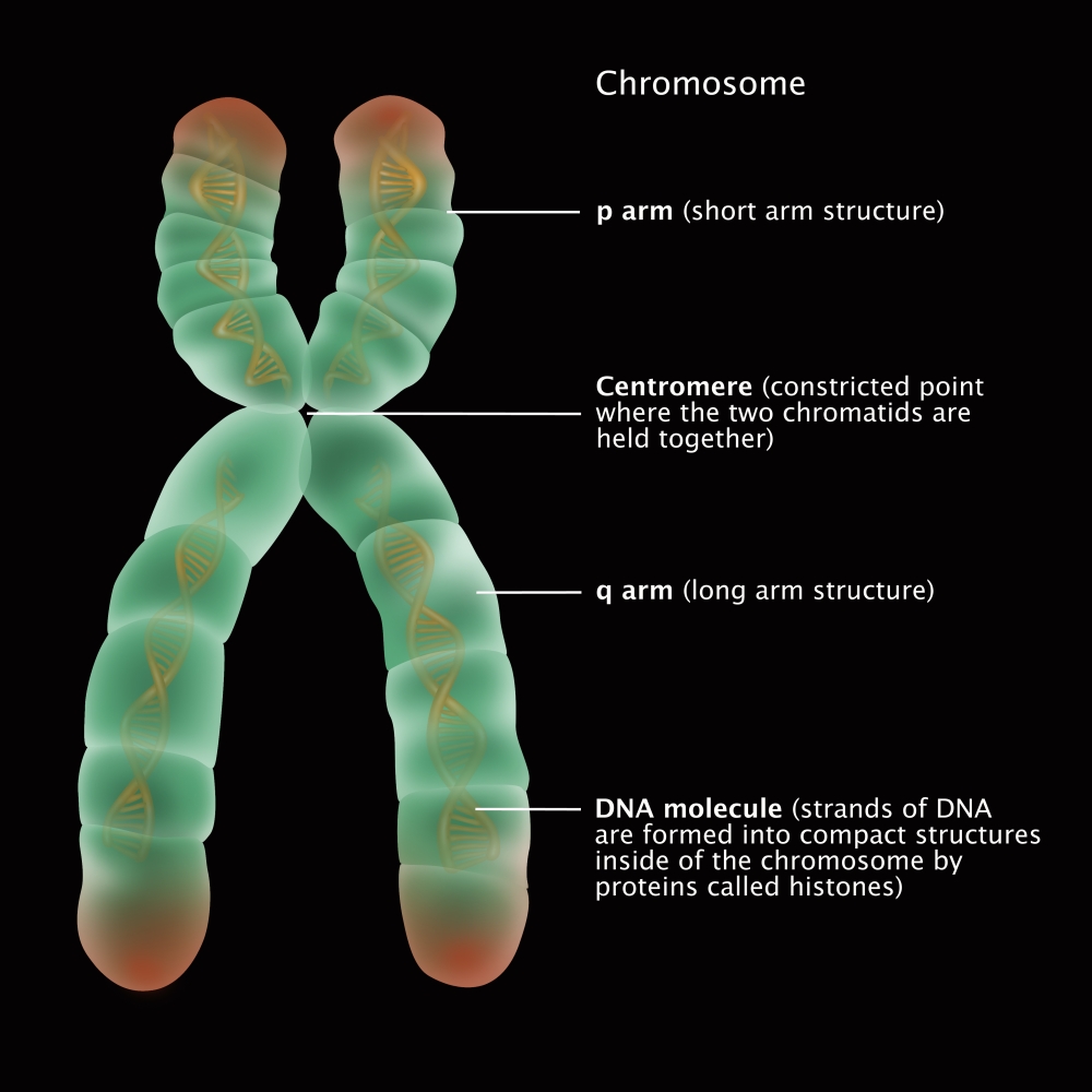
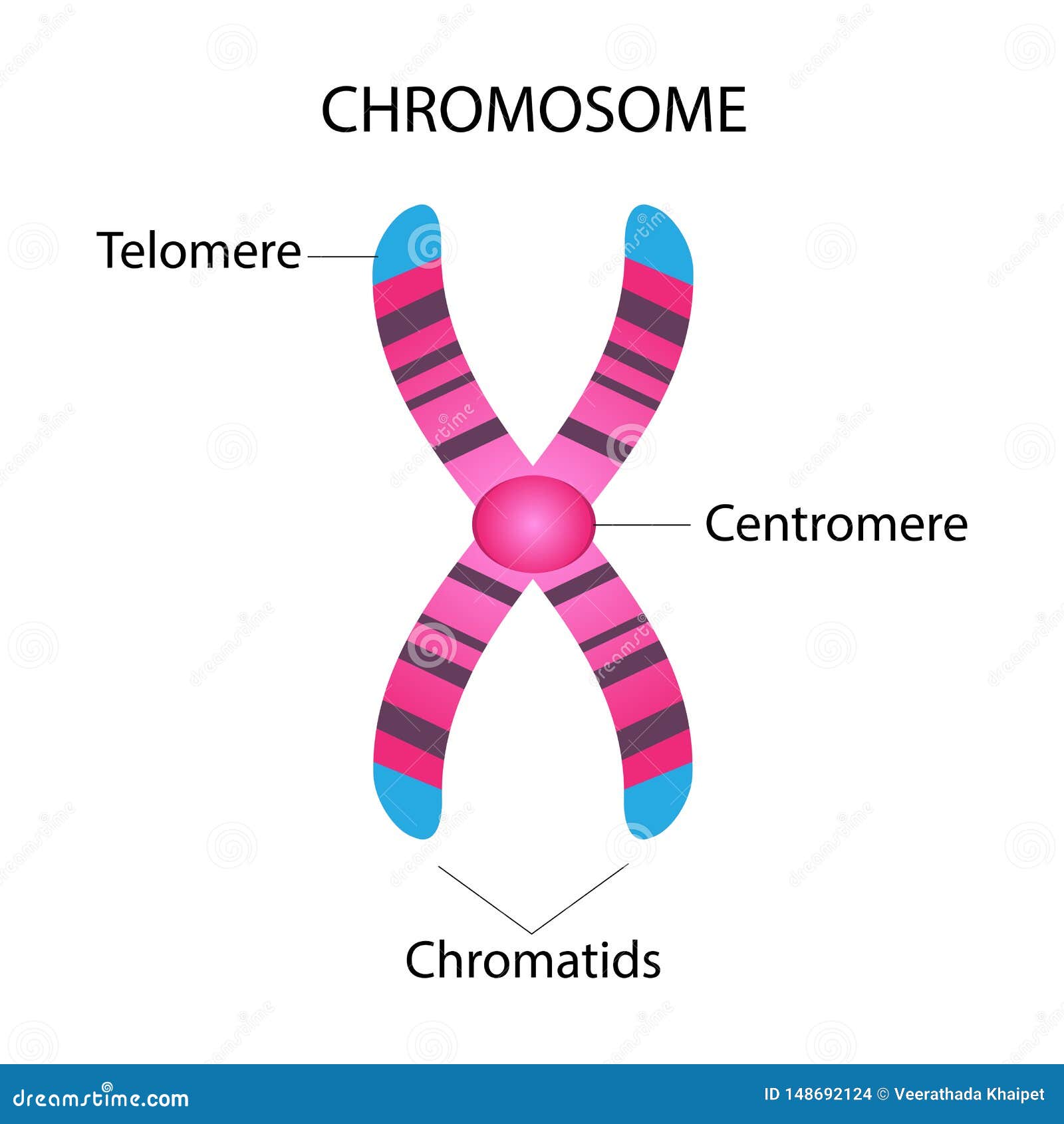
Chromosomes:a threadlike structure of nucleic acids and protein found in the nucleus of most living cells, carrying genetic information in the form of genes. Anaphase i separates homologous pairs, while telophase i forms two new cells with a. In metaphase i, chromosomes line up in the middle of the cell. Web during mitosis, chromosomes become attached to the structure known as the mitotic spindle.in the late 1800s, theodor boveri created the earliest detailed drawings of the spindle based on his. The length and linear nature of eukaryotic chromosomes increase the challenge of keeping the genetic material. Prophase i, metaphase i, anaphase i, and telophase i. 22 pairs of autosomes and 1 pair of sex chromosomes. Web model cytokinesis l by drawing the formation of a cleavage furrow to divide the cytoplasm into two and form two separate cells. Chromatid:each of the two threadlike strands into which a chromosome divides longitudinally during cell division. The position of the centromere, which separates the p and q arms, is shown by the hatched area.

Parts of Chromosome Diagram Quizlet
Web to put that another way, meiosis in humans is a division process that takes us from a diploid cell—one with two sets of chromosomes—to haploid cells—ones with a single set of chromosomes. By mapping segments of dna to chromosomes, we can begin to see which ancestors gave us which pieces of dna, and thus how new matches are related..

how to draw chromosomes in easy way drawing chromosomes step by step
Chromatid:each of the two threadlike strands into which a chromosome divides longitudinally during cell division. These paired chromosomes are called homologous. Our genetic information is stored in 23 pairs of chromosomes that vary widely in size and shape. Web drawing derivative chromosomes online. These 46 chromosomes are organized into 23 pairs:

Human Chromosome Drawing Stock Illustration Download Image Now
As a result, dna painter has quickly become an essential tool for genealogists!”. Human sperm and eggs, which have only one homologous chromosome from each pair, are said to be haploid ( 1n ). Different species have different numbers of chromosomes. The length and linear nature of eukaryotic chromosomes increase the challenge of keeping the genetic material. These chromosomes are.

Chromosome Structure
Females have a pair of x chromosomes (46, xx),. Svg container, single chromosome container, chromosome, chromosome id, gene lines, gene id, connection and scale. Chromosome 1 is the largest and is over three times bigger than chromosome 22. For example, humans have 46 chromosomes in a typical body cell. These are termed as chromomeres.
Chromosome Structure, Illustration Poster Print by Gwen Shockey/Science
Each chromosome is made of protein and a single molecule of deoxyribonucleic acid (dna). For example, humans have 46 chromosomes in a typical body cell. As a result, dna painter has quickly become an essential tool for genealogists!”. In humans, the haploid cells made in meiosis are sperm and eggs. This simple worksheet shows a diagram of a chromosome and.
Draw the structure of the chromosome and label its parts.
Long strands of dna wind around proteins called histones, giving rise to a “beads on a string” structure. The sex cells of a human are haploid (n), containing only one. Redraw the nuclear membrane around the chromosomes and draw a nucleolus inside of each nucleus. Usually, the centromere lies within the primary constriction (thinner chromosomal. During prophase i, chromosomes pair.
Parts Of A Chromosome
These two cells will now enter meiosis ll. In humans, the haploid cells made in meiosis are sperm and eggs. Svg container, single chromosome container, chromosome, chromosome id, gene lines, gene id, connection and scale. Many species have chromosomes that come in matched pairs. These paired chromosomes are called homologous.

Drawing dna molecule chromosome biology Vector Image
Web drawing a karyogram online. This simple worksheet shows a diagram of a chromosome and where it is located in the nucleus of the cell. Different species have different numbers of chromosomes. Each chromosome is made of protein and a single molecule of deoxyribonucleic acid (dna). It is crucial for sexual reproduction in eukaryotes.

How to draw TYPES OF CHROMOSOMES easily Class 11 Biology YouTube
Most prokaryotic cells contain a single circular chromosome. These processes contribute to genetic diversity by shuffling the genes found on the chromosomes. Long strands of dna wind around proteins called histones, giving rise to a “beads on a string” structure. Web the 46 chromosomes of a human cell are organized into 23 pairs, and the two members of each pair.
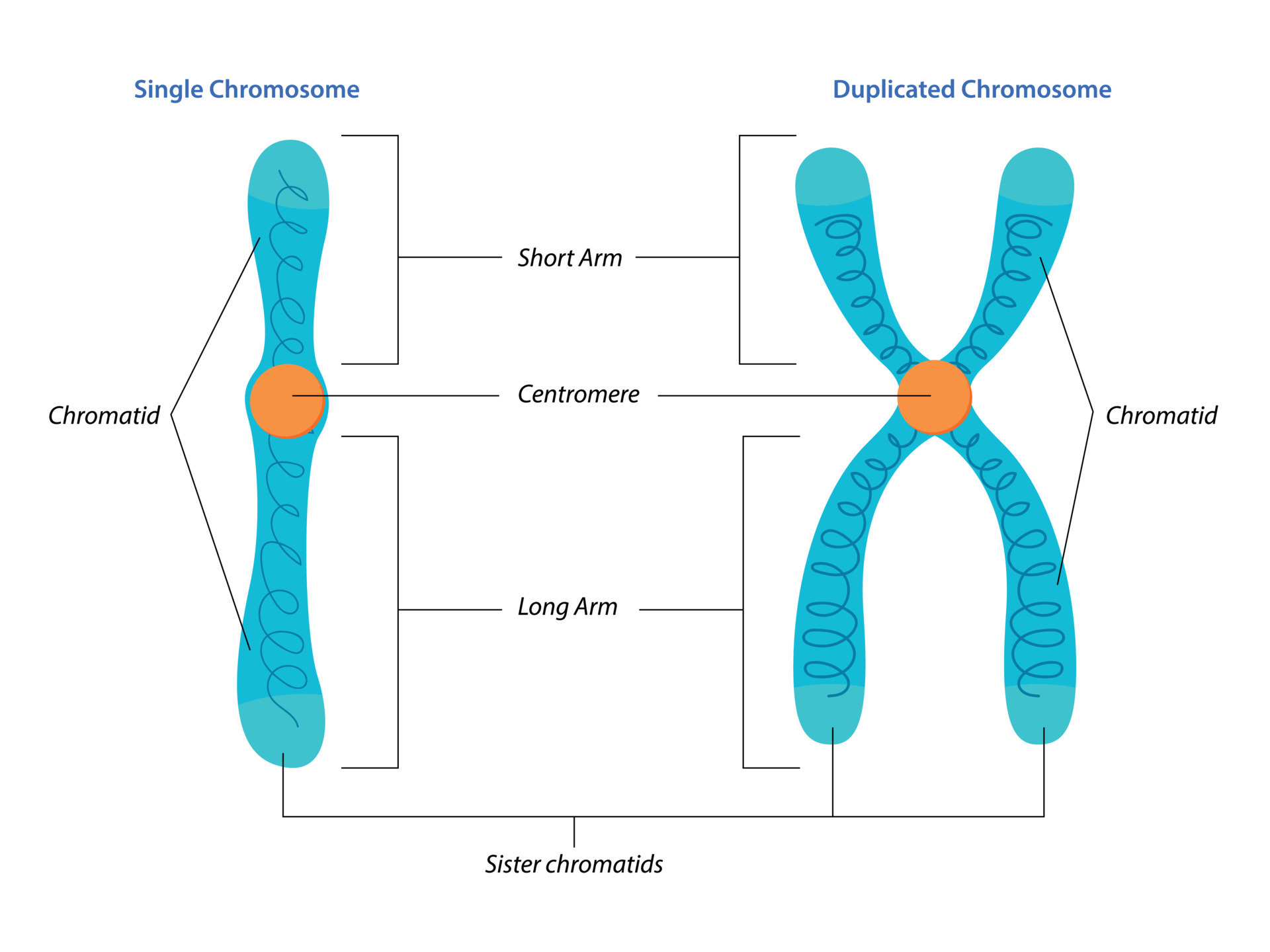
Illustration of Singel and duplicated chromosome structure 12324913
Web the 46 chromosomes of a human cell are organized into 23 pairs, and the two members of each pair are said to be homologues of one another (with the slight exception of the x and y chromosomes; Different species have different numbers of chromosomes. Web in meiosis i, cells go through four phases: Web to put that another way,.
Anaphase I Separates Homologous Pairs, While Telophase I Forms Two New Cells With A.
(3)the visualization results are finally stored into an image file and displayed on the webpage. 22 pairs of autosomes and 1 pair of sex chromosomes. Our genetic information is stored in 23 pairs of chromosomes that vary widely in size and shape. These paired chromosomes are called homologous.
The Sex Cells Of A Human Are Haploid (N), Containing Only One.
Chromosomes:a threadlike structure of nucleic acids and protein found in the nucleus of most living cells, carrying genetic information in the form of genes. For example, humans have 46 chromosomes in a typical body cell. For example, humans are diploid (2n) and have 46 chromosomes in their normal body cells. These chromosomes are made up of thousands of shorter segments of dna, called genes.
These Instructions Are Stored Inside Each Of Your Cells, Distributed Among 46 Long Structures Called Chromosomes.
Redraw the nuclear membrane around the chromosomes and draw a nucleolus inside of each nucleus. During prophase i, chromosomes pair up and exchange genetic material, creating more variation. Web dna structure and function. The position of the centromere, which separates the p and q arms, is shown by the hatched area.
Web During Mitosis, Chromosomes Become Attached To The Structure Known As The Mitotic Spindle.in The Late 1800S, Theodor Boveri Created The Earliest Detailed Drawings Of The Spindle Based On His.
In metaphase i, chromosomes line up in the middle of the cell. Usually, the centromere lies within the primary constriction (thinner chromosomal. Dna is the information molecule. Passed from parents to offspring, dna contains the specific instructions that make each type of living creature unique.