Cochlea Drawing
Cochlea Drawing - Web the schematic drawing below represents the osseous (top left) and membranous (seen by transparency in the main drawing) labyrinths. Drawing the last panel of cochlea & eustachia page 64. Cochlea diagram stock photos are available in a variety of sizes and formats to fit your needs. Audiologist gift inner ear anatomy art cochlea histology medical art doctor gift clinic wall decor. The eustachian tube (seen on the drawing above) links the middle ear cavity to the pharynx, allowing an equal pressure on both sides of the eardrum. It is a hollow, spirally coiled chamber inside the temporal bone that makes 2.75 turns around its axis, which is called the modiolus. It plays a vital role in the function of hearing rather than simply being another component of the skeletal system. Anatomy of the human eye and ear, published in 1861. The scala vestibuli, scala tympani, and scala media ( cochlear duct ). 576 views 7 years ago.
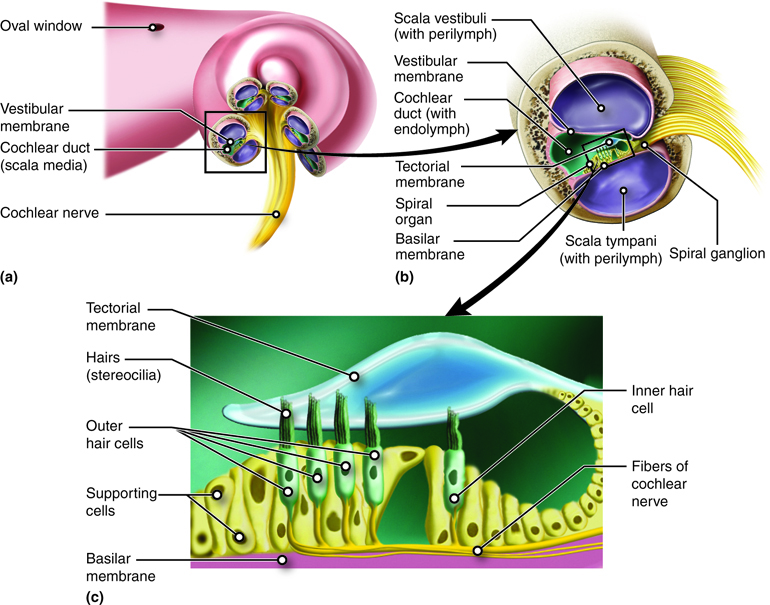
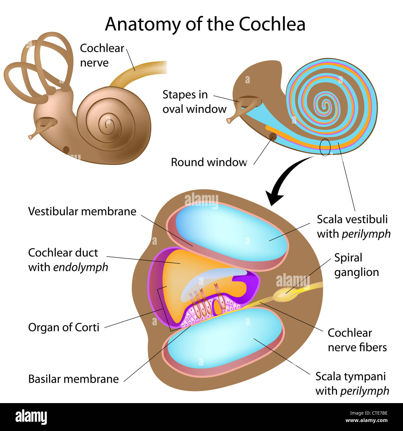
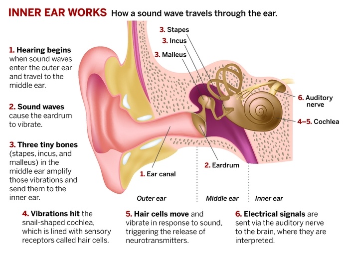
Cochleae) is a spiraled, hollow, conical chamber of bone, in which waves propagate from the base (near the middle ear and the oval window) to the apex (the top or. The cochlea contains hair cells that convert sound waves into electrical impulses that are carried along the auditory nerve to the brain. It plays a vital role in the function of hearing rather than simply being another component of the skeletal system. It is divided longitudinally into three parallel chambers or scalae: It is a hollow, spirally coiled chamber inside the temporal bone that makes 2.75 turns around its axis, which is called the modiolus. Web the cochlea is a component of the labyrinth of the internal ear that is responsible for hearing. In greek, cochlea means snail, which suites this structure that resembles a snail. Sound waves are transduced into electrical impulses that the brain can interpret as individual sound frequencies. It forms a cone approximately 9 mm (0.35 inch) in diameter at its base and 5 mm in height. Audiologist gift inner ear anatomy art cochlea histology medical art doctor gift clinic wall decor.
Cochlea) is a bony canal within the internal ear that forms a spiral shape, making 2.5 turns around its axis. It forms a cone approximately 9 mm (0.35 inch) in diameter at its base and 5 mm in height. The scala vestibuli, scala tympani, and scala media ( cochlear duct ). It is a hollow, spirally coiled chamber inside the temporal bone that makes 2.75 turns around its axis, which is called the modiolus. The cochlea contains hair cells that convert sound waves into electrical impulses that are carried along the auditory nerve to the brain. Web the cochlea is a component of the labyrinth of the internal ear that is responsible for hearing. 576 views 7 years ago. Web a similar transfer of force can be seen with a drawing pin: Web the cochlear canal begins in the floor of the vestibule. Web structural diagram of the cochlea showing how fluid pushed in at the oval window moves, deflects the cochlear partition, and bulges back out at the round window.

Inner ear cochlea crosssection of one spiral Vector Image
Drawing the last panel of cochlea & eustachia page 64. Your acoustic nerve, or cochlear nerve, processes auditory information from the inner ear to your brain. The cochlea contains hair cells that convert sound waves into electrical impulses that are carried along the auditory nerve to the brain. It is a hollow, spirally coiled chamber inside the temporal bone that.
Hearing and Equilibrium Anatomy and Physiology
Your acoustic nerve, or cochlear nerve, processes auditory information from the inner ear to your brain. Audiologist gift inner ear anatomy art cochlea histology medical art doctor gift clinic wall decor. Human ear anatomy art, medical artwork, audiology poster, watercolor drawing, inner ear. Web the schematic drawing below represents the osseous (top left) and membranous (seen by transparency in the.
Anatomy of the cochlea of human ear Stock Photo Alamy
Web the cochlear canal begins in the floor of the vestibule. It is divided longitudinally into three parallel chambers or scalae: It forms a cone approximately 9 mm (0.35 inch) in diameter at its base and 5 mm in height. The scala vestibuli, scala tympani, and scala media ( cochlear duct ). Cochleae) is a spiraled, hollow, conical chamber of.
Cochlea Key Stage Wiki
In greek, cochlea means snail, which suites this structure that resembles a snail. Drawing the last panel of cochlea & eustachia page 64. Web structural diagram of the cochlea showing how fluid pushed in at the oval window moves, deflects the cochlear partition, and bulges back out at the round window. This motion triggers hair cells, causing potassium and calcium.
The Cochlea of the Ear ClipArt ETC
Web the cochlear canal begins in the floor of the vestibule. Web browse 279 cochlea anatomy photos and images available, or start a new search to explore more photos and images. Web the schematic drawing below represents the osseous (top left) and membranous (seen by transparency in the main drawing) labyrinths. 576 views 7 years ago. It plays a vital.
Hearing, Transmission of sound waves in Cochlea, Functions of Cochlea
Cochlea) is a bony canal within the internal ear that forms a spiral shape, making 2.5 turns around its axis. Web the schematic drawing below represents the osseous (top left) and membranous (seen by transparency in the main drawing) labyrinths. Web a similar transfer of force can be seen with a drawing pin: When stretched out, the spiral tube is.

Anatomy of the Cochlea. Cartoon illustration of the cochlea. Panel a. A
Web structural diagram of the cochlea showing how fluid pushed in at the oval window moves, deflects the cochlear partition, and bulges back out at the round window. The ear (human anatomy) medical ilustration showing endolymph in the membranous labyrinth of the inner ear. Anatomy of the human eye and ear, published in 1861. Web the schematic drawing below represents.

Ear structures Cochlea, Vestibule, Semicircular canals Diagram Quizlet
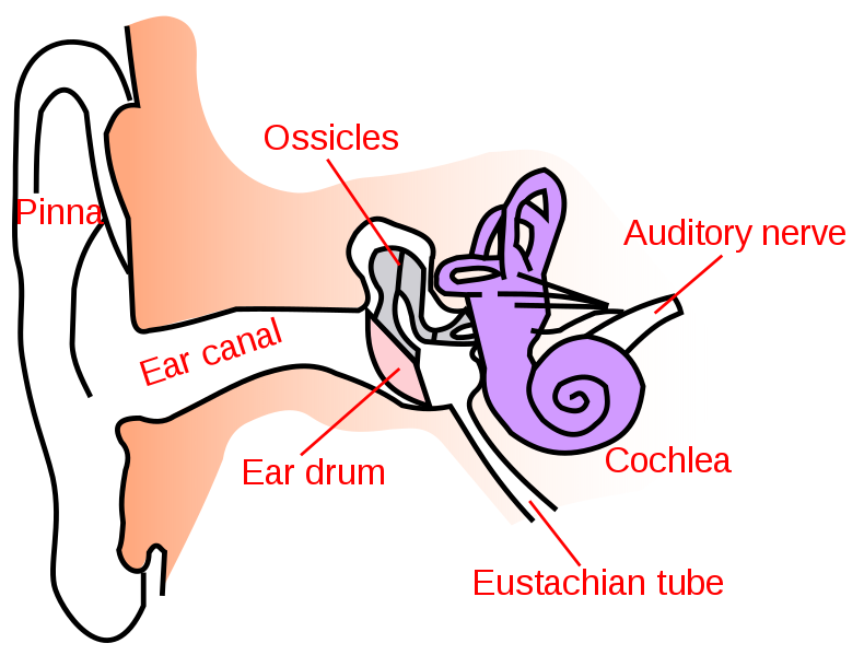
The ear (human anatomy) medical ilustration showing endolymph in the membranous labyrinth of the inner ear. Frontal section through the external, middle, and internal ear. Human ear anatomy art, medical artwork, audiology poster, watercolor drawing, inner ear. Web the schematic drawing below represents the osseous (top left) and membranous (seen by transparency in the main drawing) labyrinths. The scala vestibuli,.
Drawing shows the normal anatomy of the inner ear the cochlea (C
Damage to this nerve can cause hearing loss. This motion triggers hair cells, causing potassium and calcium to flow into the cell, firing an action potential. The cochlea contains hair cells that convert sound waves into electrical impulses that are carried along the auditory nerve to the brain. Web browse 279 cochlea anatomy photos and images available, or start a.
/Ear-GettyImages-586038190-42999e6443b441d5876c5e3c5dd640cf.jpg)
Cochlea Anatomy, Function, and Treatment
This motion triggers hair cells, causing potassium and calcium to flow into the cell, firing an action potential. Web a similar transfer of force can be seen with a drawing pin: Cochleae) is a spiraled, hollow, conical chamber of bone, in which waves propagate from the base (near the middle ear and the oval window) to the apex (the top.
Web Structural Diagram Of The Cochlea Showing How Fluid Pushed In At The Oval Window Moves, Deflects The Cochlear Partition, And Bulges Back Out At The Round Window.
The stapes bone vibrates, pushing fluid through the cochlea, which moves the organ of corti. Human ear anatomy art, medical artwork, audiology poster, watercolor drawing, inner ear. Cochlea) is a bony canal within the internal ear that forms a spiral shape, making 2.5 turns around its axis. Sound waves are transduced into electrical impulses that the brain can interpret as individual sound frequencies.
It Forms A Cone Approximately 9 Mm (0.35 Inch) In Diameter At Its Base And 5 Mm In Height.
This motion triggers hair cells, causing potassium and calcium to flow into the cell, firing an action potential. The cochlea contains hair cells that convert sound waves into electrical impulses that are carried along the auditory nerve to the brain. The scala vestibuli, scala tympani, and scala media ( cochlear duct ). It plays a vital role in the function of hearing rather than simply being another component of the skeletal system.
Damage To This Nerve Can Cause Hearing Loss.
When stretched out, the spiral tube is approximately 30 mm in length. Your acoustic nerve, or cochlear nerve, processes auditory information from the inner ear to your brain. Cochlea diagram stock photos are available in a variety of sizes and formats to fit your needs. Cochleae) is a spiraled, hollow, conical chamber of bone, in which waves propagate from the base (near the middle ear and the oval window) to the apex (the top or.
It Is Divided Longitudinally Into Three Parallel Chambers Or Scalae:
576 views 7 years ago. In greek, cochlea means snail, which suites this structure that resembles a snail. Web a similar transfer of force can be seen with a drawing pin: The eustachian tube (seen on the drawing above) links the middle ear cavity to the pharynx, allowing an equal pressure on both sides of the eardrum.