Draw A Diagram Of The Heart
Draw A Diagram Of The Heart - One chamber on the right receives blood with waste (from the body) and another chamber pumps it out toward the lungs where the waste is exhaled. The maximum induced emf generated in the coil is. This thick layer is the muscle that contracts to pump and propel blood. Includes an exercise, review worksheet, quiz, and model drawing of an anterior view (frontal section) of the heart in. Web in this lecture, dr mike shows the two best ways to draw and label the heart! Start your sketch at the edge of the superior vena cava, and work the shape down to the top edge of the heart’s body. Valves are present to prevent the backflow of blood. To find a good diagram, go to google images, and type in the internal structure of the human heart. Find an image that displays the entire heart, and click on it to enlarge it. Web heart, organ that serves as a pump to circulate the blood.
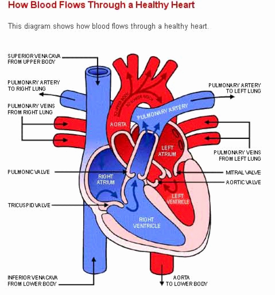
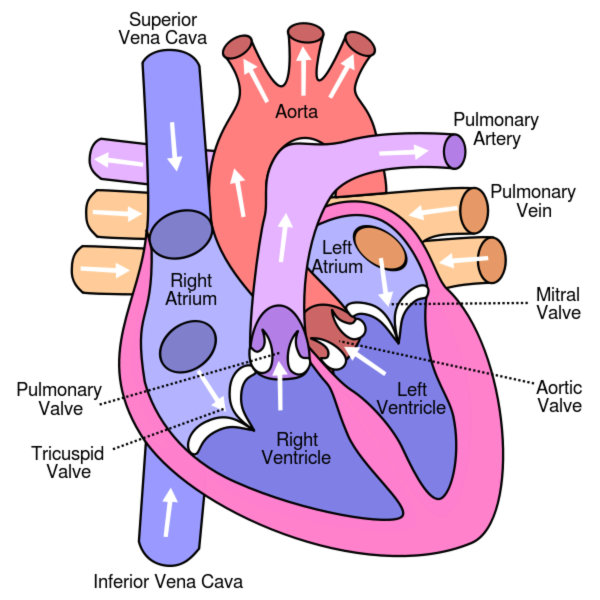
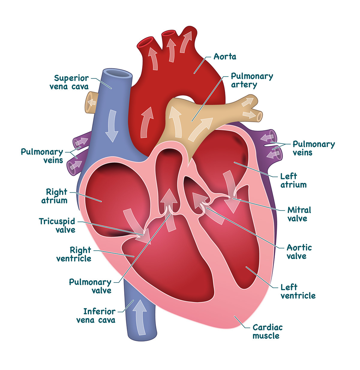
Valves are present to prevent the backflow of blood. This thick layer is the muscle that contracts to pump and propel blood. In fishes the heart is a folded tube, with three or four enlarged areas that correspond. One chamber on the right receives blood with waste (from the body) and another chamber pumps it out toward the lungs where the waste is exhaled. Web the $130,000 payment arranged by trump's personal lawyer and fixer, michael cohen, is at the heart of the first criminal trial in history against a former president. The maximum induced emf generated in the coil is. To find a good diagram, go to google images, and type in the internal structure of the human heart. Web the heart has three layers. The right margin is the small section of the right atrium that extends between the superior and inferior vena cava. Web function and anatomy of the heart made easy using labeled diagrams of cardiac structures and blood flow through the atria, ventricles, valves, aorta, pulmonary arteries veins, superior inferior vena cava, and chambers.
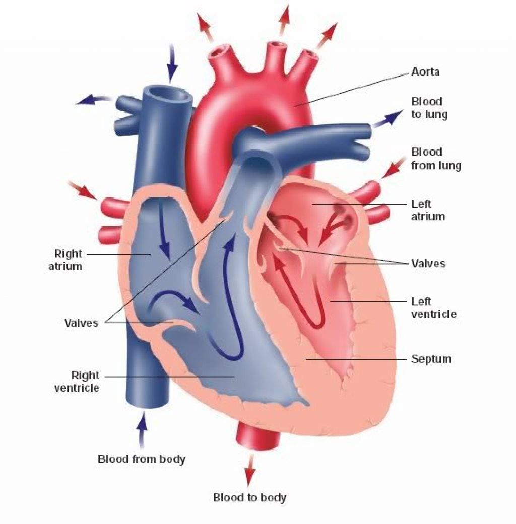
Web heart, organ that serves as a pump to circulate the blood. The aorta, vena cava, pulmonary trunk, and pulmonary veins. Web the heart blood flow diagram (flowchart) given below will help you to understand the pathway of blood through the heart.initial five points denotes impure or deoxygenated blood and the last five points denotes pure or oxygenated blood. The top of the heart, known as the heart’s base, connects to the great blood vessels of the body: Clara was 8 when she crafted the diagram in 2023. Web function and anatomy of the heart made easy using labeled diagrams of cardiac structures and blood flow through the atria, ventricles, valves, aorta, pulmonary arteries veins, superior inferior vena cava, and chambers. The diagram of heart is beneficial for class 10 and 12 and is frequently. Web the bottom tip of the heart, known as its apex, is turned to the left, so that about 2/3 of the heart is located on the body’s left side with the other 1/3 on right. Drag and drop the text labels onto the boxes next to the diagram. The maximum induced emf generated in the coil is.
Human Heart Drawing Simple at Explore collection
It may be a straight tube, as in spiders and annelid worms, or a somewhat more elaborate structure with one or more receiving chambers (atria) and a main pumping chamber (ventricle), as in mollusks. Find an image that displays the entire heart, and click on it to enlarge it. The top of the heart, known as the heart’s base, connects.

How to Draw the Internal Structure of the Heart 13 Steps
It pumps blood from the heart to different parts of the body and back to the heart. Step 1 and 6 involve a blood vessel, which makes sense as this is how blood enters and exits that side of the heart. In this easy class diagram tutorial, we’ve covered the key areas you need to know to draw class diagrams.
When one teaches, two learn. The heart and the circulatory system
Its pumping power also pushes blood through organs like the lungs to remove waste products like co2. Web heart, organ that serves as a pump to circulate the blood. Clara was 8 when she crafted the diagram in 2023. The heart is a muscular organ that pumps blood through the blood vessels of the circulatory system. In coordination with valves,.

How To Draw Human Heart Diagram
To find a good diagram, go to google images, and type in the internal structure of the human heart. In fishes the heart is a folded tube, with three or four enlarged areas that correspond. On its superior end, the base of the heart is attached to the aorta,mycontentbreak pulmonary arteries and veins, and the vena cava. Blood transports oxygen.
Learn About the Heart and Circulatory System for Kids HubPages
Find an image that displays the entire heart, and click on it to enlarge it. Web one of the first things you will notice if you look at the 12 steps is the pattern between the right and left side of the heart is similar. In this interactive, you can label parts of the human heart. To find a good.

How to Draw the Internal Structure of the Heart 14 Steps
Blood transports oxygen and nutrients to the body. One chamber on the right receives blood with waste (from the body) and another chamber pumps it out toward the lungs where the waste is exhaled. On its superior end, the base of the heart is attached to the aorta,mycontentbreak pulmonary arteries and veins, and the vena cava. Your brain and nervous.

External Structure Of Heart Anatomy Diagram
Web the $130,000 payment arranged by trump's personal lawyer and fixer, michael cohen, is at the heart of the first criminal trial in history against a former president. Web in this lecture, dr mike shows the two best ways to draw and label the heart! This key circulatory system structure is comprised of four chambers. Web function and anatomy of.
Heart And Labels Drawing at GetDrawings Free download
The most common heart attack symptoms or warning signs are chest pain, breathlessness, nausea, sweating etc. Web the heart is a unidirectional pump. In fishes the heart is a folded tube, with three or four enlarged areas that correspond. This unidirectional flow of blood through the heart shows that mammals have a double circulatory system. Your brain and nervous system.

Human heart anatomy. Vector diagram in 2021 Heart anatomy, Human
Web in this lecture, dr mike shows the two best ways to draw and label the heart! Web heart, organ that serves as a pump to circulate the blood. Start with the pulmonary veins. This shape represents the aorta. Web anatomy of the heart made easy along with the blood flow through the cardiac structures, valves, atria, and ventricles.

humanheartdiagram Tim's Printables
Web an increase in magnetic flux through a coil of 100 turns in 0.1 s is 0.001 wb. Electrical impulses make your heart beat, moving blood through these chambers. In this easy class diagram tutorial, we’ve covered the key areas you need to know to draw class diagrams without a struggle. It may be a straight tube, as in spiders.
This Interactive Atlas Of Human Heart Anatomy Is Based On Medical Illustrations And Cadaver Photography.
To find a good diagram, go to google images, and type in the internal structure of the human heart. Web anatomy of the heart: Right, left, superior, and inferior: It may be a straight tube, as in spiders and annelid worms, or a somewhat more elaborate structure with one or more receiving chambers (atria) and a main pumping chamber (ventricle), as in mollusks.
Step 1 And 6 Involve A Blood Vessel, Which Makes Sense As This Is How Blood Enters And Exits That Side Of The Heart.
In this easy class diagram tutorial, we’ve covered the key areas you need to know to draw class diagrams without a struggle. Find an image that displays the entire heart, and click on it to enlarge it. Drag and drop the text labels onto the boxes next to the diagram. The human heart is the most crucial organ of the human body.
Anatomical Illustrations And Structures, 3D Model And Photographs Of Dissection.
It also has several margins: Find a piece of paper and something to draw with. Web the heart blood flow diagram (flowchart) given below will help you to understand the pathway of blood through the heart.initial five points denotes impure or deoxygenated blood and the last five points denotes pure or oxygenated blood. Web the heart is a unidirectional pump.
The User Can Show Or Hide The Anatomical Labels Which Provide A Useful Tool To Create Illustrations Perfectly Adapted For Teaching.
In this interactive, you can label parts of the human heart. The inferior tip of the heart, known as the apex, rests just superior to the diaphragm. The diagram of heart is beneficial for class 10 and 12 and is frequently. Web the heart is located in the thoracic cavity medial to the lungs and posterior to the sternum.