Draw And Label The Human Heart
Draw And Label The Human Heart - The heart lies in the thoracic cavity between the two lungs in the mediastinal space and behind the sternum. What does the heart look like. Important questions about the human heart. Practise labelling the human heart diagram. The lower two chambers of the heart are called ventricles. Web your heart is located in the front of your chest. Web anatomy of the human heart made easy using labeled diagrams of the main cardiac structures, along with their function, blood flow through the heart, and a review with a quiz at the end to test your knowledge! Web this interactive atlas of human heart anatomy is based on medical illustrations and cadaver photography. Web to draw a realistic human heart, start by making a shape like the bottom half of an acorn. After reading this article you will learn about the structure of human heart.
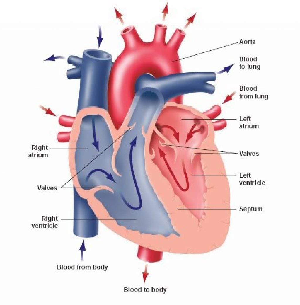
They permit blood flow in one direction only, and prevent backflow of blood. Web this interactive atlas of human heart anatomy is based on medical illustrations and cadaver photography. These layers are separated by a pericardial fluid. Then, fill in the base of the heart with the right and left ventricles and the right and left atriums. It is a muscular organ with four chambers. The user can show or hide the anatomical labels which provide a useful tool to create illustrations perfectly adapted for teaching. Draw the first construction lines. Practise labelling the human heart diagram. The heart is a mostly hollow, muscular organ composed of cardiac muscles and connective tissue that acts as a pump to distribute blood throughout the body’s tissues. Web in this interactive, you can label parts of the human heart.
Your ribcage protects your heart, everyone’s heart is a slightly different. The four types of valves are: What does the heart look like. Web this interactive atlas of human heart anatomy is based on medical illustrations and cadaver photography. This covering is like a membrane which holds all the parts of the heart. Web to draw a realistic human heart, start by making a shape like the bottom half of an acorn. The outer layer is associated with the major blood vessels whereas the inner layer is attached to the cardiac muscles. This will form the main part of the heart. Web to draw the internal structure of the heart, start by sketching the 2 pulmonary veins to the lower left of the aorta and the bottom of the inferior vena cava slightly to the right of that. Drawing a human heart is easier than you may think.
Heart Diagram with Labels and Detailed Explanation
What does the heart look like. The outer layer is associated with the major blood vessels whereas the inner layer is attached to the cardiac muscles. The heart lies in the thoracic cavity between the two lungs in the mediastinal space and behind the sternum. These valves have been clearly shown in the labeled diagram of the heart. They permit.

How to Draw the Internal Structure of the Heart 13 Steps
These valves have been clearly shown in the labeled diagram of the heart. The middle layer of the heart wall is called myocardium. After reading this article you will learn about the structure of human heart. Then, fill in the base of the heart with the right and left ventricles and the right and left atriums. Drawing a human heart.
Heart And Labels Drawing at GetDrawings Free download
These valves have been clearly shown in the labeled diagram of the heart. Cropped by ~~~ to remove white space (this cropping is not the same as wapcaplet's original crop). The heart wall is made up of three layers: If you want to redo an answer, click on the box and the answer will go back to the top so.

heart anatomy labeling
What does the heart look like. They permit blood flow in one direction only, and prevent backflow of blood. The heart is a muscle. These valves have been clearly shown in the labeled diagram of the heart. The outer layer of the heart wall is called epicardium.

How To Draw Human Heart Diagram
Human heart is covered by a double layered structure which is known as pericardium. Web your heart is located in the front of your chest. The heart wall is made up of three layers: Web one idea you could go with would be to look up a diagram of a human heart and label the different parts of the heart..
Labeled Drawing Of The Heart at GetDrawings Free download
Begin this tutorial, by drawing the main shape of the human heart represented by a tilted triangle. Plus, you may just learn something new along the way. These valves have been clearly shown in the labeled diagram of the heart. The lower two chambers of the heart are called ventricles. Then, fill in the base of the heart with the.
Anatomy of the human heart. Cross sectional diagram of the heart with
Web your heart is located in the front of your chest. Web diagram of the human heart, created by wapcaplet in sodipodi. Anatomical illustrations and structures, 3d model and photographs of dissection. Within the triangle, draw a horizontal and vertical centerline to split the triangle into four pieces. Web in this interactive, you can label parts of the human heart.

31 Human Heart To Label Labels Design Ideas 2020
Draw the main shape of your human heart drawing. The human heart is one of the most important organs responsible for sustaining life. The user can show or hide the anatomical labels which provide a useful tool to create illustrations perfectly adapted for teaching. It's situated a little to the left of your chest center, and it's around your fist.
When one teaches, two learn. The heart and the circulatory system
Drawing a human heart is easier than you may think. It is a muscular organ with four chambers. Your ribcage protects your heart, everyone’s heart is a slightly different. Within the triangle, draw a horizontal and vertical centerline to split the triangle into four pieces. Web diagram of the human heart, created by wapcaplet in sodipodi.

humanheartdiagram Tim's Printables
Web one idea you could go with would be to look up a diagram of a human heart and label the different parts of the heart. Plus, you may just learn something new along the way. Introduction to the human heart. Web your heart sure does work hard, but that doesn’t mean you have to work hard to draw it!.
Draw The First Construction Lines.
Web the heart is shaped as a quadrangular pyramid, and orientated as if the pyramid has fallen onto one of its sides so that its base faces the posterior thoracic wall, and its apex is pointed toward the anterior thoracic wall. After reading this article you will learn about the structure of human heart. Draw the main shape of your human heart drawing. The four types of valves are:
It Sits Slightly Behind And To The Left Of Your Sternum (Breastbone).
Web one idea you could go with would be to look up a diagram of a human heart and label the different parts of the heart. Web this interactive atlas of human heart anatomy is based on medical illustrations and cadaver photography. The heart wall is made up of three layers: Human heart is covered by a double layered structure which is known as pericardium.
The Heart Is A Mostly Hollow, Muscular Organ Composed Of Cardiac Muscles And Connective Tissue That Acts As A Pump To Distribute Blood Throughout The Body’s Tissues.
The upper two chambers of the heart are called auricles. This will also help you to draw the structure and diagram of human heart. Drawing a human heart is easier than you may think. Important questions about the human heart.
Anatomical Illustrations And Structures, 3D Model And Photographs Of Dissection.
Introduction to the human heart. It's situated a little to the left of your chest center, and it's around your fist size. The heart is a muscle. Your ribcage protects your heart, everyone’s heart is a slightly different.