Draw Plasma Membrane
Draw Plasma Membrane - For students doing ib biology. Web the fluid mosaic model of the cell membrane is how scientists describe what the cell membrane looks and functions like, because it is made up of a bunch of different. How to draw plasma membrane step by step. For comparison, human red blood cells, visible via light microscopy, are approximately 8 µm wide, or approximately 1,000. Web the principal components of the plasma membrane are lipids (phospholipids and cholesterol), proteins, and carbohydrate groups that are attached to some of the lipids and proteins. Requiem for a fish by. Describe the structure of cell membranes. A phospholipid is a lipid made of glycerol, two fatty acid tails, and a phosphate. Identify components of the cell membrane, including phospholipids, cholesterol, proteins, and carbohydrates. Web the cell membrane, also called the plasma membrane, is a thin layer that surrounds the cytoplasm of all prokaryotic and eukaryotic cells, including plant and animal cells.
Requiem for a fish by. For students doing ib biology. Web the plasma membrane (also known as the cell membrane or cytoplasmic membrane) is a biological membrane that separates the interior of a cell from its outside environment. How to draw cell membrane easily/cell membrane drawing. Like all other cellular membranes, the plasma membrane consists of both lipids and proteins. A phospholipid is a lipid made of glycerol, two fatty acid tails, and a phosphate. Web plasma membrane is selectively permeable to organic molecules and ions, it regulates the movement of particles in and out of organelles and cells. How to draw plasma membrane step by step. Basic cell and molecular biology (bergtrom) 16: As a comparison, human red blood cells, visible via light microscopy, are approximately 8 μm thick, or approximately 1,000 times.
How to draw cell membrane easily/cell membrane drawing. Web the fluid mosaic model of the cell membrane is how scientists describe what the cell membrane looks and functions like, because it is made up of a bunch of different. Web the principal components of the plasma membrane are lipids (phospholipids and cholesterol), proteins, and carbohydrate groups that are attached to some of the lipids and proteins. Follow along and draw the plasma membrane. Describe the structure of cell membranes. Identify components of the cell membrane, including phospholipids, cholesterol, proteins, and carbohydrates. 2.4.2 explain how the hydrophobic and. 86k views 11 years ago. Requiem for a fish by. A phospholipid is a lipid made of glycerol, two fatty acid tails, and a phosphate.

How to Draw Cell Membrane Fluid Mosaic Model Diagram Step by Step
Web the principal components of the plasma membrane are lipids (phospholipids and cholesterol), proteins, and carbohydrate groups that are attached to some of the lipids and proteins. Like all other cellular membranes, the plasma membrane consists of both lipids and proteins. 67 views 3 days ago #howtodraweasy #plasmamembrane. 1 view 1 minute ago #howtodraw. Web plasma membrane is selectively permeable.

How to Draw Fluid Mosaic Model of Plasma Membrane (3D) Simplified
As a comparison, human red blood cells, visible via light microscopy, are approximately 8 μm thick, or approximately 1,000 times. This video shows scientists how to draw cell membrane structure, so scientists like you can make professional scientific illustration. How to draw plasma membrane step by step. Web plasma membrane is selectively permeable to organic molecules and ions, it regulates.

How to Draw Cell Membrane Fluid mosaic Model Of Plasma Membrane in
How to draw plasma membrane step by step. Web the fluid mosaic model of the cell membrane is how scientists describe what the cell membrane looks and functions like, because it is made up of a bunch of different. 86k views 11 years ago. The plasma membrane of a cell is a network of lipids and proteins that forms the.
STRUCTURE of PLASMA MEMBRANE
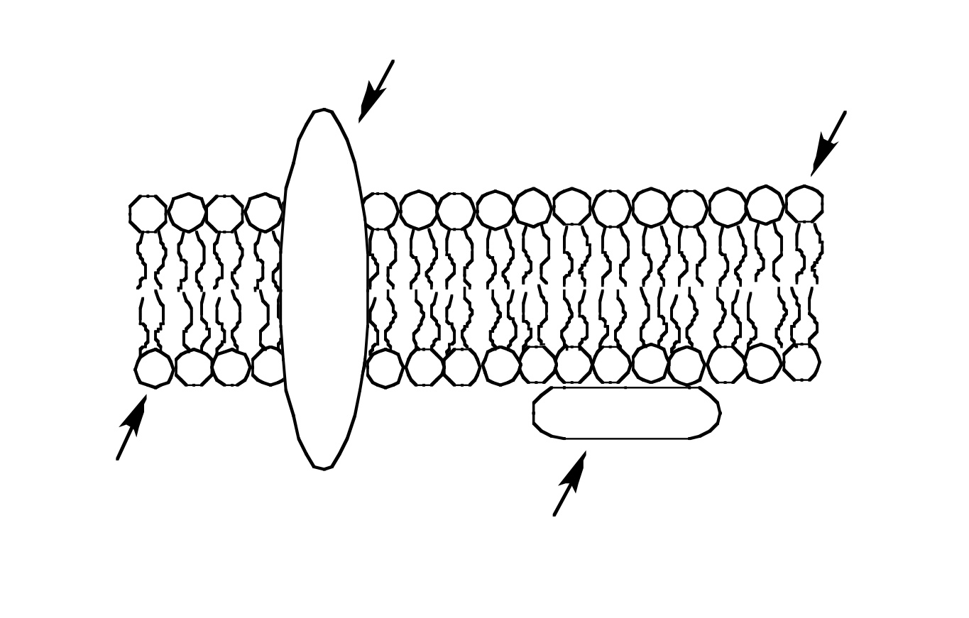
Follow along and draw the plasma membrane. As a comparison, human red blood cells, visible via light microscopy, are approximately 8 μm thick, or approximately 1,000 times. How to draw plasma membrane step by step. Web tutorial for drawing lipid bilayer membrane. 2.4.2 explain how the hydrophobic and.

Plasma membrane. Molecular structure of plasma membrane, eps8 , Ad,
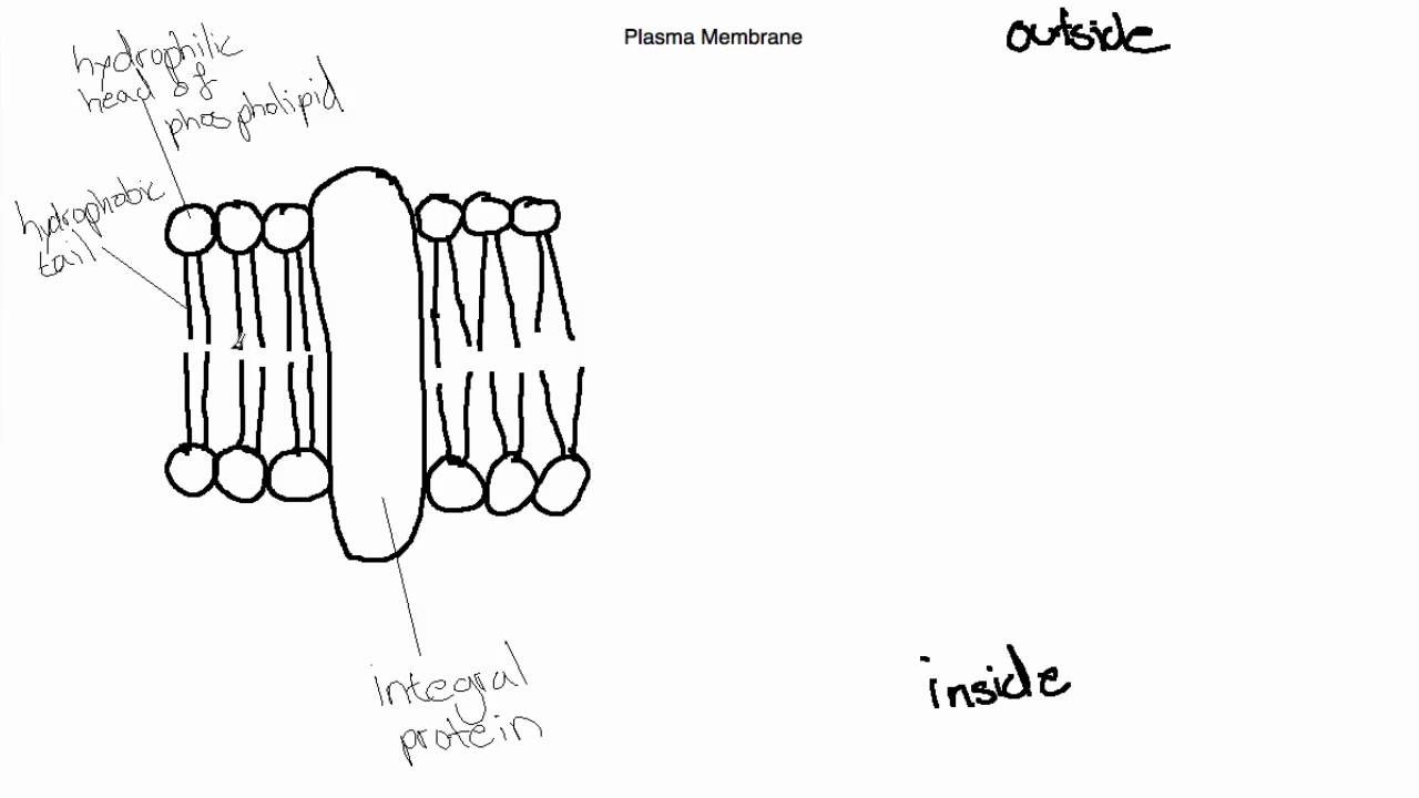
The fundamental structure of the. Web the plasma membrane (also known as the cell membrane or cytoplasmic membrane) is a biological membrane that separates the interior of a cell from its outside environment. These membranes are composed of phospholipids, forming a bilayer with hydrophilic heads. Identify components of the cell membrane, including phospholipids, cholesterol, proteins, and carbohydrates. How to draw.
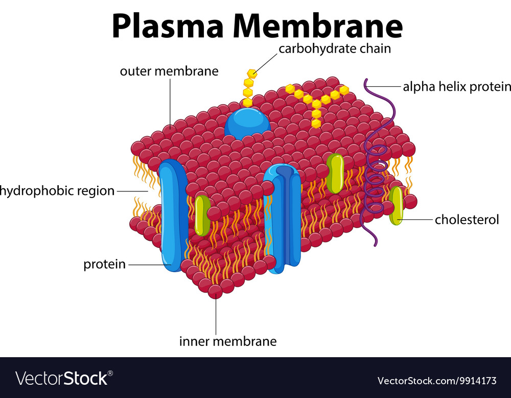
Diagram with plasma membrane Royalty Free Vector Image
Web the cell membrane, also called the plasma membrane, is a thin layer that surrounds the cytoplasm of all prokaryotic and eukaryotic cells, including plant and animal cells. The fundamental structure of the. How to draw plasma membrane step by step. Web plasma membranes range from 5 to 10 nm in thickness. For students doing ib biology.

Labeled Diagram Of Plasma Membrane Best Of Plasma Membrane Diagrams
Web 2.4.1 draw and label a diagram to show the structure of membranes. Describe the structure of cell membranes. For students doing ib biology. As a comparison, human red blood cells, visible via light microscopy, are approximately 8 μm thick, or approximately 1,000 times. It is very easy drawing detailed method to help you.
Use the simplified diagram of the plasma membrane to
2.4.2 explain how the hydrophobic and. Web the plasma membrane is a selectively permeable membrane, which permits the movement of only certain molecules both in and out of the cell. 86k views 11 years ago. Web the plasma membrane, which is also called the cell membrane, has many functions, but the most basic one is to define the borders of.
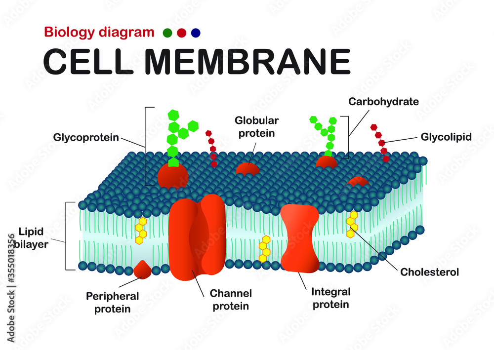
Biology diagram show structure of cell membrane (or plasma membrane
Web the plasma membrane, which is also called the cell membrane, has many functions, but the most basic one is to define the borders of the cell and keep the cell functional. 2.4.2 explain how the hydrophobic and. As a comparison, human red blood cells, visible via light microscopy, are approximately 8 μm thick, or approximately 1,000 times. 9k views.
IB Biology Topic 2.4.1 Draw and Label the Plasma Membrane YouTube
Describe the structure of cell membranes. 1 view 1 minute ago #howtodraw. 9k views 11 months ago easy science drawing. For comparison, human red blood cells, visible via light microscopy, are approximately 8 µm wide, or approximately 1,000. 67 views 3 days ago #howtodraweasy #plasmamembrane.
Web The Plasma Membrane (Also Known As The Cell Membrane Or Cytoplasmic Membrane) Is A Biological Membrane That Separates The Interior Of A Cell From Its Outside Environment.
For students doing ib biology. Basic cell and molecular biology (bergtrom) 16: Follow along and draw the plasma membrane. 67 views 3 days ago #howtodraweasy #plasmamembrane.
Requiem For A Fish By.
Identify components of the cell membrane, including phospholipids, cholesterol, proteins, and carbohydrates. Web plasma membranes range from 5 to 10 nm in thickness. The fundamental structure of the. Web the cell membrane, also called the plasma membrane, is a thin layer that surrounds the cytoplasm of all prokaryotic and eukaryotic cells, including plant and animal cells.
Web The Plasma Membrane Is A Selectively Permeable Membrane, Which Permits The Movement Of Only Certain Molecules Both In And Out Of The Cell.
9k views 11 months ago easy science drawing. The fluid mosaic model explains the structure of cell membranes. The plasma membrane of a cell is a network of lipids and proteins that forms the boundary between a cell’s contents and the outside of the cell. 2.4.2 explain how the hydrophobic and.
This Video Shows Scientists How To Draw Cell Membrane Structure, So Scientists Like You Can Make Professional Scientific Illustration.
Like all other cellular membranes, the plasma membrane consists of both lipids and proteins. Web tutorial for drawing lipid bilayer membrane. It is very easy drawing detailed method to help you. As a comparison, human red blood cells, visible via light microscopy, are approximately 8 μm thick, or approximately 1,000 times.