Drawing Of Cervix
Drawing Of Cervix - Similar to cervical cancer, cervical dysplasia is often related to hpv infection. Web the cervix is the lower part of the uterus situated between the external os (external orifice) and internal os (internal orifice). Web the cervical portion of the spine is an important one anatomically and clinically. The uterus and vagina are also shown. The cervical canal has two orifices: Anatomically and histologically, the cervix is distinct from the uterus, and hence we consider it as a separate anatomical structure. Getting vaccinated for hpv and having regular pap smears can help prevent. Your cervix is also vulnerable to hpv infections that can cause cervical cancer. Existing staining standardization networks often fail to achieve structure preservation and style approximation. The long axis of the.
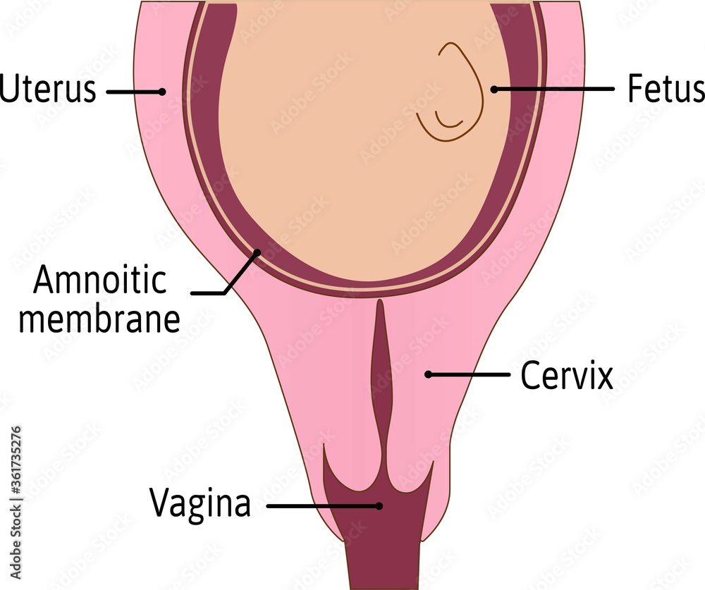
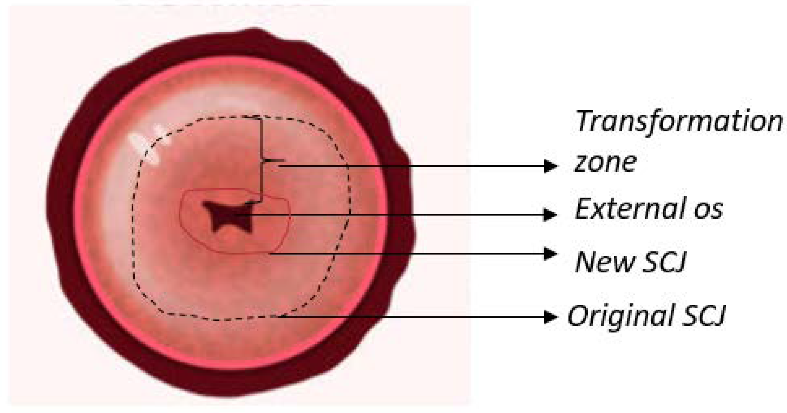
Although it is described as being cylindrical in shape, the anterior and posterior walls are more often ordinarily apposed. The isthmus is an about 1 cm long narrow passage. Getting vaccinated for hpv and having regular pap smears can help prevent. Web the uterus is divided into three parts: Drawing of the anatomy of the cervix showing the internal os, endocervical canal, endocervix, ectocervix, and external os. The internal orifice to the isthmus and the external orifice to the vagina. Web the cervix is a fibromuscular organ that forms a canal between the lower, narrow end of the uterus and the vagina. The cervical spine also allows passage of important vasculature to reach. Cervix during missed abortion.png 750 × 1,334; Similar to cervical cancer, cervical dysplasia is often related to hpv infection.
Web the uterus is divided into three parts: Web the cervix is a fibromuscular organ that links the uterine cavity to the vagina. The internal orifice to the isthmus and the external orifice to the vagina. Web anatomy of the female reproductive system. Web cervical cell image styles may vary due to factors such as specimen preparation methods and staining schemes. Web the uterine cervix produces a mucus that aids in carrying sperm from the vagina to the uterus, where it can fertilize an egg if the woman is ovulating. Cervix during missed abortion.png 750 × 1,334; Your cervix is also vulnerable to hpv infections that can cause cervical cancer. Anatomically and histologically, the cervix is distinct from the uterus, and hence we consider it as a separate anatomical structure. Web cervical dysplasia is a condition in which abnormal cells grow on the surface of the cervix.
Cervix Cartoons, Illustrations & Vector Stock Images 3789 Pictures to
Web the cervix is the lower part of the uterus situated between the external os (external orifice) and internal os (internal orifice). Web cervical cell image styles may vary due to factors such as specimen preparation methods and staining schemes. The cervix is approximately 4 cm in length and 3 cm in diameter. The isthmus is an about 1 cm.

Free Vector Images, Vector Free, Png Images, Female Reproductive System
Owing to its relationships, it is less freely movable than the body, so that the latter may bend on it. The cervix is approximately 4 cm in length and 3 cm in diameter. Cervical cancer awareness month card, january. Web the uterine cervix produces a mucus that aids in carrying sperm from the vagina to the uterus, where it can.
Cervix diagram hires stock photography and images Alamy
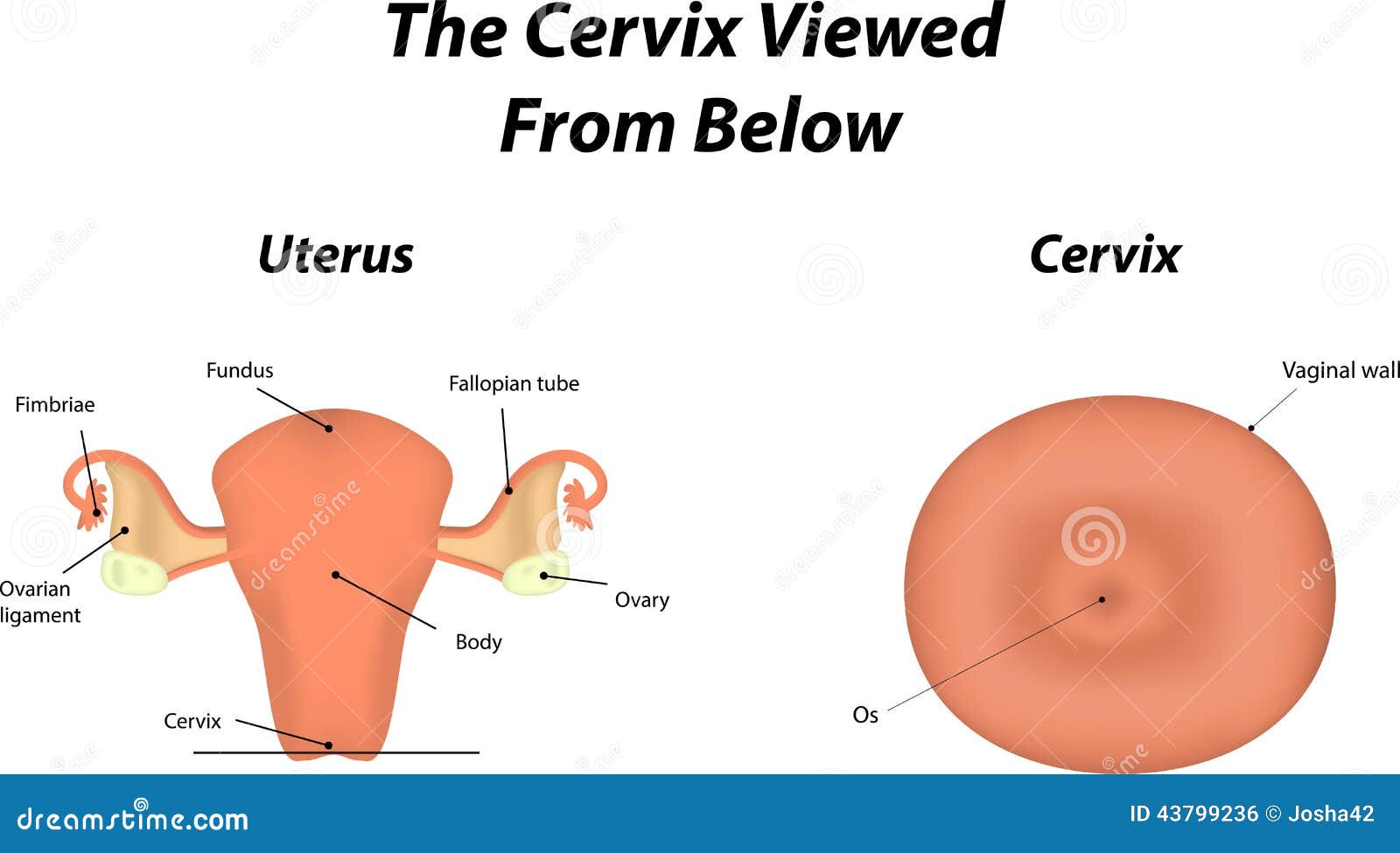
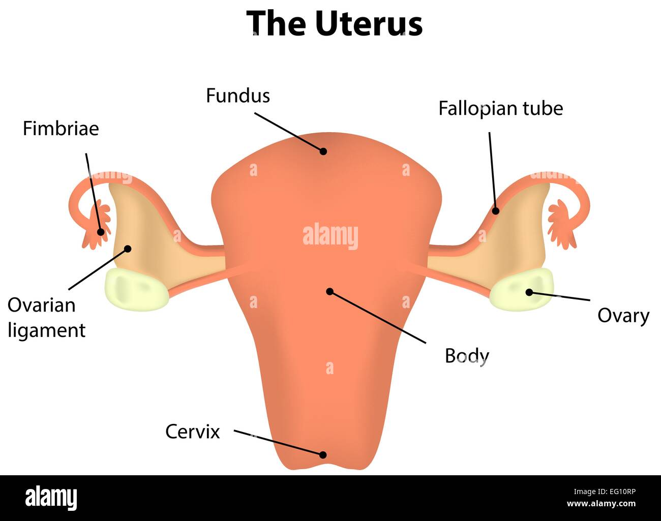
Web the uterus is divided into three parts: The organs in the female reproductive system include the uterus, ovaries, fallopian tubes, cervix, and vagina. Web the cervical portion of the spine is an important one anatomically and clinically. Web the internal sex organs are the vagina, uterus, fallopian tubes, and ovaries. Web cervical cell image styles may vary due to.

Image result for parts of cervix Anatomy, Human body anatomy, Body
3d illustartion of human femail reproductive system anatomy. Although it is described as being cylindrical in shape, the anterior and posterior walls are more often ordinarily apposed. Anatomically and histologically, the cervix is distinct from the uterus, and hence we consider it as a separate anatomical structure. Web squamous epithelial cells of human cervix under the microscope view. It is.

Cervix Bloody Marvellous
Web cervical dysplasia is a condition in which abnormal cells grow on the surface of the cervix. The functions of these organs. The following 18 files are in this category, out of 18 total. It is somewhat conical in shape, with its truncated apex directed downward and backward, but is slightly wider in the middle than either above or below..
Cervix is not effaced. First stage of delivery process. cervix is
Unusual vaginal bleeding, such as after sex, between periods, after menopause, or after a pelvic exam. It is within this region that the nerves to the arms arise via the brachial plexus, and where the cervical plexus forms providing innervation to the diaphragm among other structures. Web the cervix is the lower fibromuscular portion of the uterus that projects within.
Cervical Imaging in the Low Resource Setting Encyclopedia MDPI
These variations can cause inconsistencies among pathologists and degrade the model performance. Your cervix is also vulnerable to hpv infections that can cause cervical cancer. Web early signs of cervical cancer include: It is an anatomical component exclusive to the female in mammals. Web your cervix is a small canal that connects your uterus and vagina.

Printable Cervix Diagrams HD 101 Diagrams
Web the uterus is divided into three parts: When the woman isn’t ovulating, the. It connects the vagina with the main body of the uterus, acting as a gateway between them. Unusual vaginal bleeding, such as after sex, between periods, after menopause, or after a pelvic exam. Pain when you have sex.
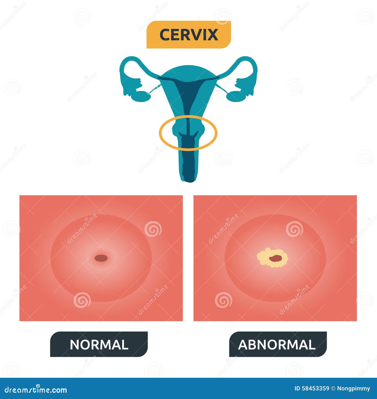
Cervix stock vector. Illustration of normal, smear, medical 58453359
The organs in the female reproductive system include the uterus, ovaries, fallopian tubes, cervix, and vagina. Cervical cancer awareness month card, january. The isthmus is an about 1 cm long narrow passage. During childbirth, your cervix widens so that a baby can be born. Web the cervix is the lower portion (or the neck) of the uterus.it is approximately 1.
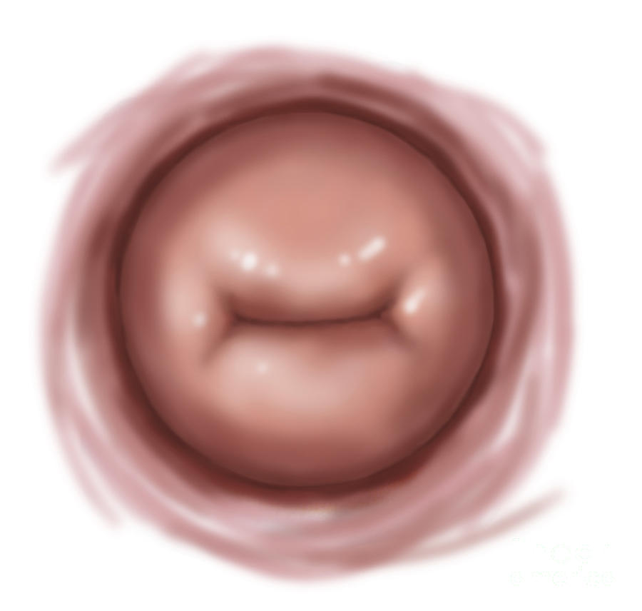
Vaginal View Of Cervix Digital Art by TriFocal Communications
The cervical spine also allows passage of important vasculature to reach. Web the cervix is a fibromuscular organ that links the uterine cavity to the vagina. It allows fluids to leave and enter your uterus. Pain when you have sex. Existing staining standardization networks often fail to achieve structure preservation and style approximation.
Normal Female Reproductive System Anatomy.
Web the cervix is the lower part of the uterus situated between the external os (external orifice) and internal os (internal orifice). Web the uterine cervix produces a mucus that aids in carrying sperm from the vagina to the uterus, where it can fertilize an egg if the woman is ovulating. During childbirth, your cervix widens so that a baby can be born. The organs in the female reproductive system include the uterus, ovaries, fallopian tubes, cervix, and vagina.
The Uterus Has A Muscular Outer Layer Called The Myometrium And An Inner Lining Called The Endometrium.
Unusual vaginal bleeding, such as after sex, between periods, after menopause, or after a pelvic exam. Web the cervix (neck) lies subperitoneally and consists of a part projecting into the vagina (vaginal portion or portio) and a part fixed in the parametrium (supravaginal portion). Fast shippingshop our huge selectionshop best sellersread ratings & reviews Web the internal sex organs are the vagina, uterus, fallopian tubes, and ovaries.
The Cervical Spine Also Allows Passage Of Important Vasculature To Reach.
Although it is described as being cylindrical in shape, the anterior and posterior walls are more often ordinarily apposed. Web the cervix is the lower portion (or the neck) of the uterus.it is approximately 1 inch long and 1 inch wide and opens into the vagina.the cervix functions as the entrance for sperm to enter the uterus. Web the cervix is a fibromuscular organ that links the uterine cavity to the vagina. Anatomically and histologically, the cervix is distinct from the uterus, and hence we consider it as a separate anatomical structure.
The Cervix Is Approximately 4 Cm In Length And 3 Cm In Diameter.
When the woman isn’t ovulating, the. The cervical canal has two orifices: Drawing of the anatomy of the cervix showing the internal os, endocervical canal, endocervix, ectocervix, and external os. It is an anatomical component exclusive to the female in mammals.