Drawing Of Foot Bones
Drawing Of Foot Bones - Front, back and side view. One continuous line drawing of bare foot. Illustration of men's body and male anatomy. The muscle that causes this development goes from the external to the internal side of the leg. Helping the foot withstand the weight of the body whilst standing and in motion. Web bones of the foot and ankle joint medical vector illustration. Elegance female leg in simple linear style. To explain the term in layman’s language, it is the heel bone in the skeletal system. Its main function is to transfer most of the body weight from the legs to the ground. A stress fracture is a very small, fine break in the bone caused by continuous overuse.
Helping the foot withstand the weight of the body whilst standing and in motion. , sometimes known as the wear. The bones in the midfoot (metatarsals) in runners are especially at risk for. One continuous line drawing of bare foot. Web within the foot bones, both the calcaneus and the talus bones form the proximal row of the tarsals. May 13, 2024 | 00:00:27. The forefoot consists of 19 bones; A good understanding of foot and ankle anatomy is necessary for the. All artwork this article collection the artist. Study the bones, muscles, tendons, and ligaments of the foot to gain insight into its intricate design and functionality.
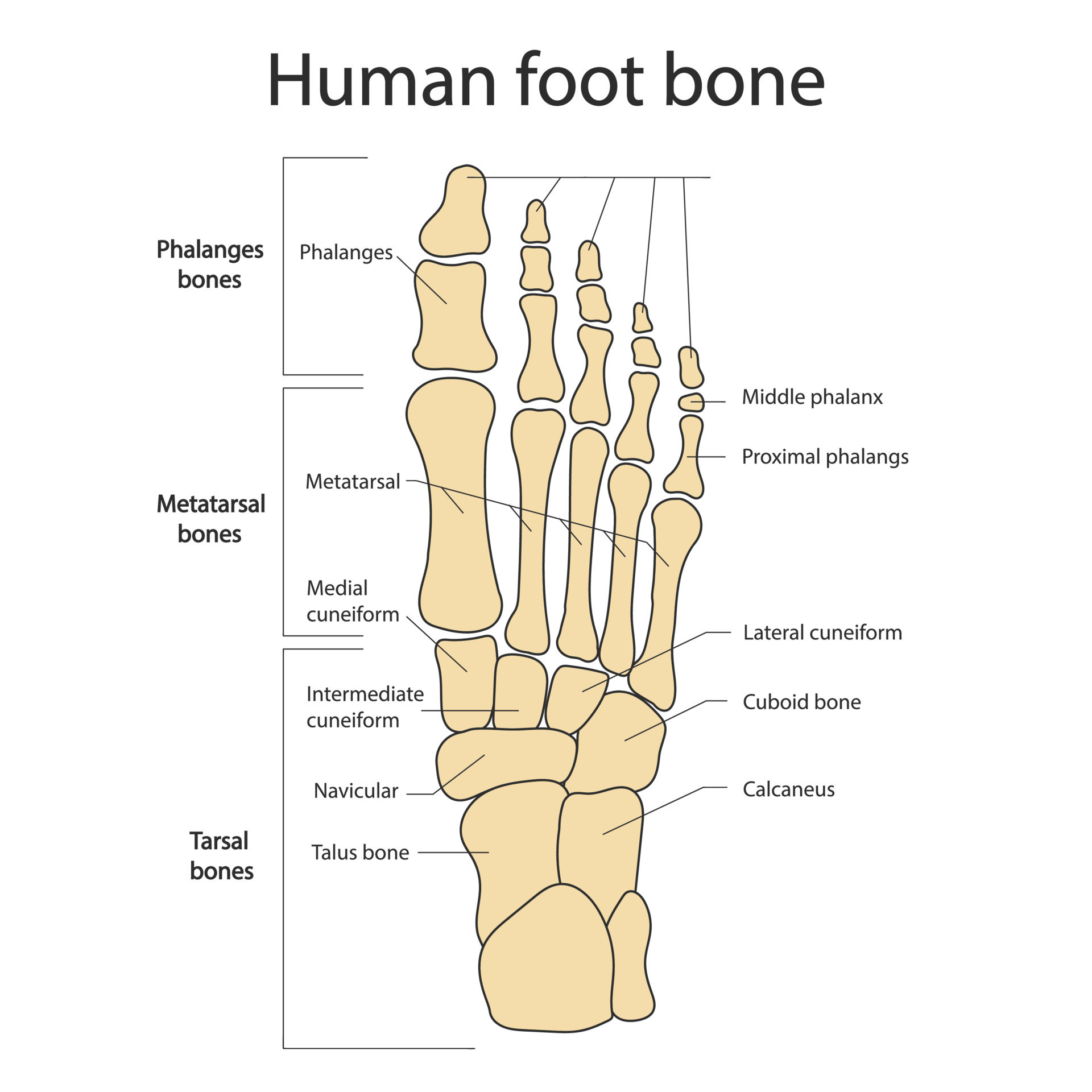
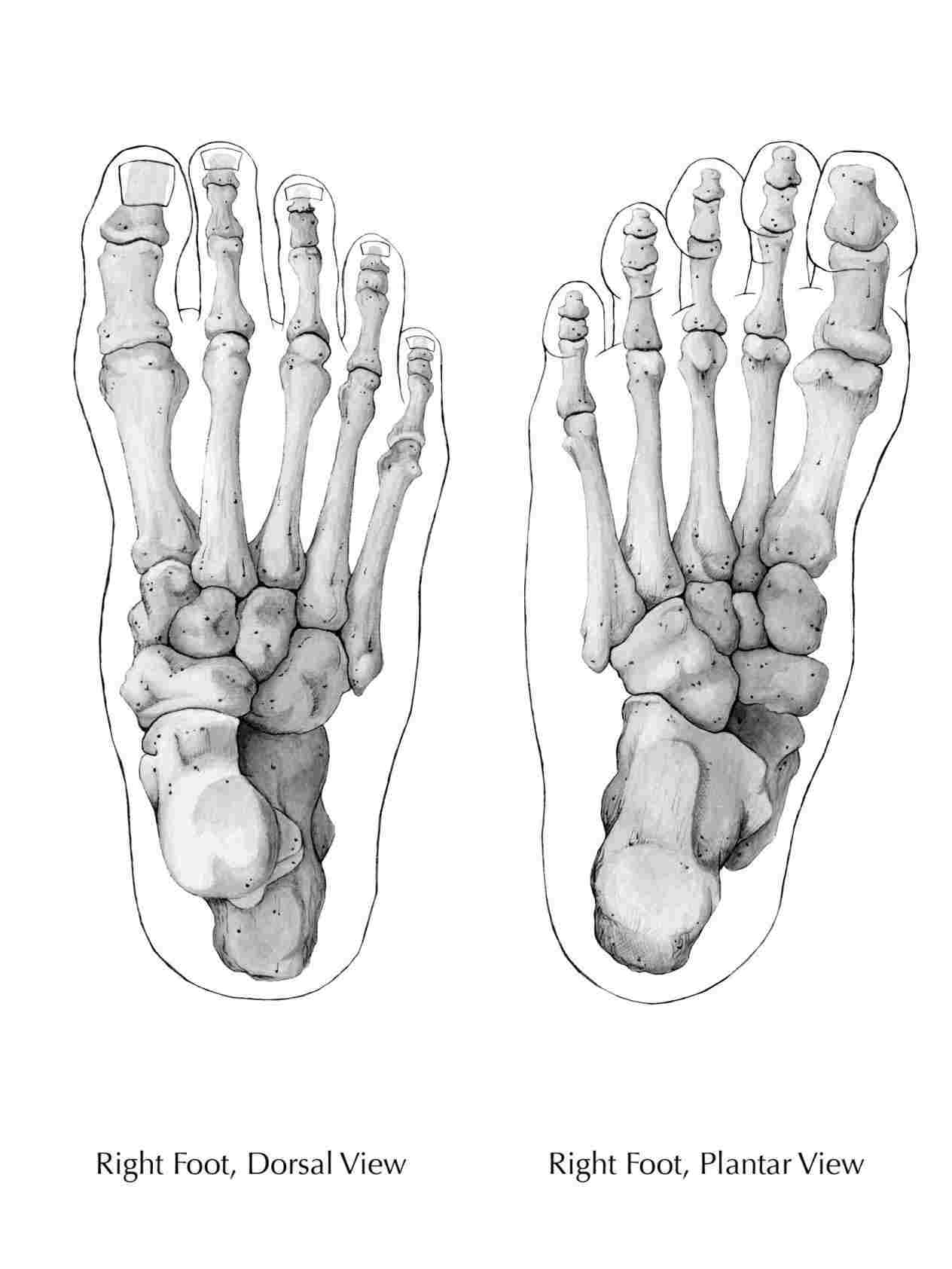
May 13, 2024 | 00:00:27. Web bone outstep by david jon kassan, 2008, graphite drawing on bristol board, 11 x 17. 5 metatarsal bones and 14 phalanges. Web the bones of the foot are arranged to form 3 arches that give it the strength to support our bodies. The upper ankle joint is formed by the inferior surfaces of tibia and. Web the tarsal bones in the foot are located amongst tibia, metatarsal bones, and fibula. Observe real feet in various positions and angles. Illustration of men's body and male anatomy. No toes, no arches, just the basic shape. Upper ankle joint (tibiotarsal), talocalcaneonavicular, and subtalar joints.the last two together are called the lower ankle joint.
.jpg)
Foot Bone Diagram resource Imageshare
Skull, spine, rib cage, pelvis, joints anatomy and medicine, vector icon set drawing of a foot bones stock. These bones are divided into three main regions: The bones in the midfoot (metatarsals) in runners are especially at risk for. A simple way to draw feet is to begin by drawing the sole of the foot. Web the bones of the.
Foot bones. Anatomy of the skeletal system of the human legs and feet
The tendons that pass around the internal lower leg bone turn the foot in. One continuous line drawing of bare foot. It helps transfer weight and pressure across the ankle joint. The muscle that causes this development goes from the external to the internal side of the leg. Web ankle and foot injuries are common musculoskeletal injuries with a high.

Bone Structure Of Foot
With the lower body bones in place, our skeleton drawing is nearly complete. This drawing shows where one of the bones of the lower leg (the tibia) meets the ankle (the talus) and how this sits on top of the heel bone (the calcaneus). In humans, the foot is one of the most complex structures in the body. Web the.
Bones of the Feet ClipArt ETC
Web bones of foot. Let's move on to the hands and feet to finish our masterpiece. The foot is additionally fit for turning and raising its internal outskirt. All artwork this article collection the artist. [1] typical injuries include sprains, fractures, tears, and inflammation.

Foot Bone Anatomy Vector Illustration 539973 Vector Art at Vecteezy
With the lower body bones in place, our skeleton drawing is nearly complete. Observe real feet in various positions and angles. Web the bones of the foot are arranged to form 3 arches that give it the strength to support our bodies. Web with “hot dog in the city,” the artists jen catron and paul outlaw question the lore and.

Foot bones anatomy Royalty Free Vector Image VectorStock
Together, these foot bones form the distal. These bones are divided into three main regions: It is situated between talus and cuboid. The hindfoot, midfoot, and forefoot. To explain the term in layman’s language, it is the heel bone in the skeletal system.
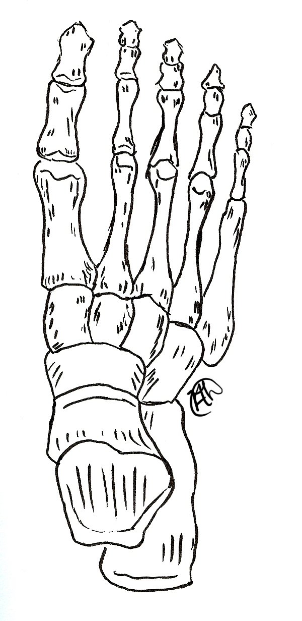
Foot Skeleton Drawing at GetDrawings Free download
5 metatarsal bones and 14 phalanges. Observe real feet in various positions and angles. This is the large bone at the heel of the foot (heel bone). It is made up of three joints: One continuous line drawing of bare foot.
.jpg)
Foot Bone Diagram resource Imageshare
The hindfoot, midfoot, and forefoot. A stress fracture is a very small, fine break in the bone caused by continuous overuse. Upper ankle joint (tibiotarsal), talocalcaneonavicular, and subtalar joints.the last two together are called the lower ankle joint. Together, these foot bones form the distal. In humans, the foot is one of the most complex structures in the body.

Foot & Ankle Bones
Web osteoblastomas (obs) are benign neoplasms constituting approximately 1% of primary bone tumors with a predilection for the spine and sacrum. Bones of the foot and ankle joint medical vector illustration isolated on white background eps 10 human skeleton structure. Web all 26 bones of the foot are described generally for drawing purposes. Helping the foot withstand the weight of.
Skeleton Feet Drawing at Explore collection of
Web the legend of crick foot (review) by blacktooth may 12, 2024, 12:20 pm 0. It is made up of three joints: Web ankle and foot injuries are common musculoskeletal injuries with a high prevalence among professional athletes, but they also occur in recreational sports and as a result of routine daily activities. Web what are stress fractures of the.
Web The Tendons That Go Round The External Lower Leg Bone Draw The Foot In An Outward Course.
Web osteoblastomas (obs) are benign neoplasms constituting approximately 1% of primary bone tumors with a predilection for the spine and sacrum. The tendons that pass around the internal lower leg bone turn the foot in. One continuous line drawing of bare foot. Web the anatomy of feet:
5 Metatarsal Bones And 14 Phalanges.
Web bone outstep by david jon kassan, 2008, graphite drawing on bristol board, 11 x 17. The remaining 5 tarsal foot bones are: This drawing shows where one of the bones of the lower leg (the tibia) meets the ankle (the talus) and how this sits on top of the heel bone (the calcaneus). Web [spoken outro] yeah i'm not gonna lie, this shit was some, some good exercise, like it's good to get out, get the pen workin' you would be a worthy competitor if i was really a predator and you.
The Hind Foot Consists Of The Talus And Calcaneus Bones, Which Form The Ankle Joint And Provide Stability For Weight.
I always feel like i. Web bones of foot. While stress fractures can occur in many bones that are subjected to repetitive activities, the bones of the legs and feet are at greatest risk. One continuous line drawing of bare foot.
May 13, 2024 | 00:00:27.
With the lower body bones in place, our skeleton drawing is nearly complete. A stress fracture is a very small, fine break in the bone caused by continuous overuse. The muscle that causes this development goes from the external to the internal side of the leg. Observe real feet in various positions and angles.