Drawing Of Human Heart With Label
Drawing Of Human Heart With Label - Your heart sure does work hard, but that doesn’t mean you have to work hard to draw it! Do you want a fun way to learn the structure of the heart? January 29, 2024 | published on: The heart has five surfaces: Base (posterior), diaphragmatic (inferior), sternocostal (anterior), and left and right pulmonary surfaces. 14 views 1 year ago. Discussed in this video is how to draw and label. For this first step of our guide on how to draw a human heart, we will start with some outlines for the heart. Click to view large image. Heart diagram with labels in english.
For more help, like including how to give your drawing more details, read on. Web heart, organ that serves as a pump to circulate the blood. New 3d rotate and zoom. April 25, 2024 fact checked. For this first step of our guide on how to draw a human heart, we will start with some outlines for the heart. Web cardiovascular heart diagram: 1.1m views 3 years ago drawing tutorials. After all, we know that stress is bad for the heart! Make sure to write “the human heart” above your drawing as a title when you’re finished. Web medically reviewed by the healthline medical network — by the healthline editorial team — updated on january 20, 2018.
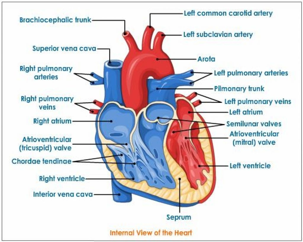
Web the heart is located in the thoracic cavity medial to the lungs and posterior to the sternum. The right margin is the small section of the right atrium that extends between the superior and inferior vena cava. The upper two chambers of the heart are called auricles. Make sure to write “the human heart” above your drawing as a title when you’re finished. For this first step of our guide on how to draw a human heart, we will start with some outlines for the heart. The heart has five surfaces: Web the human heart, comprises four chambers: Web in this lecture, dr mike shows the two best ways to draw and label the heart! After all, we know that stress is bad for the heart! Hi everyone, in this video i.
Heart And Labels Drawing at GetDrawings Free download
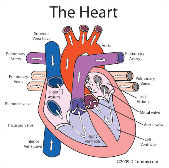
Selecting or hovering over a box will highlight each area in the diagram. Web the cardiovascular system. Right atrium, left atrium, right ventricle and left ventricle. Web the human heart, comprises four chambers: Do you want a fun way to learn the structure of the heart?

How to Draw the Internal Structure of the Heart 13 Steps
The upper two chambers of the heart are called auricles. 336k views 1 year ago easy diagrams drawings. Web the diagram of heart is beneficial for class 10 and 12 and is frequently asked in the examinations. Right, left, superior, and inferior: Web how to draw a human heart step by step 🤎 human heart drawing easy.

Human heart anatomy. Vector diagram in 2021 Heart anatomy, Human
Includes an exercise, review worksheet, quiz, and model drawing of an anterior vi Web medically reviewed by the healthline medical network — by the healthline editorial team — updated on january 20, 2018. Selecting or hovering over a box will highlight each area in the diagram. Learn all about the heart, blood vessels, and composition of blood itself with our.

humanheartdiagram Tim's Printables
Includes an exercise, review worksheet, quiz, and model drawing of an anterior vi Web in this lecture, dr mike shows the two best ways to draw and label the heart! Right atrium, left atrium, right ventricle and left ventricle. Web the diagram of heart is beneficial for class 10 and 12 and is frequently asked in the examinations. Learn all.
Heart Parts! Mr.Kubiak's Blasttastic Science Blog!
On its superior end, the base of the heart is attached to the aorta,mycontentbreak pulmonary arteries and veins, and the vena cava. Right, left, superior, and inferior: January 29, 2024 | published on: Right atrium, left atrium, right ventricle and left ventricle. The innermost layer, the endocardium, lines the interior structures of the heart.
Heart And Labels Drawing at GetDrawings Free download
The heart is a mostly hollow, muscular organ composed of cardiac muscles. Drag and drop the text labels onto the boxes next to the diagram. On its superior end, the base of the heart is attached to the aorta,mycontentbreak pulmonary arteries and veins, and the vena cava. Web how to draw a human heart step by step 🤎 human heart.

Basic Anatomy of the Human Heart Cardiology Associates of Michigan
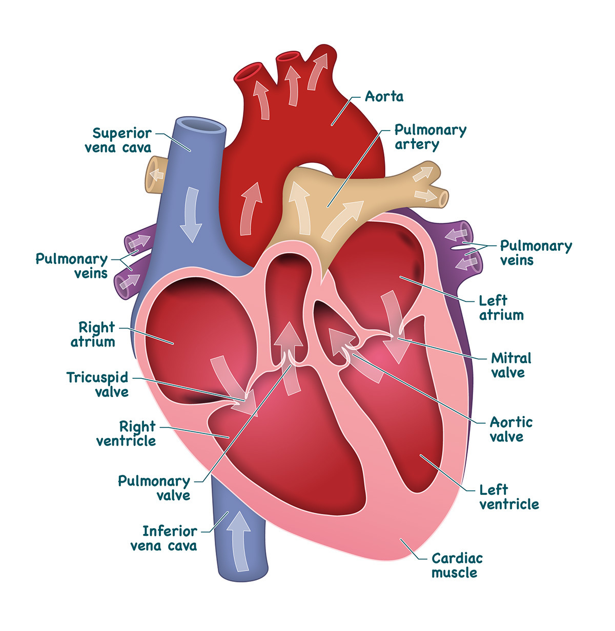
Web the human heart, comprises four chambers: Web this interactive atlas of human heart anatomy is based on medical illustrations and cadaver photography. Base (posterior), diaphragmatic (inferior), sternocostal (anterior), and left and right pulmonary surfaces. New 3d rotate and zoom. Right, left, superior, and inferior:

Drawing Of Heart With Labels at Explore collection
Make sure to write “the human heart” above your drawing as a title when you’re finished. Web after you’ve drawn the structure, color the different sections of the heart distinct colors and appropriately label them. Selecting or hovering over a box will highlight each area in the diagram. Selecting or hovering over a box will highlight each area in the.

31 Human Heart To Label Labels Design Ideas 2020
Discussed in this video is how to draw and label. Click to view large image. Drag and drop the text labels onto the boxes next to the diagram. The innermost layer, the endocardium, lines the interior structures of the heart. Web the epicardium covers the heart, wraps around the roots of the great blood vessels, and adheres the heart wall.

How to Draw the Internal Structure of the Heart 14 Steps
Web medically reviewed by the healthline medical network — by the healthline editorial team — updated on january 20, 2018. Web the epicardium covers the heart, wraps around the roots of the great blood vessels, and adheres the heart wall to a protective sac. The middle layer is the myocardium. 14 views 1 year ago. Use some curved lines for.
Web After You’ve Drawn The Structure, Color The Different Sections Of The Heart Distinct Colors And Appropriately Label Them.
Selecting or hovering over a box will highlight each area in the diagram. 1.1m views 3 years ago drawing tutorials. 14 views 1 year ago. Drag and drop the text labels onto the boxes next to the diagram.
336K Views 1 Year Ago Easy Diagrams Drawings.
The two upper chambers are called the left and the right atria, and the two lower chambers are known as the left and the right ventricles. By following the simple steps, you too can easily draw a perfect human heart. Hi everyone, in this video i. Web this interactive atlas of human heart anatomy is based on medical illustrations and cadaver photography.
The Inferior Tip Of The Heart, Known As The Apex, Rests Just Superior To The Diaphragm.
Web the epicardium covers the heart, wraps around the roots of the great blood vessels, and adheres the heart wall to a protective sac. January 29, 2024 | published on: The two atria and ventricles are separated from each other by a muscle wall called ‘septum’. This strong muscle tissue powers the heart’s pumping action.
In This Interactive, You Can Label Parts Of The Human Heart.
Make sure to write “the human heart” above your drawing as a title when you’re finished. New 3d rotate and zoom. Web how to draw a human heart step by step 🤎 human heart drawing easy. For more help, like including how to give your drawing more details, read on.