Drawing Of Knee Anatomy
Drawing Of Knee Anatomy - We will cover anatomy and then attempt to draw the knee with a few easy to follow examples. Knee anatomy involves more than just muscles and bones. When drawing a knee, we have two big volumes—the upper and lower leg. Web atlas of the anatomy of the joint of the knee on a ct arthrogram in axial, coronal, and sagittal sections, on a 3d images and on conventional athrogram. Web the knee joint is a synovial joint which connects the femur (thigh bone), the longest bone in the body, to the tibia (shin bone). The patella (or kneecap, as it is commonly called) is made of bone and sits in front of. Web to start drawing a knee, begin by drawing a basic oval shape for the kneecap. Web how does the knee joint work? Web the knee is the meeting point of the femur (thigh bone) in the upper leg and the tibia (shinbone) in the lower leg. Cartoon illustration of the human knee joint anatomy.
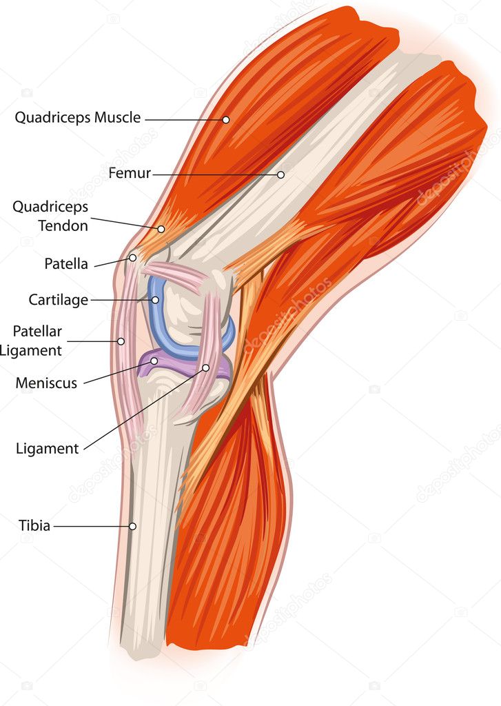
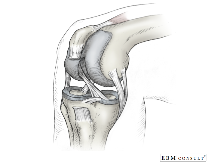
So how to draw the knee? Ligaments, tendons, and cartilage work together to connect the thigh bone, shin bone, and knee cap and allow the leg to bend back and forth like a hinge. It allows the lower leg to move relative to the thigh while supporting the body’s weight. The largest joint in the body, the knee is also one of the most easily injured. Web to start drawing a knee, begin by drawing a basic oval shape for the kneecap. Radiological anatomy of the knee on a ct arthrogram. The knee is a complex joint that flexes, extends, and twists slightly from. Illustration of the human knee joint anatomy. Which ligaments keep it stable? Web the knee joint is a synovial joint which connects the femur (thigh bone), the longest bone in the body, to the tibia (shin bone).
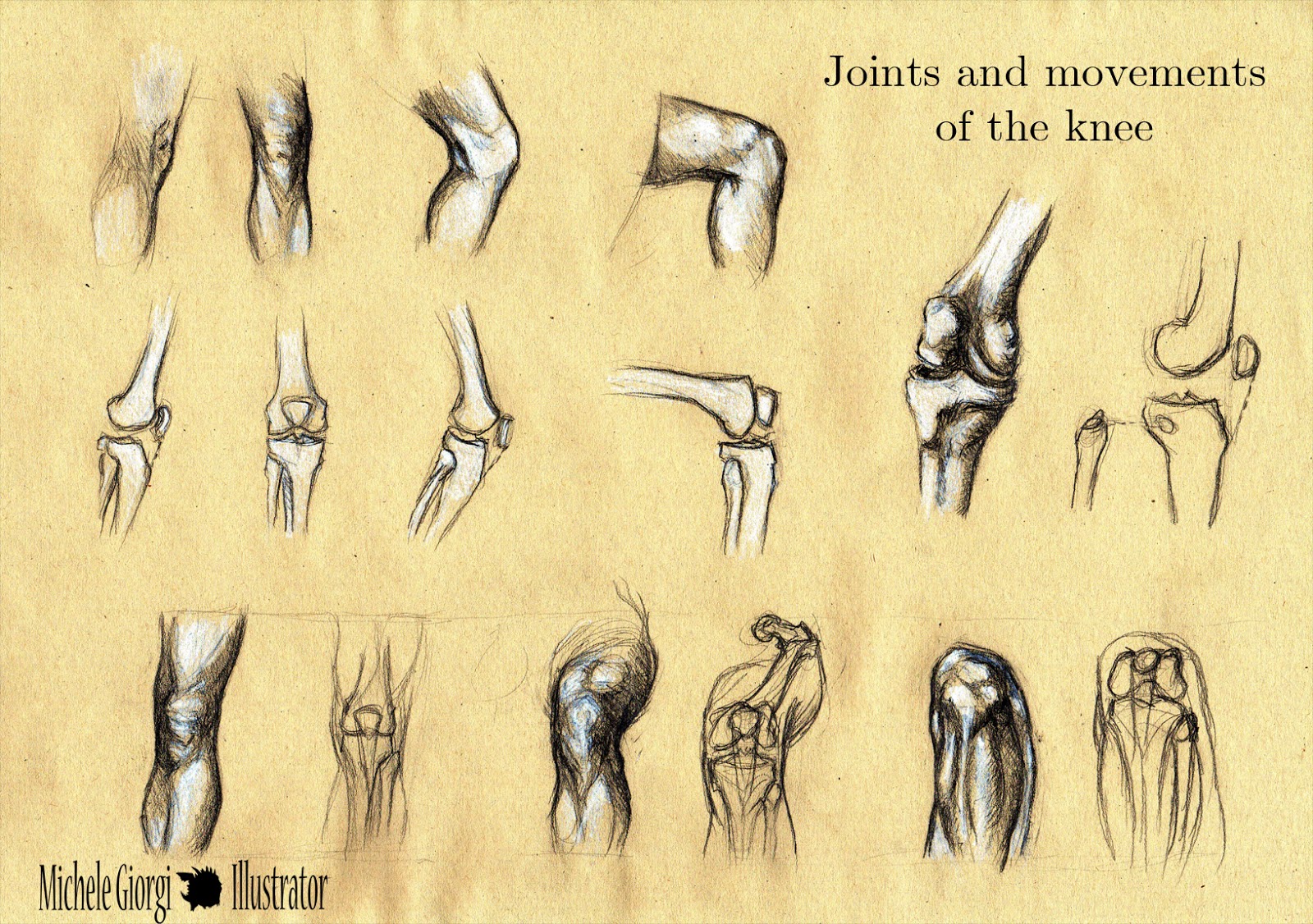
Web an overview of the anatomy of the knee joint including bony articulations, ligaments, menisci, arterial supply, innervation and relevant muscles. Radiological anatomy of the knee on a ct arthrogram. Web the knee joint is the largest synovial joint in the body and it’s these articulations between the femur and the tibia and also between the patella and the femur. So how to draw the knee? Cartoon illustration of the human knee joint anatomy. Web the knee joint is one of the strongest and most important joints in the human body. Web anatomy of the knee. In this video lesson, you will discover how to draw knees from life with the necessary knowledge of this joint's proportions and anatomy. It allows the lower leg to move relative to the thigh while supporting the body’s weight. To draw the knee, begin by visualizing the bones and tendons underneath to help with the placement of landmarks.
Vector drawing of knee diagram Free SVG
Web the knee joint is one of the strongest and most important joints in the human body. Web in this video lesson, you will learn how to draw realistic knees from life with the necessary knowledge of this joint's proportions and anatomy. Finally, draw a thicker oval shape for the upper leg bone, making sure that it is angled slightly.
Michele Illustrator Anatomy Sketches Joints and movements of
When drawing a knee, we have two big volumes—the upper and lower leg. Web in this video lesson, you will learn how to draw realistic knees from life with the necessary knowledge of this joint's proportions and anatomy. Knee anatomy for figurative artists. Ligaments, tendons, and cartilage work together to connect the thigh bone, shin bone, and knee cap and.
Knee injuries causes, types, symptoms, knee injuries prevention & treatment
Learn everything about the anatomy and function of the knee now at kenhub! Web in this video lesson, you will learn how to draw realistic knees from life with the necessary knowledge of this joint's proportions and anatomy. Radiological anatomy of the knee on a ct arthrogram. These illustrations review the basics of the anatomy of the knee and make.

Radiopaedia Drawing Bones of the knee joint English labels
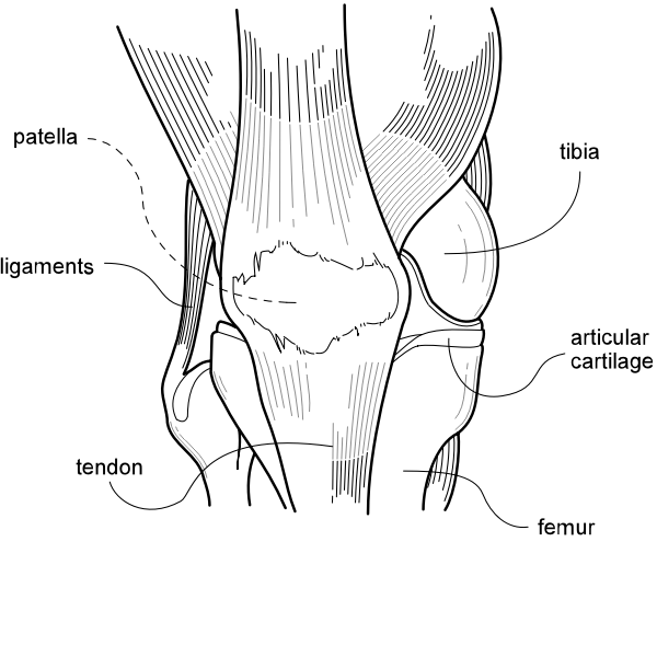
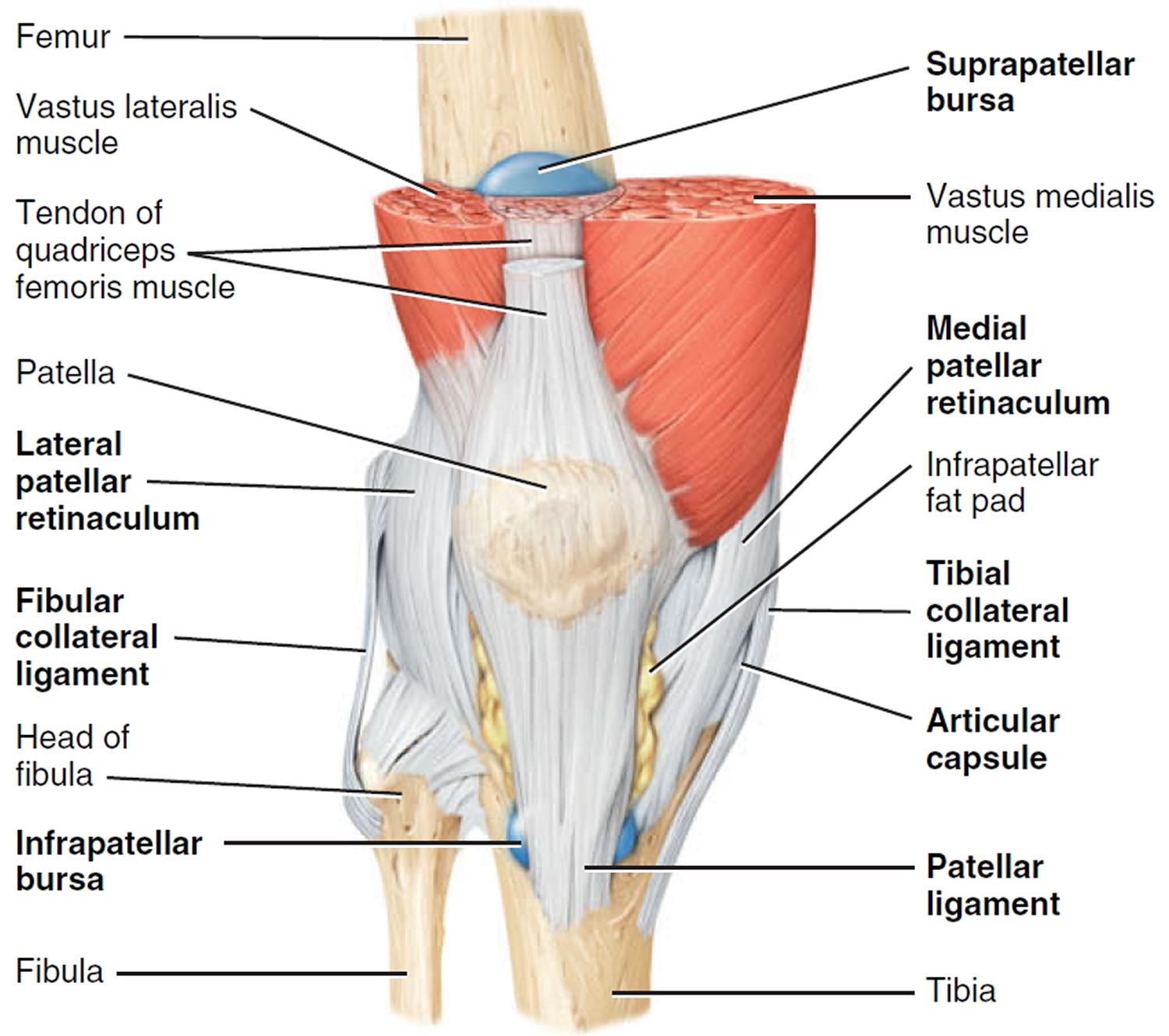
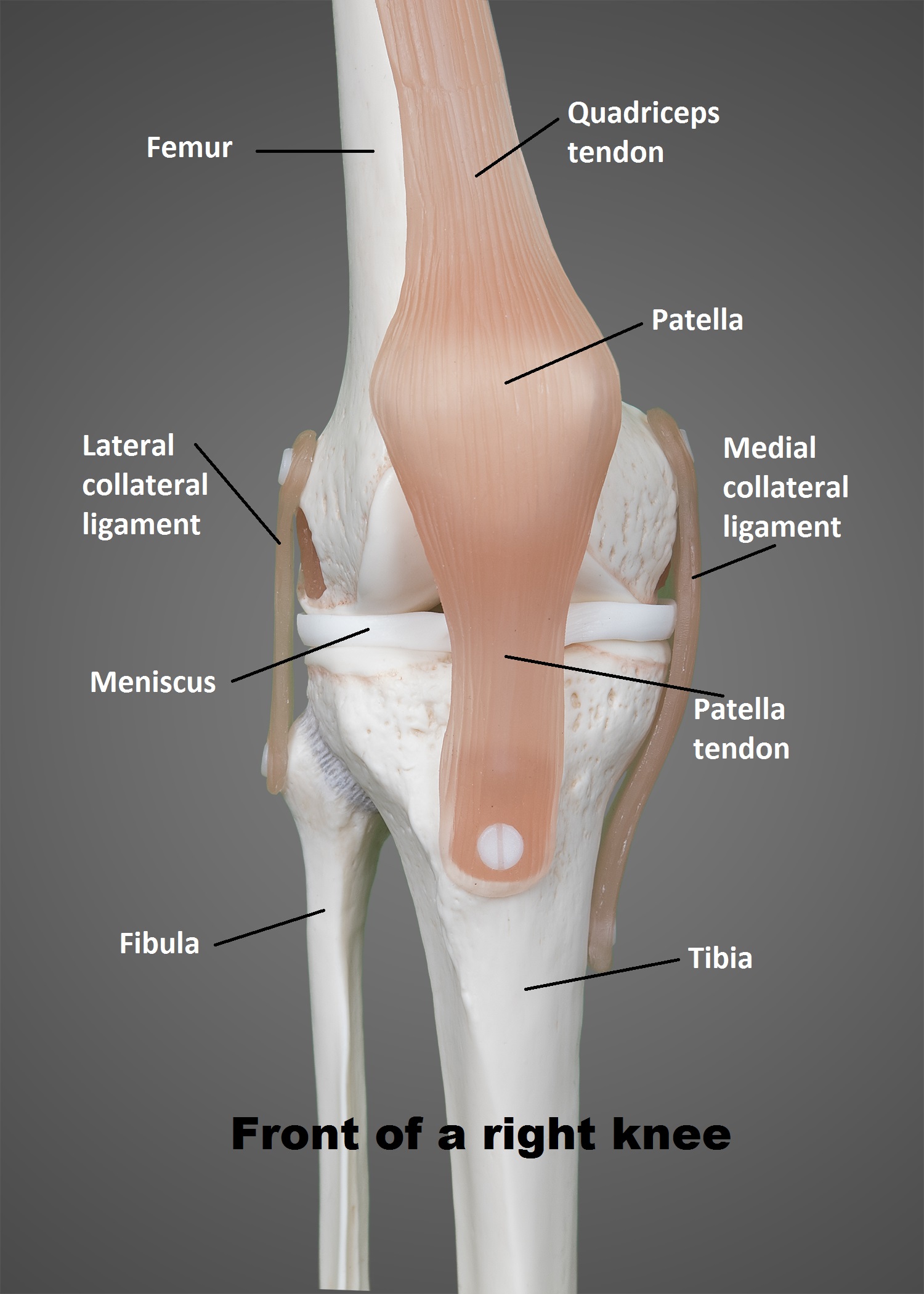
The most basic component of knee joint anatomy are the bones which provide the structure to the knee. The patella (or kneecap, as it is commonly called) is made of bone and sits in front of. These illustrations review the basics of the anatomy of the knee and make it easier to place them on an mri by using the.

FileKnee diagram.svg Wikimedia Commons
Knee anatomy involves more than just muscles and bones. Finally, draw a thicker oval shape for the upper leg bone, making sure that it is angled slightly forward. Learning about knee anatomy can help the students to know about the structure and function of the knee. Below are the muscle groups we will go over. It’s a hinge joint and.

Schematic illustration of the knee joint anatomy. Download
Then, draw a longer oval shape for the lower leg bone, making sure that it is angled slightly backward. Learning about knee anatomy can help the students to know about the structure and function of the knee. We will cover anatomy and then attempt to draw the knee with a few easy to follow examples. Web the knee is the.
Knee anatomy Stock Vector Image by ©Lukaves 18341225
Illustration of the human knee joint anatomy. Knee anatomy involves more than just muscles and bones. It allows the lower leg to move relative to the thigh while supporting the body’s weight. Web the knee joint is one of the strongest and most important joints in the human body. The upper leg is facing the viewer and can be simplified.

Anatomy of Knee
Learn everything about the anatomy and function of the knee now at kenhub! The largest joint in the body, the knee is also one of the most easily injured. The patella (or kneecap, as it is commonly called) is made of bone and sits in front of. There are two main joints in the knee: When drawing a knee, we.
Anatomy Knee
Web in this video lesson, you will learn how to draw realistic knees from life with the necessary knowledge of this joint's proportions and anatomy. Web to start drawing a knee, begin by drawing a basic oval shape for the kneecap. Web atlas of the anatomy of the joint of the knee on a ct arthrogram in axial, coronal, and.
The Knee UT Health San Antonio
Knee anatomy for figurative artists. There are four knee bones that fit together to make two different knee joints: Ligaments, tendons, and cartilage work together to connect the thigh bone, shin bone, and knee cap and allow the leg to bend back and forth like a hinge. In this video lesson, you will discover how to draw knees from life.
Which Ligaments Keep It Stable?
Before we go over simplified anatomy of the leg, let’s look at where we want to end up. We will cover anatomy and then attempt to draw the knee with a few easy to follow examples. So how to draw the knee? Web we added 3d illustrations of the bones of the knee (femur, tibia, fibula (peroneal) and patella (kneecap)) which we then labeled.
To Draw The Knee, Begin By Visualizing The Bones And Tendons Underneath To Help With The Placement Of Landmarks.
Web the knee joint is the largest synovial joint in the body and it’s these articulations between the femur and the tibia and also between the patella and the femur. There are two main joints in the knee: Movements at the knee joint are essential to many everyday activities, including walking, running, sitting and standing. When drawing a knee, we have two big volumes—the upper and lower leg.
It’s A Hinge Joint And The Main Movements You Get At This Joint Are Flexion And Extension.
The knee joint is a complex hinge joint. Learn everything about the anatomy and function of the knee now at kenhub! Damage in even one part can hinder the functioning of the knee. Then, draw a longer oval shape for the lower leg bone, making sure that it is angled slightly backward.
Radiological Anatomy Of The Knee On A Ct Arthrogram.
Arthrogram (arthrogram) knee joint (knee joint, femorotibial joint): All these parts combine and work together. Web in this tutorial on drawing the knee, i’ll show you exactly what all those bumps are. When drawing a knee, we have two big volumes—the upper and lower leg.