Drawing Of Lysosomes
Drawing Of Lysosomes - Web how to draw lysosomes | structure of lysosome | step by step👉how to draw diagrams? To have finer control over how your membrane brush looks, you can separate a brush into editable icons (1:47). They were discovered and named by christian de duve in 1955. Its size ranges from 0.2 to 0.5 μm. There is variation in the shape and size from one cell to another and from time to time. Carcinoma cell, colored transmission electron micrograph (tem) cell. Illustration of human cell anatomy. Lysosomes are critical for cellular. It breaks down old and unnecessary structures so their molecules can be reused. Peroxisomes protect cells by isolating and breaking down harmful hydrogen peroxide into water and oxygen.
Web the structure of lysosomes. Carcinoma cell, colored transmission electron micrograph (tem) cell. 7.4k views 3 years ago uploaded videos. The diagram below shows the lysosome structure within a cell. To have finer control over how your membrane brush looks, you can separate a brush into editable icons (1:47). Lysosomes are specialized vesicles within cells that digest large molecules through the use of hydrolytic enzymes. It's an educational video from 9th biology ptb. Illustration of human cell anatomy. How to draw structure of lysosomes step by step for beginners hi viewers in this video i will tell you about how can be draw diagram of. Web how to draw lysosomes | structure of lysosome | step by step👉how to draw diagrams?
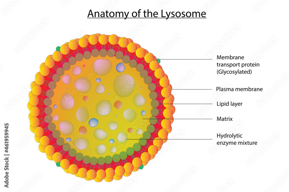
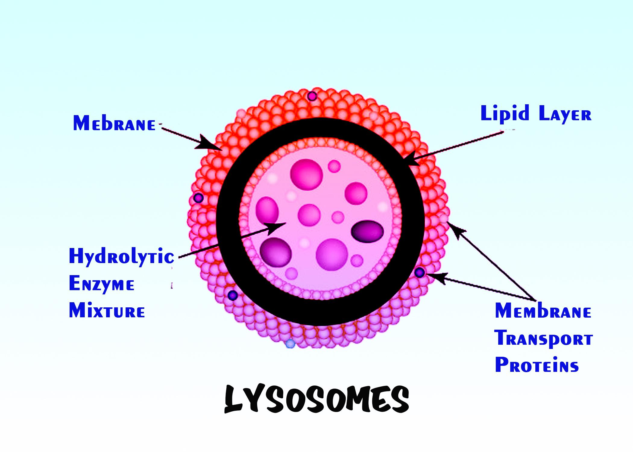
They were discovered and named by christian de duve in 1955. Web how to draw lysosomes | structure of lysosome | step by step👉how to draw diagrams? Illustration of human cell anatomy. Web in the biosynthetic pathway, lysosomes are formed and acquire their necessary components, such as newly synthesized proteins. Each lysosome is surrounded by a membrane that maintains an acidic environment within the interior via a proton pump. To have finer control over how your membrane brush looks, you can separate a brush into editable icons (1:47). Its size ranges from 0.2 to 0.5 μm. There is variation in the shape and size from one cell to another and from time to time. Lysosomes are part of the endomembrane system, and some vesicles that leave the golgi are bound for the lysosome. It is spherical and has the lipid bilayer, i.e., phospholipids.

Anatomy of the Lysosome. Vector Diagram for Medical Use Stock Vector
There is variation in the shape and size from one cell to another and from time to time. Changes in lysosome function are essential to support cellular adaptation to multiple signals and stimuli. Lysosomes, the cell's recycling centers, use acid hydrolases to break down waste into reusable parts through autophagy and crinophagy. These organelles ensure efficient and safe cellular function..
Biological illustration of lysosome (Structure of cell lysosome) Stock
[1] [2] they are spherical vesicles that contain hydrolytic enzymes that digest many kinds of biomolecules. Its size ranges from 0.2 to 0.5 μm. Web in the biosynthetic pathway, lysosomes are formed and acquire their necessary components, such as newly synthesized proteins. They also resemble lysosomes in being filled with enzymes. 17k views 4 years ago educational videos.
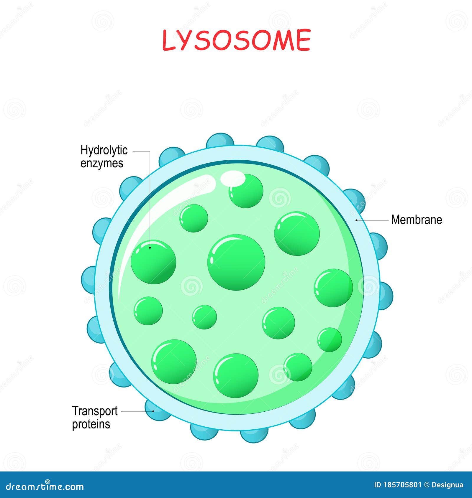
Lysosome anatomy stock vector. Illustration of structure 185705801
17k views 4 years ago educational videos. There is variation in the shape and size from one cell to another and from time to time. Each lysosome is surrounded by a membrane that maintains an acidic environment within the interior via a proton pump. Web in the biosynthetic pathway, lysosomes are formed and acquire their necessary components, such as newly.

Structure lysosomes infographics Royalty Free Vector Image
Presence of lysosomes in the animal tissue. They also resemble lysosomes in being filled with enzymes. It is spherical and has the lipid bilayer, i.e., phospholipids. Web structure of lysosome. Lysosome is round, vacuolar, and filled with dense material.
Lysosome Structure
These organelles ensure efficient and safe cellular function. Its size ranges from 0.2 to 0.5 μm. The breakdown/digestion of macromolecules (carbohydrates, lipids, proteins, and nucleic acids), cell membrane repairs, and responses against foreign substances such as bacteria, viruses and other antigens. It's an educational video from 9th biology ptb. It varies in shape and density.
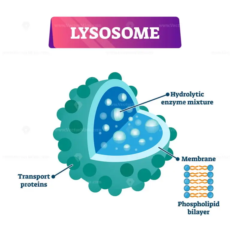
Lysosome cell organelle vector illustration labeled cross section
They were discovered and named by christian de duve in 1955. In this short tutorial, learn how to draw an endosome fusing with a lysosome. To have finer control over how your membrane brush looks, you can separate a brush into editable icons (1:47). A lysosome has a specific composition, of both its membrane proteins and its lumenal proteins. Web.

Lysosome In Plant Cell Description Raine AP Biology Chapter 6 / To
Web lysosome, subcellular organelle that is found in nearly all types of eukaryotic cells (cells with a clearly defined nucleus) and that is responsible for the digestion of macromolecules, old cell parts, and microorganisms. Web how to draw lysosomes | structure of lysosome | step by step👉how to draw diagrams? Web in the biosynthetic pathway, lysosomes are formed and acquire.

Structure of lysosomes infographics Royalty Free Vector
Web lysosomes are degradation centers and signaling hubs in cells and play important roles in cellular homeostasis, development, and aging. It is spherical and has the lipid bilayer, i.e., phospholipids. The membrane surrounding the lysosome is vital to ensure these enzymes do not leak out into the cytoplasm and damage the cell. These organelles ensure efficient and safe cellular function..

Lysosomes are membraneenclosed organelles. Lysosomes in cell. 8143455
The lysosomes then fuse with membrane vesicles from one of three routes: Web lysosome, subcellular organelle that is found in nearly all types of eukaryotic cells (cells with a clearly defined nucleus) and that is responsible for the digestion of macromolecules, old cell parts, and microorganisms. It's an educational video from 9th biology ptb. [1] [2] they are spherical vesicles.
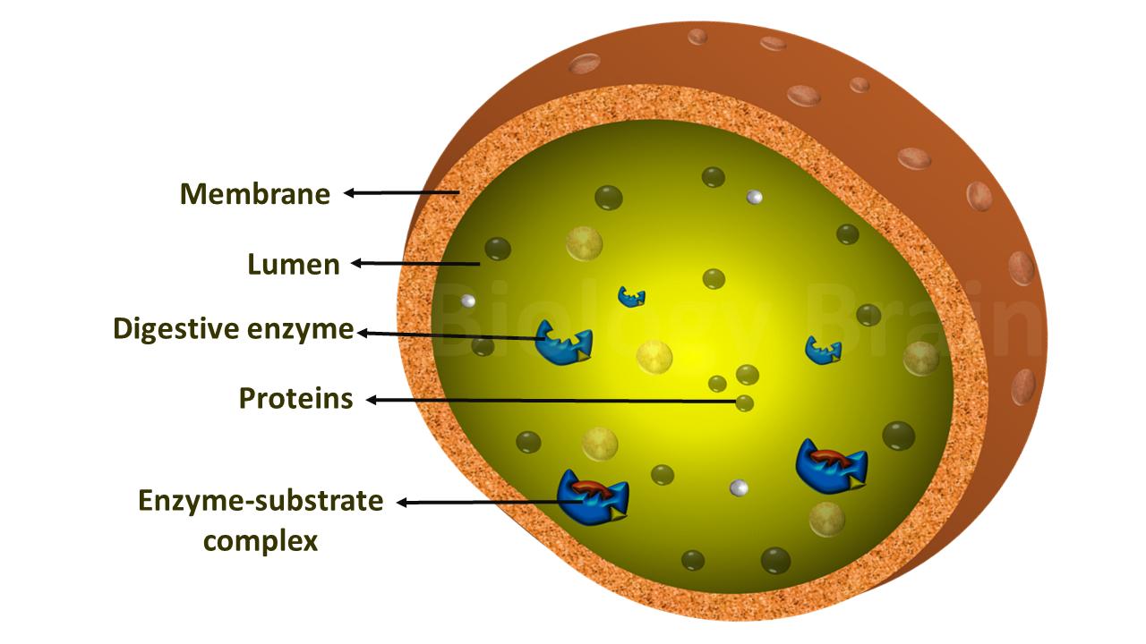
Diagram of Lysosomes and Types Biology Brain
Carcinoma cell, colored transmission electron micrograph (tem) cell. They also resemble lysosomes in being filled with enzymes. Its size ranges from 0.2 to 0.5 μm. [1] [2] they are spherical vesicles that contain hydrolytic enzymes that digest many kinds of biomolecules. Vesicles are small spheres of fluid surrounded by a lipid bilayer membrane, and they have roles in.
[1] [2] They Are Spherical Vesicles That Contain Hydrolytic Enzymes That Digest Many Kinds Of Biomolecules.
7.4k views 3 years ago uploaded videos. Illustration of human cell anatomy. De duve used lysosomes to describe an organelle containing hydrolytic enzymes capable of breaking down biological polymers such as nucleic acid, proteins, carbohydrates, and lipids. Web lysosomes are degradation centers and signaling hubs in cells and play important roles in cellular homeostasis, development, and aging.
Lysosomes Are Specialized Vesicles Within Cells That Digest Large Molecules Through The Use Of Hydrolytic Enzymes.
To have finer control over how your membrane brush looks, you can separate a brush into editable icons (1:47). Burnetii to replicate and achieve successful intracellular life in the cell cytosol is vastly dependent. Carcinoma cell, colored transmission electron micrograph (tem) cell. It is spherical and has the lipid bilayer, i.e., phospholipids.
Lysosomes, The Cell's Recycling Centers, Use Acid Hydrolases To Break Down Waste Into Reusable Parts Through Autophagy And Crinophagy.
Lysosomes contain numerous hydrolytic enzymes which catalyse hydrolysis reactions. Its size ranges from 0.2 to 0.5 μm. There is variation in the shape and size from one cell to another and from time to time. 17k views 4 years ago educational videos.
How To Draw Structure Of Lysosomes Step By Step For Beginners Hi Viewers In This Video I Will Tell You About How Can Be Draw Diagram Of.
The breakdown/digestion of macromolecules (carbohydrates, lipids, proteins, and nucleic acids), cell membrane repairs, and responses against foreign substances such as bacteria, viruses and other antigens. Qiuyuan yin et al, exploring lysosomal biology: The diagram below shows the lysosome structure within a cell. In this video, you will learn how to draw diagram of.