Drawing Of Paramecium
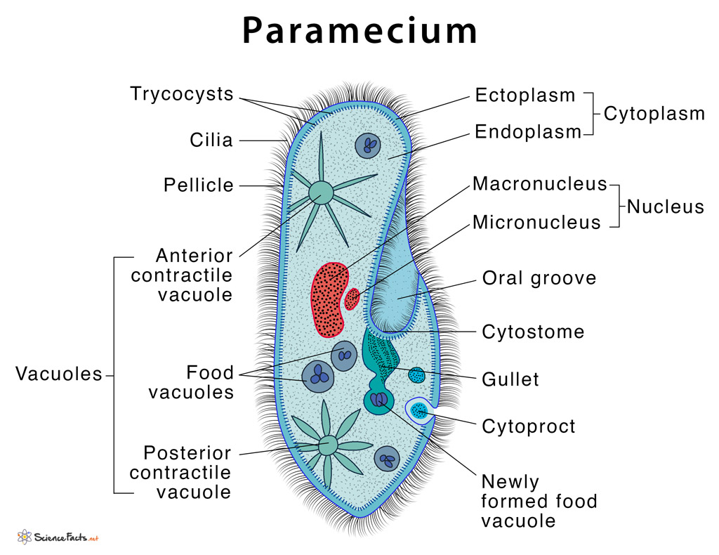
Drawing Of Paramecium - Web panel 1 (a,b): In 1703 an anonymous writer wrote a description of and sketched out illustrations of paramecium that was published in the philosophical transactions of the royal society of london. Fresh water, free living, omnipresent and is found in stagnant water. Light microscopic appearance of paramecium caudatum.panel 1 (c): Web fast forward in time a bit and there is some mystery around who might have published the first drawings of the paramecium. The body of the paramecium cell is enclosed by a stiff but elastic membrane, called pellicle. 1,142 × 1,007 (149 kb) Web #paramecium #howtodraw #biologythis is a diagram of the paramecium. This video helps you to draw science diagrams with great ease and. Paramecium vary in length from about 0.05 to 0.32 mm (0.002 to 0.013 inch).
To gather the food it makes use of its cilia, making quick movements with cilia to draw the water along with its prey organisms inside the mouth opening through its oral groove. Web the drawing also shows food vacuoles and cilia. They are the most common of all ciliate organisms that are characterized by the presence of cilia all along their. It is a prey for other microorganisms such as didinium. Paramecium vary in length from about 0.05 to 0.32 mm (0.002 to 0.013 inch). Web panel 1 (a,b): Binary fission divides a cell transversely and followed by mitotic division in the micronucleus. Web diagram of paramecium. They have a lifespan of a hundred, a thousand or even a million years. Ralph wichterman, the biology of paramecium, 2nd edition, 1986 (fig.
Web the drawing also shows food vacuoles and cilia. Web fast forward in time a bit and there is some mystery around who might have published the first drawings of the paramecium. Besides that, they are unique in having contractile vacuoles, mouth pore and anal pore. Draw a neat labelled diagram of pbr322. Higher magnification of the metachronal. Paramecium are widespread in freshwater, brackish, and marine environments and are. Paramecium swims in fresh water by beating its thousands of cilia, and feeds on smaller microorganisms such as bacteria and algae. Paramecium structure consists of trichocysts, contractile vacuoles, and cilia among other specialized organelles. Under favourable conditions, paramecium multiplies rapidly up to three times a day. Draw a neat labelled diagram of paramecium.

How TO Draw paramecium easy with pencil YouTube
The name “paramecium” was given to the. Web the drawing also shows food vacuoles and cilia. Web paramecium wears a soft armor, called pellicle. Paramecium swims in fresh water by beating its thousands of cilia, and feeds on smaller microorganisms such as bacteria and algae. Binary fission divides a cell transversely and followed by mitotic division in the micronucleus.

DRAW IT NEAT How to draw Paramecium
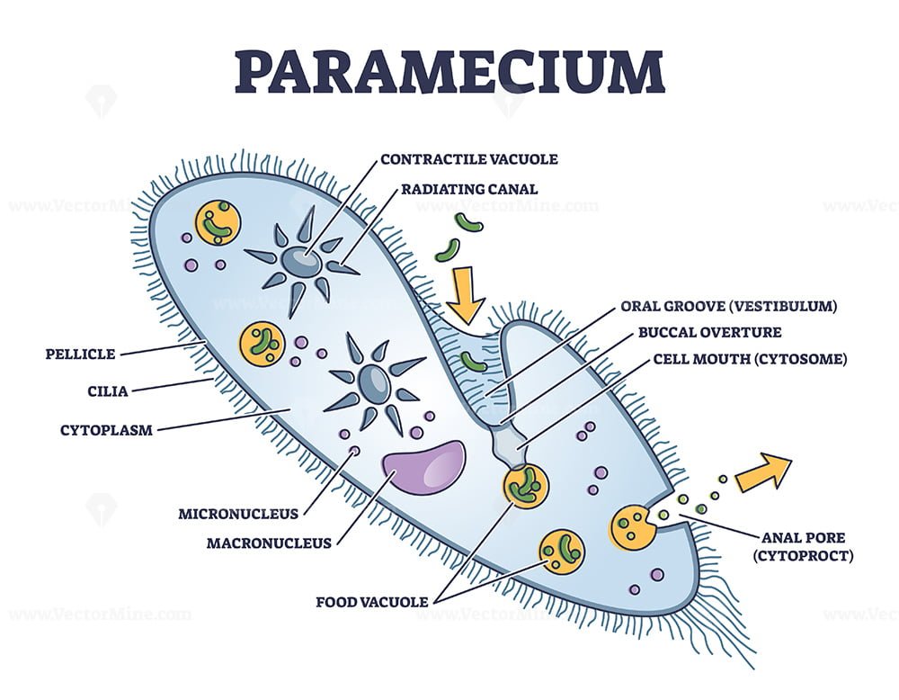
It is the pencil diagram of paramecium for class 10, 11 and 12. This will also help you to draw the structure and diagram of paramecium. Draw a neat labelled diagram of euglena. Web added a label for the buccal overture, a structure frequently mislabeled as the cytostome on diagrams of paramecium. Sience biology in easy steps and compact way.

How to draw paramecium step by step easy paramecium diagram YouTube
Web this video explains how to draw paramecium : For an accurate representation of these structures, see: Cv contractile vacuoles, fv food vacuoles, manu macronucleus, mino micronucleus, pe peristome, tr trichocysts and ve vestibulum.panel 1 (d): Paramecium are widespread in freshwater, brackish, and marine environments and are. The food further passes into the gullet through the mouth.
Paramecium Definition, Structure, Characteristics and Diagram
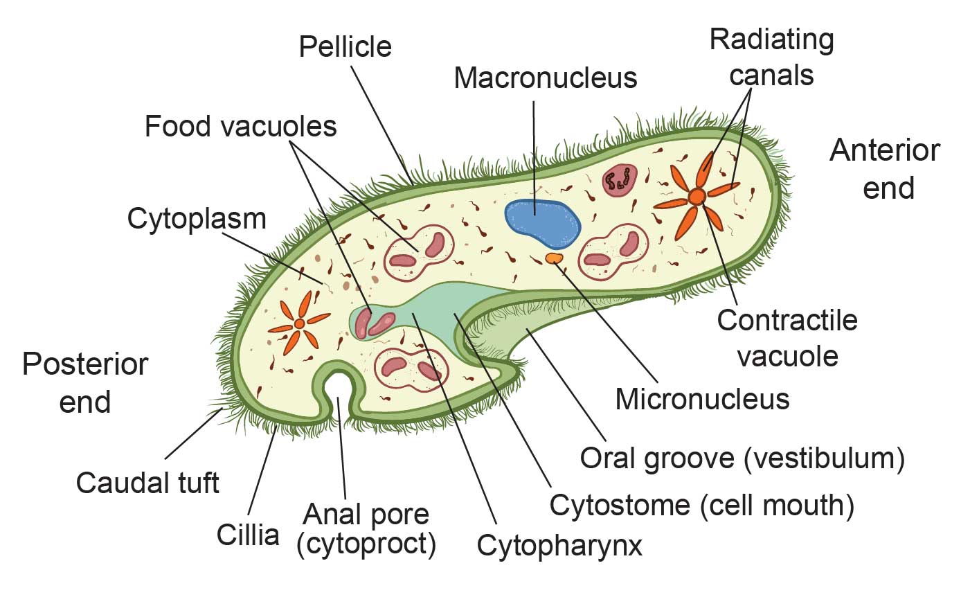
Binary fission divides a cell transversely and followed by mitotic division in the micronucleus. Unlike amoeba, paramecium has a distinct and permanent shape and certain areas of cytoplasm, (cell organelles), are specialised to carry out specific functions. Sience biology in easy steps and compact way. Paramecium is found in freshwater habitats and is used as a model organism in scientific.

How To Draw Paramecium Diagram YouTube
Ralph wichterman, the biology of paramecium, 2nd edition, 1986 (fig. Web added a label for the buccal overture, a structure frequently mislabeled as the cytostome on diagrams of paramecium. Web in this article we will discuss about the structure of paramecium. Web fast forward in time a bit and there is some mystery around who might have published the first.

Paramecium Diagrams to Print 101 Diagrams
Web in this article we will discuss about the structure of paramecium. Draw a neat labelled diagram of human sperm. To gather the food it makes use of its cilia, making quick movements with cilia to draw the water along with its prey organisms inside the mouth opening through its oral groove. Pellicle is made up of a thin, gelatinous.
Paramecium microscopic closeup structure with anatomical outline
Cv contractile vacuoles, fv food vacuoles, manu macronucleus, mino micronucleus, pe peristome, tr trichocysts and ve vestibulum.panel 1 (d): It is a prey for other microorganisms such as didinium. The layer of the pellicle gives the paramecium a definite shape and good protection of its cell content. Their basic shape is an elongated oval with rounded or pointed ends, such.

What is Paramecium? (with pictures)
They are the most common of all ciliate organisms that are characterized by the presence of cilia all along their. Unlike amoeba, paramecium has a distinct and permanent shape and certain areas of cytoplasm, (cell organelles), are specialised to carry out specific functions. Paramecium contains organelles necessary to sustain life, including. Web added a label for the buccal overture, a.

DRAW IT NEAT How to draw Paramecium
Paramecium is found in freshwater habitats and is used as a model organism in scientific studies. Paramecium contains organelles necessary to sustain life, including. Web hello friends in this video i will tell you about how to draw labelled diagram of paramecium step by step for beginners in easy wayso friends if you have pro. To gather the food it.
The Structure of Paramecium Cell Rs' Science
Draw a neat labelled diagram of pbr322. Besides that, they are unique in having contractile vacuoles, mouth pore and anal pore. Web asexual reproduction in paramecium is by binary fission. The name “paramecium” was given to the. Web diagram of paramecium.
Paramecium Vary In Length From About 0.05 To 0.32 Mm (0.002 To 0.013 Inch).
Uniform ciliation all over body except at post, end. Web in this article we will discuss about the structure of paramecium. The mature cell divides into two cells and each grows rapidly and develops into a new organism. Web diagram of paramecium.
Web Next Drawing > Paramecium Is A Ciliate Protozoan.
For an accurate representation of these structures, see: 1,142 × 1,007 (149 kb) Web drawing paramecium and lab. Ralph wichterman, the biology of paramecium, 2nd edition, 1986 (fig.
Pellicle Is Made Up Of A Thin, Gelatinous Substance Produced By The Cell.
Sience biology in easy steps and compact way. Paramecium is found in freshwater habitats and is used as a model organism in scientific studies. They have a lifespan of a hundred, a thousand or even a million years. Paramecium comprises organelles common to eukaryotes.
Web #Paramecium #Howtodraw #Biologythis Is A Diagram Of The Paramecium.
Higher magnification of the metachronal. Light microscopic appearance of paramecium caudatum.panel 1 (c): The name “paramecium” was given to the. Draw a neat labelled diagram of human sperm.