Drawing Of Prokaryotic Cell
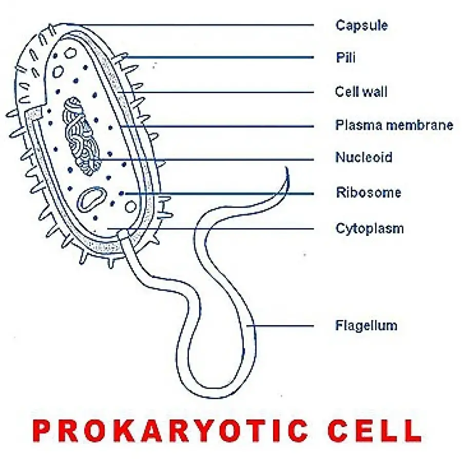
Drawing Of Prokaryotic Cell - The composition of their cell walls also differs from the eukaryotic cell walls. General science, 7th standard text book.this video explains how to draw pro. This scaffolding provides structural support to the cell and plays a role in cell division. Web it is useful to draw a prokaryotic cell easily.#cell #pactriotic Web a prokaryotic cell structure is as follows: As i go, i give tips on drawing the various structures. Web diagram and micrograph of intestinal cells, showing the protruding fingers of plasma membrane—called microvilli—that contact the fluid inside the small intestine. Web bacterial cell anatomy and internal structure. Watch this video tutorial and follow the step by step instructions to create a realistic and colorful diagram of a bacterial. The following image is a diagram of a prokaryotic cell;
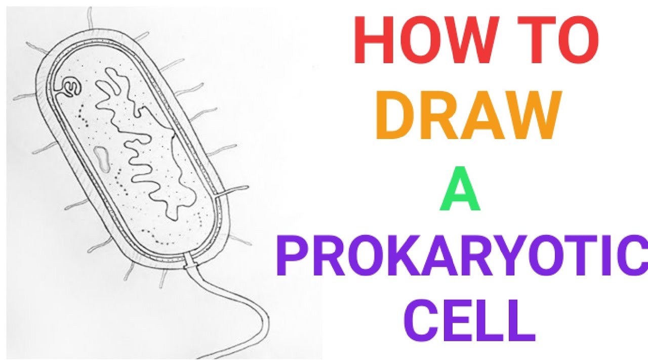
Genetic material is not enclosed by a nuclear membrane. Web do you want to learn how to draw a prokaryotic cell in an easy way? Web i draw a bacterial cell to show you how to make an accurate biological drawing of a prokaryotic cell. This is a good example of how scientific knowledge is revised as. Web diagram and micrograph of intestinal cells, showing the protruding fingers of plasma membrane—called microvilli—that contact the fluid inside the small intestine. The anatomy of a bacterial cell prokaryotic cell structure. The figure below shows the sizes of prokaryotic, bacterial, and eukaryotic, plant and animal, cells as well as other molecules and organisms on a. General science, 7th standard text book.this video explains how to draw pro. Our body has over 100 trillion bacterial cells. Watch this video tutorial and follow the step by step instructions to create a realistic and colorful diagram of a bacterial.
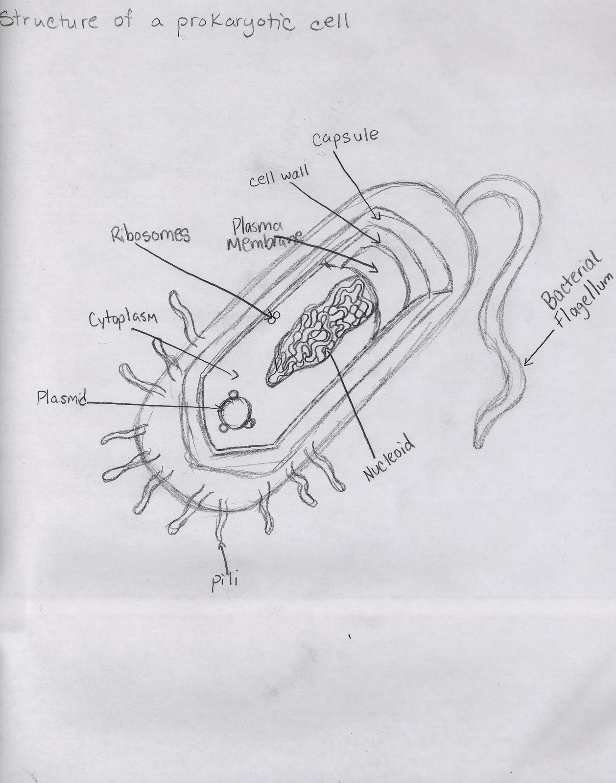
These cells are very minute in size 0.1 to 5.0 μ m. Recall that prokaryotes are divided into two different domains, bacteria and archaea, which together with eukarya, comprise the three domains of life (figure 27.2.3 27.2. The features of a typical prokaryotic cell are shown. The composition of their cell walls also differs from the eukaryotic cell walls. Prokaryotic cell size ranges from 0.1 to 5.0 μm in diameter. Evidence from fossil studies shows that prokaryotes exist on earth since 3.5 billion years ago. Prokaryotic cells do not have a true nucleus that contains their genetic material as eukaryotic cells do. In this case, a bacterium. We will shortly come to see that this is significantly different in eukaryotes. In prokaryotes, which lack a nucleus, cytoplasm.
Simple Prokaryotic Cell Diagram
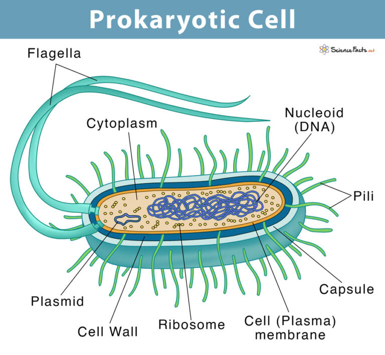
Web hello friends!!!!in this video, i will be showing you that how to draw a prokaryotic cell very easily.please like, share and subscribe!!! Web prokaryotic cell diagram and facts. Prokaryotic cells do not have a true nucleus that contains their genetic material as eukaryotic cells do. Typical prokaryotic cells range from 0.1 to 5.0 micrometers (μm) in diameter and are.
2.3 Unique Characteristics of Prokaryotic Cells Allied Health
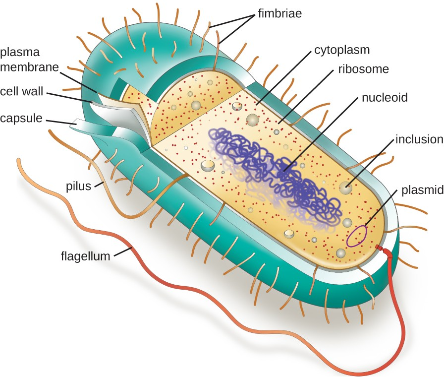
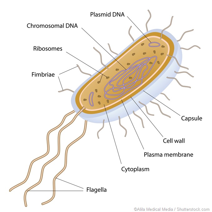
Web the diagram of prokaryotic cell show that it contains genetic material in a nucleoid region, have a cell wall, and may possess flagella or pili for movement and attachment. Many also have a capsule or slime layer made of polysaccharide. Web prokaryotic cell diagram. These cells are very minute in size 0.1 to 5.0 μ m. The features of.

Prokaryotic Cell Diagram and Facts
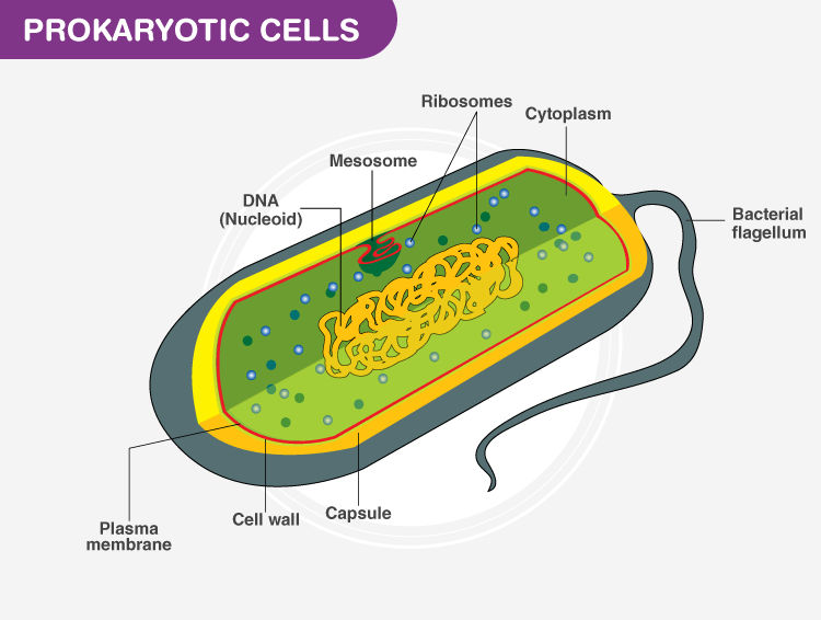
This is a good example of how scientific knowledge is revised as. Diagram of a typical prokaryotic cell. Prokaryotic cells do not have a true nucleus that contains their genetic material as eukaryotic cells do. The composition of their cell walls also differs from the eukaryotic cell walls. Recall that prokaryotes are divided into two different domains, bacteria and archaea,.
Prokaryotic Cells Definition, Structure, Characteristics, and Examples
Common prokaryotic cell is a bacterial cell. In eukaryotic cells, which have a nucleus, the cytoplasm is everything between the plasma membrane and the nuclear envelope. This scaffolding provides structural support to the cell and plays a role in cell division. Our body has over 100 trillion bacterial cells. These neat, well labelled and.

Prokaryote Wikipedia
Web hello friends!!!!in this video, i will be showing you that how to draw a prokaryotic cell very easily.please like, share and subscribe!!! To help you remember prokaryotes parts and pieces. It helps in moisture retention, protects the cell when engulfed, and helps in the attachment of cells to nutrients and surfaces. The cell wall of a prokaryote acts as.
Prokaryotic Cell Definition, Examples, & Structure
Web it is useful to draw a prokaryotic cell easily.#cell #pactriotic Prokaryotic dna is found in a central part of the cell: To help you remember prokaryotes parts and pieces. Prokaryotic cell size ranges from 0.1 to 5.0 μm in diameter. The composition of their cell walls also differs from the eukaryotic cell walls.
How to draw a prokaryotic cell prokaryotic organism Bacterial cell
Genetic material is not enclosed by a nuclear membrane. General science, 7th standard text book.this video explains how to draw pro. Web prokaryotic cell diagram. The features of a typical prokaryotic cell are shown. Web thanks for watching!i am demonstrating the colorful diagram of prokaryotic cells step by step which you can draw very easily.
Prokaryotes
Recall that prokaryotes are divided into two different domains, bacteria and archaea, which together with eukarya, comprise the three domains of life (figure 27.2.3 27.2. The structure called a mesosome was once thought to be an organelle. ️ ️ ️ and do tell. In prokaryotes, which lack a nucleus, cytoplasm. The main components of a prokaryotic cell are the plasma.
Cell Types and Structure Structure of Prokaryotic Cell
We will shortly come to see that this is significantly different in eukaryotes. The anatomy of a bacterial cell prokaryotic cell structure. In eukaryotic cells, which have a nucleus, the cytoplasm is everything between the plasma membrane and the nuclear envelope. These cells are very minute in size 0.1 to 5.0 μ m. Common prokaryotic cell is a bacterial cell.

prokaryotic cell parts and functions
Prokaryotic cell size ranges from 0.1 to 5.0 μm in diameter. Web i draw a bacterial cell to show you how to make an accurate biological drawing of a prokaryotic cell. As i go, i give tips on drawing the various structures. Typical prokaryotic cells range from 0.1 to 5.0 micrometers (μm) in diameter and are significantly smaller than eukaryotic.
Common Prokaryotic Cell Is A Bacterial Cell.
General science, 7th standard text book.this video explains how to draw pro. Web a prokaryotic cell structure is as follows: ️ ️ ️ and do tell. Web diagram and micrograph of intestinal cells, showing the protruding fingers of plasma membrane—called microvilli—that contact the fluid inside the small intestine.
Our Body Has Over 100 Trillion Bacterial Cells.
These neat, well labelled and. The anatomy of a bacterial cell prokaryotic cell structure. Web hello friends!!!!in this video, i will be showing you that how to draw a prokaryotic cell very easily.please like, share and subscribe!!! Web bacterial cell anatomy and internal structure.
Prokaryotes Are Smaller Than Eukaryotic Cells And Include Bacteria And Archaea.
Web do you want to learn how to draw a prokaryotic cell in an easy way? Web i draw a bacterial cell to show you how to make an accurate biological drawing of a prokaryotic cell. In this case, a bacterium. Evidence from fossil studies shows that prokaryotes exist on earth since 3.5 billion years ago.
Typical Prokaryotic Cells Range From 0.1 To 5.0 Micrometers (Μm) In Diameter And Are Significantly Smaller Than Eukaryotic Cells, Which Usually Have Diameters Ranging From 10 To 100 Μm.
Watch this video tutorial and follow the step by step instructions to create a realistic and colorful diagram of a bacterial. Prokaryotes can be found almost anywhere on earth, from land to water bodies, atmosphere to hydrothermal vents, and even inside living organisms, including humans. Cells vary regarding other components. The structure called a mesosome was once thought to be an organelle.