Drawing Of The Eye Anatomy
Drawing Of The Eye Anatomy - Start with a basic sketch. At the center of the iris is a clear opening, the pupil. The clear watery fluid in the front of the eyeball. The optic nerve is the largest sensory nerve of the eye. Contrary to popular belief, the eyes are not perfectly spherical; The eye is the organ that allows sight. Eyeball [25:37] structure of the eyeball seen in a transverse section. How to learn the parts of the eye. Web 6 min read. Tubes (arteries and veins) that carry blood to and from the eye.
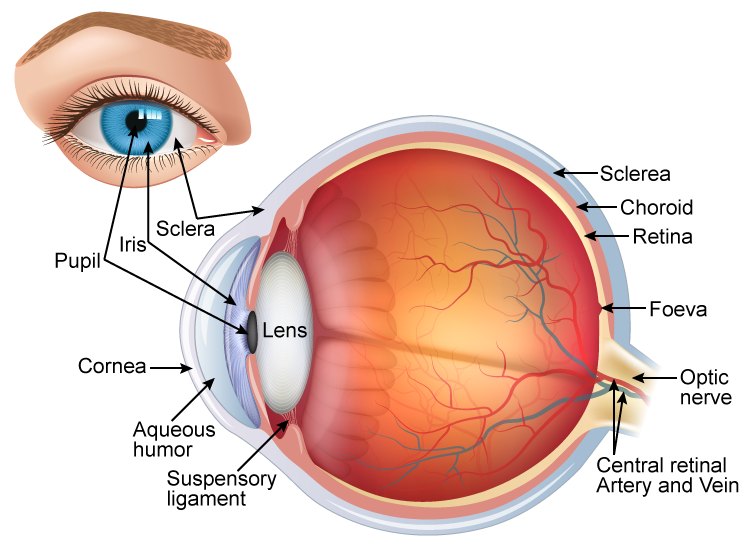
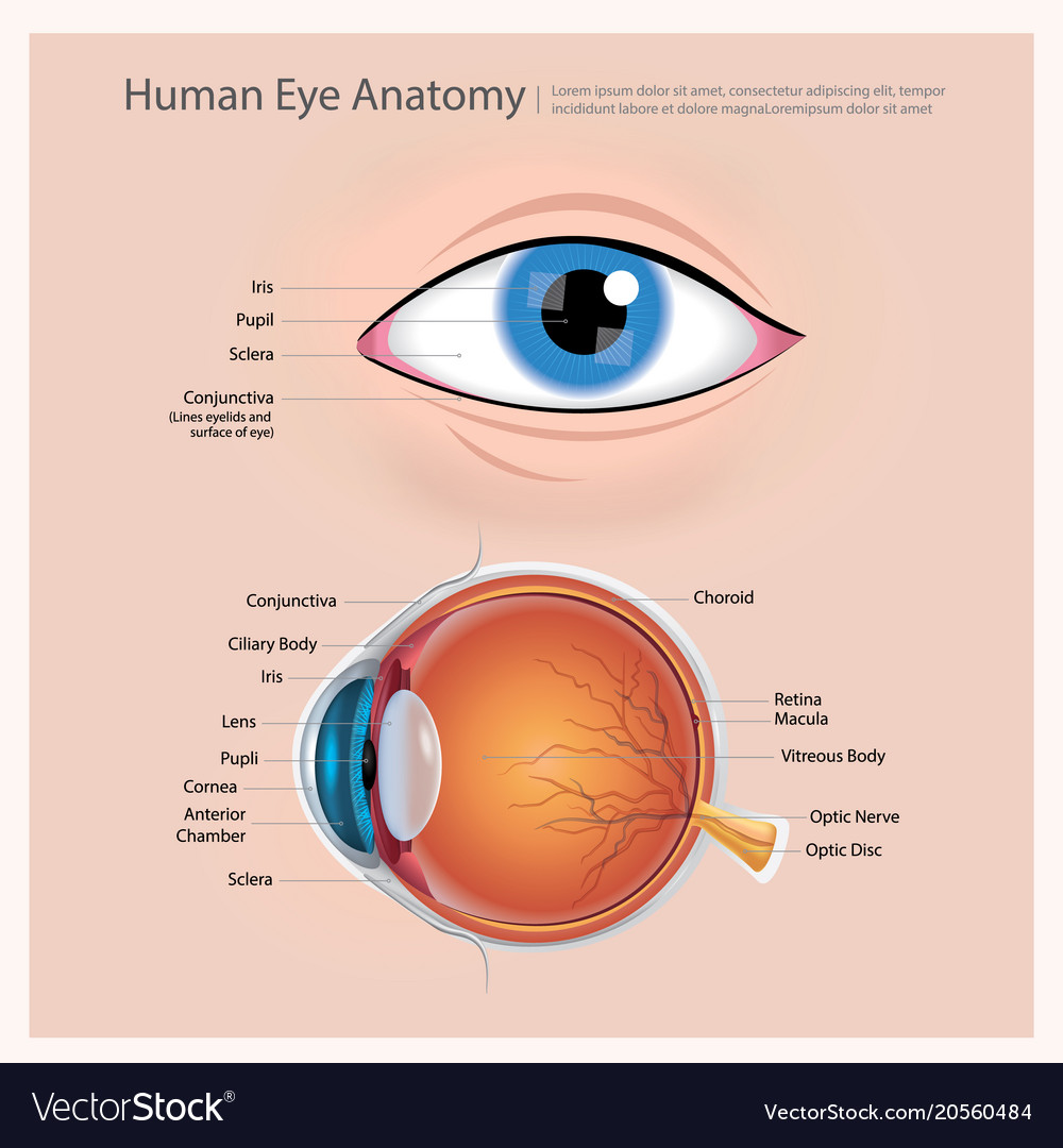
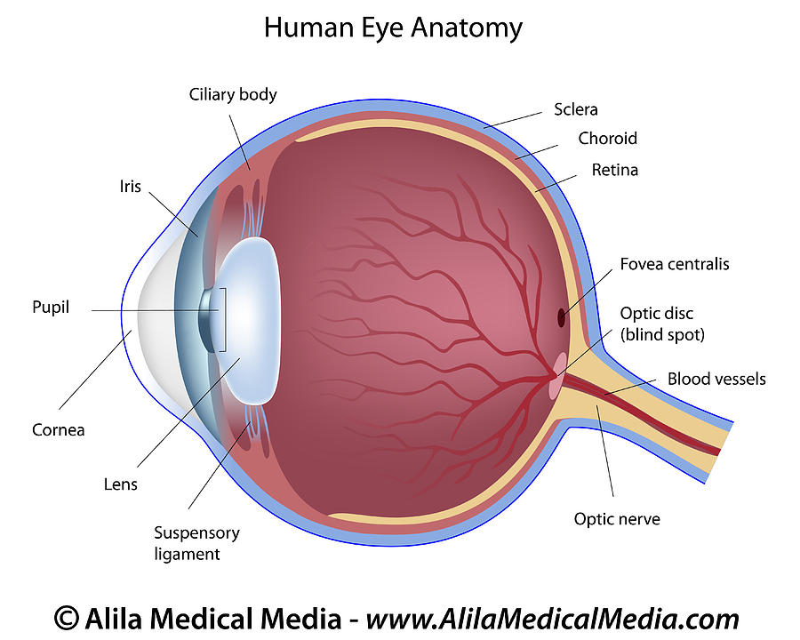
The medical information on this site is provided as an information resource only, and is. Cornea, anterior chamber, lens, vitreous chamber and retina: The eyeball, eye socket, brow ridge, eyelids, tear duct, sclera, iris, pupil, cornea, glabella, and epicanthic fold. The front part (what you see in the mirror) includes: Behind the cornea, the colored iris forms an incomplete partition within the eye. These interactive figures are provided for use in medical student education. Eyeball (bulbus oculi) the eye is a highly specialized sensory organ located within the bony orbit. Web anatomy of the eye. In this tutorial i cover how to draw the structure of the eye and it’s anatomy. External landmarks and extraocular muscles.
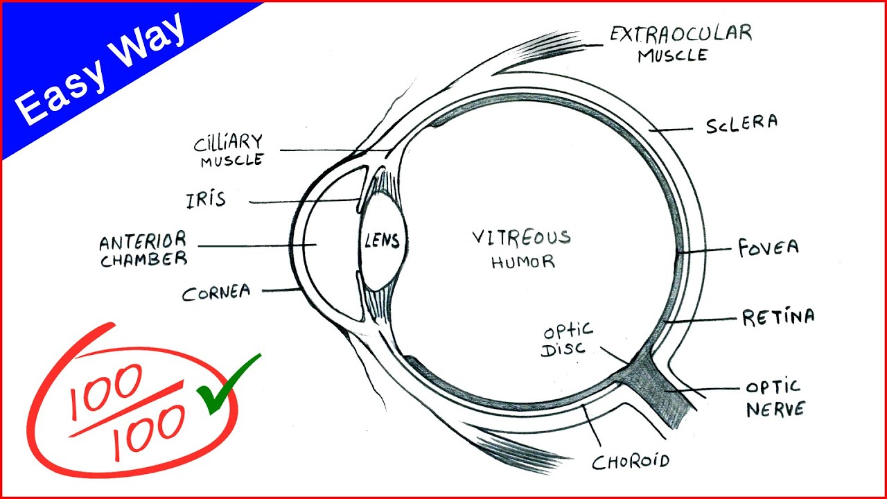
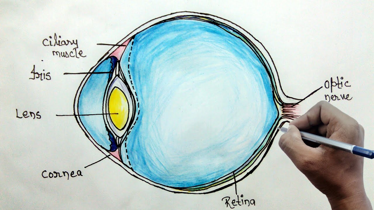
The lens is a clear part of the eye behind the iris that helps to focus light, or an image, on the retina. In this tutorial i cover how to draw the structure of the eye and it’s anatomy. Web anatomy for artists: Dec 26, 2023 3:25 pm est. Your eye is a slightly asymmetrical globe, about an inch in diameter. Web the eye's structure includes the sclera, cornea, conjunctiva, aqueous humour, lens, ciliary body, iris, pupil, vitreous humour, retina, optic nerve, choroid, fovea, and macula. Web 6 min read. The clear watery fluid in the front of the eyeball. The conjunctiva is the membrane covering the sclera (white portion of your eye). For more video tutorials visit www.proko.com and subscribe to the newsletter.
draw a neat and labelled diagram of structure of the human eye slwbyx77
Web structure of the eye: Web human eye, specialized sense organ in humans that is capable of receiving visual images, which are relayed to the brain. Here is a tour of the eye starting from the outside, going in through the front and working to the back. Anatomy of the human eye. The lens is a clear part of the.
Human eye anatomy Royalty Free Vector Image VectorStock
The conjunctiva is the membrane covering the sclera (white portion of your eye). Web labeled diagram of the eye. Web the cornea is the transparent window that allows light to enter the eye. How to learn the parts of the eye. The lens is a clear part of the eye behind the iris that helps to focus light, or an.
/GettyImages-1128675065-e4bac15b0f39449dba31f25f1020bc8a.jpg)
An Overview of Eye Anatomy
The front part (what you see in the mirror) includes: Here is a tour of the eye starting from the outside, going in through the front and working to the back. Web of light entering the eye. Web 6 min read. Web labeled diagram of the eye.
Eye Diagram drawing CBSE easy way draw Human eye anatomy Step
Behind the cornea, the colored iris forms an incomplete partition within the eye. Web the cornea is the transparent window that allows light to enter the eye. Web reviewed by ninel z gregori, md. The eyeball, eye socket, brow ridge, eyelids, tear duct, sclera, iris, pupil, cornea, glabella, and epicanthic fold. Bhavin shah, neurodevelopmental and behavioral optometrist specializing in myopia.

Human Eye Anatomy Parts of the Eye Explained Eye anatomy, Basic
A clear dome over the iris. Web anatomy for artists: Web this article uses anatomical terminology. It is located in the center of the retina. Web human eye, specialized sense organ in humans that is capable of receiving visual images, which are relayed to the brain.
How to draw human eye diagram for beginners YouTube
These interactive figures are provided for use in medical student education. The top panel shows outside of the eye including the eyelid, pupil, sclera, and iris; The medical information on this site is provided as an information resource only, and is. Dec 26, 2023 3:25 pm est. Tubes (arteries and veins) that carry blood to and from the eye.

File1413 Structure of the Eye.jpg Wikimedia Commons
In this tutorial i cover how to draw the structure of the eye and it’s anatomy. The front part (what you see in the mirror) includes: Web of light entering the eye. Start with a basic sketch. The cornea is more sharply curved than the sclera:

Diagram human eye anatomy with label Royalty Free Vector
The lens is a clear part of the eye behind the iris that helps to focus light, or an image, on the retina. A clear dome over the iris. Web please use one of the following formats to cite this article in your essay, paper or report: Quiz on the 5 layers of the cornea. It is located in the.

Anatomy of the Human Eye
Web structure of the eye: Get to know the eye structure. Superior rectus, inferior rectus, medial rectus, lateral rectus, superior oblique, inferior oblique, levator palpebrae superioris intrinsic: Tubes (arteries and veins) that carry blood to and from the eye. It's made up of many parts—each with specific names and functions.
Eye Anatomy Labeled Drawing
Web human eye anatomy (seen from above) for more details about specific structures of the eye and how they function, visit these pages: The macula is the small, sensitive area of the retina that gives central vision. The cornea is more sharply curved than the sclera: For more video tutorials visit www.proko.com and subscribe to the newsletter. 2.9m views 11.
A Clear Dome Over The Iris.
Web this article uses anatomical terminology. Tubes (arteries and veins) that carry blood to and from the eye. Web the eye's structure includes the sclera, cornea, conjunctiva, aqueous humour, lens, ciliary body, iris, pupil, vitreous humour, retina, optic nerve, choroid, fovea, and macula. The clear watery fluid in the front of the eyeball.
The Conjunctiva Is The Membrane Covering The Sclera (White Portion Of Your Eye).
Cornea, anterior chamber, lens, vitreous chamber and retina: Anatomy of the human eye. Unlabeled diagram of the eye. The white of the eye.
It's Made Up Of Many Parts—Each With Specific Names And Functions.
Here is a tour of the eye starting from the outside, going in through the front and working to the back. External landmarks and extraocular muscles. These parts work together to capture light and convert it into images. The macula is the small, sensitive area of the retina that gives central vision.
The Lens Is A Clear Part Of The Eye Behind The Iris That Helps To Focus Light, Or An Image, On The Retina.
The eyeball, eye socket, brow ridge, eyelids, tear duct, sclera, iris, pupil, cornea, glabella, and epicanthic fold. 445k views 3 years ago. Part of the teachme series. Web of light entering the eye.