Drawing Of The Spinal Cord
Drawing Of The Spinal Cord - Introduction spinal cord injury (sci). Web choose from drawing of spinal cord stock illustrations from istock. Web this article looks at the spinal cord’s function and anatomy and includes an interactive diagram. The spinal cord is divided into five different parts. The spinal cord begins at the base of the brain and extends into the pelvis. Solidify your knowledge about the spinal cord structures with our interactive study materials. Web the spinal cord is a cylinder that is roughly 45 cm long and 1 cm wide. Representation in 3/4 front view of the stucture of the spinal cord, and rachidian nerves. The vertebral arteries are the main source of blood to the spinal cord. The last ten years the research has been absolutely amazing, said dr.
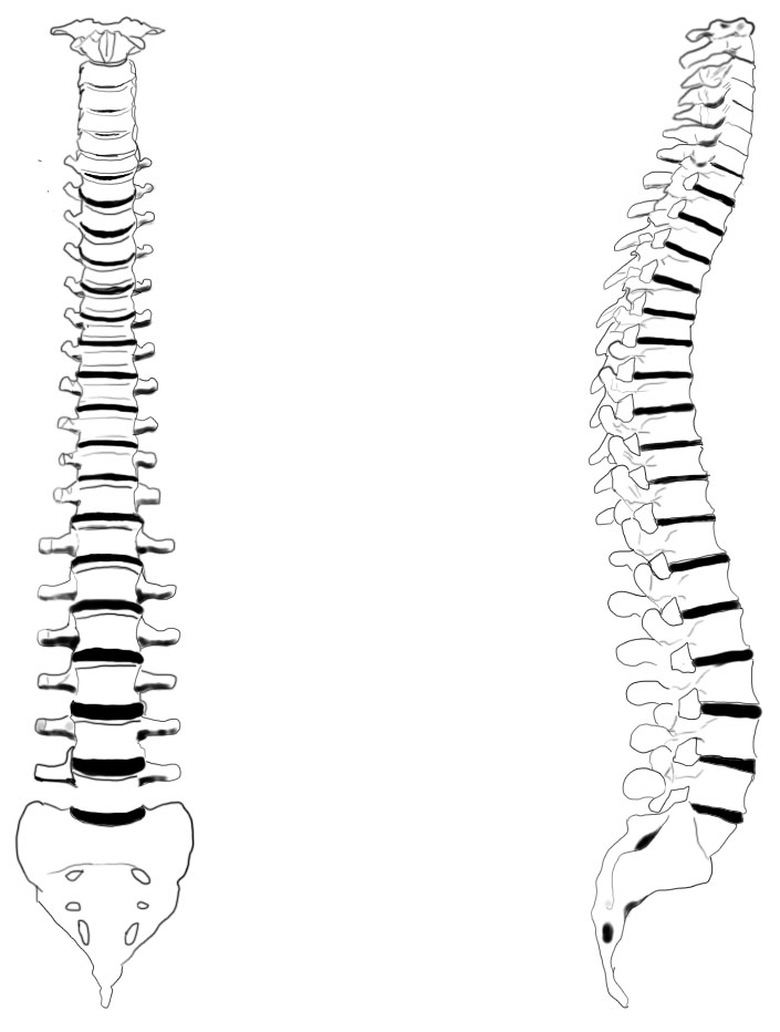
The spinal cord begins at the base of the brain and extends into the pelvis. In adults, the spinal cord is usually 40cm long and 2cm wide. It forms a vital link between the brain and the body. Spinal cord drawing stock illustrations. Web in summary, the descending tracts of the spinal cord are: Web the spinal cord is a cylinder that is roughly 45 cm long and 1 cm wide. Your spinal cord is a cylindrical structure that runs through the center of your spine, from your brainstem to your low back. The vertebral arteries are the main source of blood to the spinal cord. Web this article looks at the spinal cord’s function and anatomy and includes an interactive diagram. However, the following arteries branch from the vertebral arteries to directly supply the spinal cord itself:
Web the nervous system is divided into two main parts: 94k views 4 years ago. Web the feat of implanting the electrodes was accomplished by jocelyne bloch, a neurosurgeon at lausanne university hospital (chuv). Web this article looks at the spinal cord’s function and anatomy and includes an interactive diagram. It then travels inferiorly within the vertebral canal, surrounded by the spinal meninges containing cerebrospinal fluid. However, the following arteries branch from the vertebral arteries to directly supply the spinal cord itself: Web how to draw t.s. Web old engraved illustration of human skeletons. Concept of health care technology, parts of skeleton in anatomical science. Web spinal cord, drawing the spinal cord.

Cross section of 4 of the spinal cord's 31 segments. Source Pearson
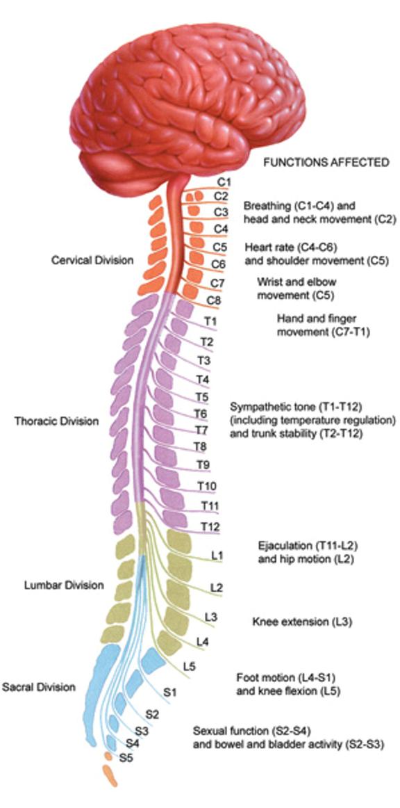
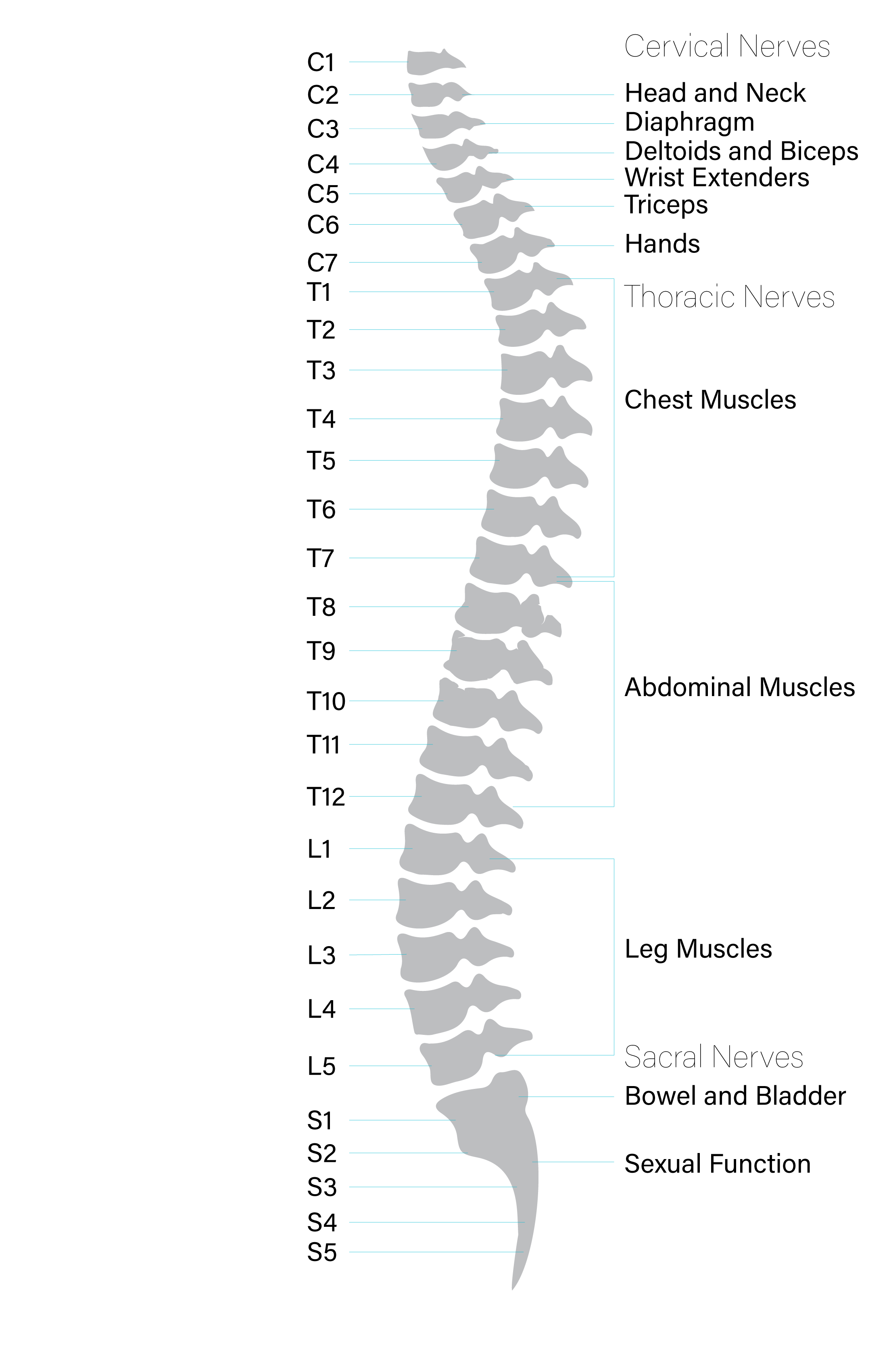
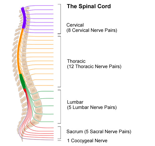
Ventral funiculus, cuneate fasciculus, gracile fasciculus. View spinal cord drawing videos. It then travels inferiorly within the vertebral canal, surrounded by the spinal meninges containing cerebrospinal fluid. Your spinal cord is a cylindrical structure that runs through the center of your spine, from your brainstem to your low back. Diagram of the spinal cord showing segments.

Spinal Cord Diagram with Detailed Illustrations and Clear Labels
Ventral funiculus, cuneate fasciculus, gracile fasciculus. Web spinal cord, drawing the spinal cord. However, the following arteries branch from the vertebral arteries to directly supply the spinal cord itself: Spinal cord drawing stock photos are available in a variety of sizes and formats to fit your needs. The spinal cord is part of the central nervous system (cns), which extends.

Spinal Cord Neurology Medbullets Step 1
However, the following arteries branch from the vertebral arteries to directly supply the spinal cord itself: Introduction spinal cord injury (sci). Diagram of the spinal cord showing segments. It forms a vital link between the brain and the body. Ventral funiculus, cuneate fasciculus, gracile fasciculus.
Human Spinal Cord Drawing Sketch Coloring Page
Lateral and ventral (anterior) corticospinal tracts deal with voluntary, discrete, skilled motor activities. Web in summary, the descending tracts of the spinal cord are: View spinal cord drawing videos. Web structure [ edit] parts of human spinal cord. However, the following arteries branch from the vertebral arteries to directly supply the spinal cord itself:

Anatomy of the spinal cord. Download Scientific Diagram
Web the nervous system is divided into two main parts: It's a delicate structure that contains nerve bundles and cells that carry messages from your brain to the rest of your body. Web anatomy of the spinal cord. Web the spinal cord is a cylinder that is roughly 45 cm long and 1 cm wide. Web old engraved illustration of.

Spinal Cord, Drawing Stock Image C017/1520 Science Photo Library
What is the spinal cord? Concept of health care technology, parts of skeleton in anatomical science. Web spine anatomy, diagram & pictures | body maps. Spinal cord drawing stock illustrations. Blood vessels of the spinal cord [12:23] arteries and veins of the spinal cord.
Spinal Cord Diagram
Web the nervous system is divided into two main parts: It has a relatively simple anatomical course: It then travels inferiorly within the vertebral canal, surrounded by the spinal meninges containing cerebrospinal fluid. Web anatomy of the spinal cord. Web spine anatomy, diagram & pictures | body maps.
Anatomy of the Spinal Cord Praxis Spinal Cord Institute
Spinal cord, funiculi of spinal cord, tectospinal tract, anterior funiculus; The spinal cord is divided into five different parts. Lateral and ventral (anterior) corticospinal tracts deal with voluntary, discrete, skilled motor activities. Web anatomy of the spinal cord. Blood supply of the spinal cord.

The Spinal Cord Neurologic Clinics
Introduction spinal cord injury (sci). Web anatomy of the spinal cord. Diagram of the spinal cord showing segments. Web in summary, the descending tracts of the spinal cord are: Web this article looks at the spinal cord’s function and anatomy and includes an interactive diagram.
Anatomy of the Spinal Cord Stanford Medicine Children's Health
Lateral and ventral (anterior) reticulospinal tracts provide excitatory or inhibitory regulation of voluntary movements and reflexes. Web the nervous system is divided into two main parts: Representation in 3/4 front view of the stucture of the spinal cord, and rachidian nerves. Your spinal cord is a cylindrical structure that runs through the center of your spine, from your brainstem to.
The Spinal Cord Is A Long Bundle Of Nerves And Cells That Carries Signals Between The.
94k views 4 years ago. Step by step, he said, it has. The spinal cord arises cranially as a continuation of the medulla oblongata (part of the brainstem). Web spinal cord, drawing the spinal cord.
Web The Spinal Cord Is A Cylinder That Is Roughly 45 Cm Long And 1 Cm Wide.
It has a relatively simple anatomical course: Web spine anatomy, diagram & pictures | body maps. Concept of health care technology, parts of skeleton in anatomical science. Lateral and ventral (anterior) corticospinal tracts deal with voluntary, discrete, skilled motor activities.
It's A Delicate Structure That Contains Nerve Bundles And Cells That Carry Messages From Your Brain To The Rest Of Your Body.
Web how to draw t.s. Web both agree that research on spinal cord injury treatment is more promising than most people realize. Representation in 3/4 front view of the stucture of the spinal cord, and rachidian nerves. Web old engraved illustration of human skeletons.
It Forms A Vital Link Between The Brain And The Body.
Web structure [ edit] parts of human spinal cord. Spinal cord drawing stock illustrations. Vector isolated set of spine pelvis, shoulder scapula or elbow, leg knee and foot ankle, arm and hand wrist with fingers for medical anatomy or surgery. Web keep learning about the white and grey matter of the spinal cord using our spinal cord diagram labeling exercises and quizzes!