Ecg Drawing
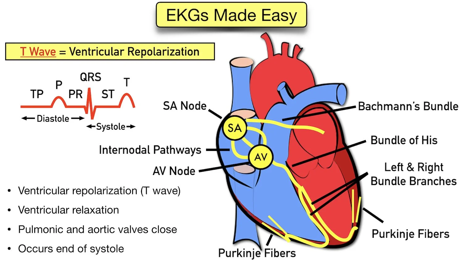
Ecg Drawing - Build your own ecgs rhythms, modify segments, and explore variations. Electrodes are placed on different parts of a patient’s limbs and chest to record the electrical activity. Web an ecg measures these changes in electrical signals (or, in fact, voltage) on different areas of skin and plots them as a graph. Packed with vital information, sparkson’s illustrated guide to ecg interpretation is an electrocardiography reference unlike any other. Web unique animated representation of the interplay of ecg image and visualisation of cardiac conduction, atrial, chamber, valve and ventricular function in 29 ecg findings relevant for practice! Web ecg is the abbreviated term for an electrocardiogram. Master the visual identification of normal and abnormal rhythms. The main components of an ekg wave include the following: The earliest manifestation of hyperkalaemia is an increase in t wave amplitude. Web learn how to draw a typical ecg by tracing out the shape of the electrocardiogram based on blood pressure and volume changes in the cardiac cycle.
It looks at how electrical impulses travel through the heart from various angles. Ecg changes generally do not manifest until there is a moderate degree of hyperkalaemia (≥ 6.0 mmol/l). When a depolarization current travels towards the electrode, it gets recorded as a positive deflection, and when it moves away from the electrode, it appears as a negative deflection. Web in order to successfully read an ekg, you must first understand the basics of an ekg waveform. Are you learning to interpret ecgs? Hyperkalaemia is defined as a serum potassium level of > 5.2 mmol/l. Web the ecg is one of the most useful investigations in medicine. It is used clinically to identify and locate pathology within the cardiac conducting system and within cardiac muscle. This applet lets you see the changes in blood pressure and volume curves, and explains them with popup windows, as you drag the mouse across the screen. Web learn how to draw a typical ecg by tracing out the shape of the electrocardiogram based on blood pressure and volume changes in the cardiac cycle.
It is used clinically to identify and locate pathology within the cardiac conducting system and within cardiac muscle. Learn for free about math, art, computer programming, economics, physics, chemistry, biology, medicine, finance, history, and more. Um diese website zu betreiben, ist es für uns notwendig cookies zu verwenden. Uses 3 electrodes (ra, la and ll) monitor displays the bipolar leads (i, ii and iii) Having a good system will avoid making errors. When a depolarization current travels towards the electrode, it gets recorded as a positive deflection, and when it moves away from the electrode, it appears as a negative deflection. The ekg/ecg is a printed capture of a brief moment in time. Cardiac axis represents the sum of depolarisation vectors generated by individual cardiac myocytes. Packed with vital information, sparkson’s illustrated guide to ecg interpretation is an electrocardiography reference unlike any other. The earliest manifestation of hyperkalaemia is an increase in t wave amplitude.

5Lead ECG Interpretation (Electrocardiogram) Tips for Nurses FRESHRN
It is used to record the electrical activity of the heart from different angles to both identify and locate pathology. Web the ecg is one of the most useful investigations in medicine. Diagram of very simple and easy standard ecg. The main components of an ekg wave include the following: It depicts if the heart has enlarged due to hypertension.

The Electrocardiogram explained What is an ECG?
Control the simulator remotely for impactful presentations and group learning. Learn for free about math, art, computer programming, economics, physics, chemistry, biology, medicine, finance, history, and more. It is an electrogram of the heart which is a graph of voltage versus time of the electrical activity of the heart using electrodes placed on the skin. Having a good system will.
ECG Waveform Explained EKG Labeled Diagrams and Components — EZmed
Uses 3 electrodes (ra, la and ll) monitor displays the bipolar leads (i, ii and iii) Web the electrocardiogram (ecg) is used to trace the electrical activity in cardiac tissue. Ecg changes generally do not manifest until there is a moderate degree of hyperkalaemia (≥ 6.0 mmol/l). Web comprehensive tutorial on ecg interpretation, covering normal waves, durations, intervals, rhythm and.
![[Solved] How to draw an EKG tracing with TIkZ 9to5Science](https://i.stack.imgur.com/KnZVz.jpg)
[Solved] How to draw an EKG tracing with TIkZ 9to5Science
Web an ecg measures these changes in electrical signals (or, in fact, voltage) on different areas of skin and plots them as a graph. Hyperkalaemia is defined as a serum potassium level of > 5.2 mmol/l. Web in order to successfully read an ekg, you must first understand the basics of an ekg waveform. Packed with vital information, sparkson’s illustrated.

The Electrocardiogram explained What is an ECG?
Web the electrocardiogram (ecg) is used to trace the electrical activity in cardiac tissue. When a depolarization current travels towards the electrode, it gets recorded as a positive deflection, and when it moves away from the electrode, it appears as a negative deflection. Control the simulator remotely for impactful presentations and group learning. Web comprehensive tutorial on ecg interpretation, covering.
The Normal ECG Trace ECG Basics MedSchool
Web the electrocardiogram (ecg) is used to trace the electrical activity in cardiac tissue. Web an ekg/ecg is a representation of the electrical activity of the heart muscle as it changes with time, usually printed on paper for easier analysis. Web learn how to draw a typical ecg by tracing out the shape of the electrocardiogram based on blood pressure.
A Basic Guide to ECG/EKG Interpretation First Aid for Free
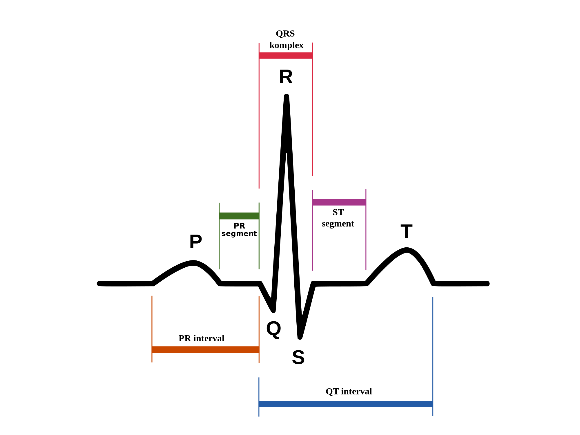
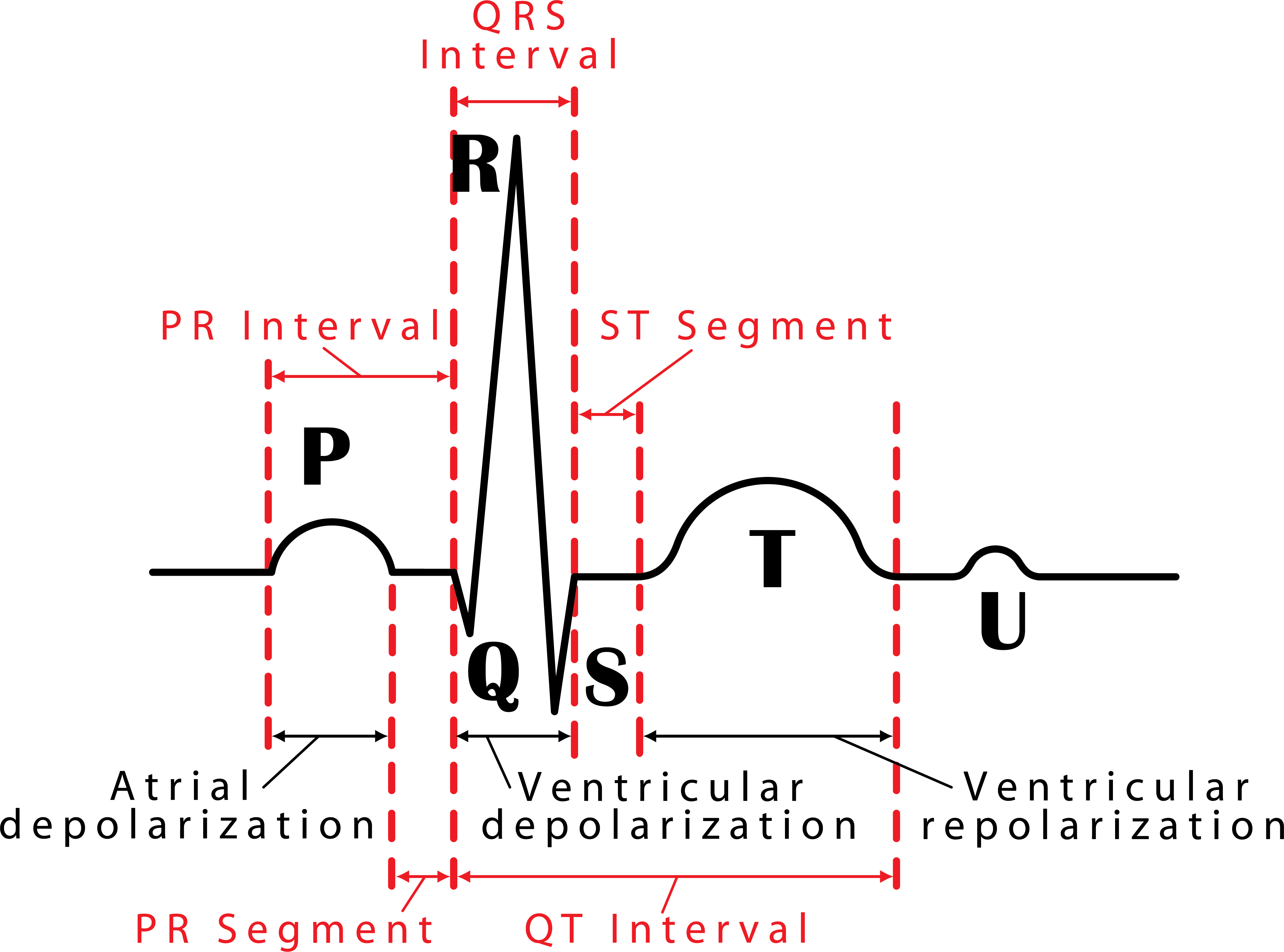
Web an electrocardiogram (ecg) is a graphic record produced by an electrocardiograph that provides details about one’s heart rate and rhythm and any other related abnormalities; This is the well labelled diagram of standard ecg. Learn for free about math, art, computer programming, economics, physics, chemistry, biology, medicine, finance, history, and more. Clinically is is reflected by the ventricular axis,.

12Lead ECG Placement Guide with Illustrations
Hyperkalaemia is defined as a serum potassium level of > 5.2 mmol/l. There are also intervals that will be discussed: The book’s format is both innovative and captivating, ensuring that readers retain a wealth of practical knowledge for accurate ecg interpretation. Web in order to successfully read an ekg, you must first understand the basics of an ekg waveform. Failure.
048 How to Read an Electrocardiogram (ECG/EKG) Interactive Biology
To start with we will cover the basics of the ecg, how it is recorded and the basic physiology. It is used clinically to identify and locate pathology within the cardiac conducting system and within cardiac muscle. Um diese website zu betreiben, ist es für uns notwendig cookies zu verwenden. Are you learning to interpret ecgs? Having a good system.
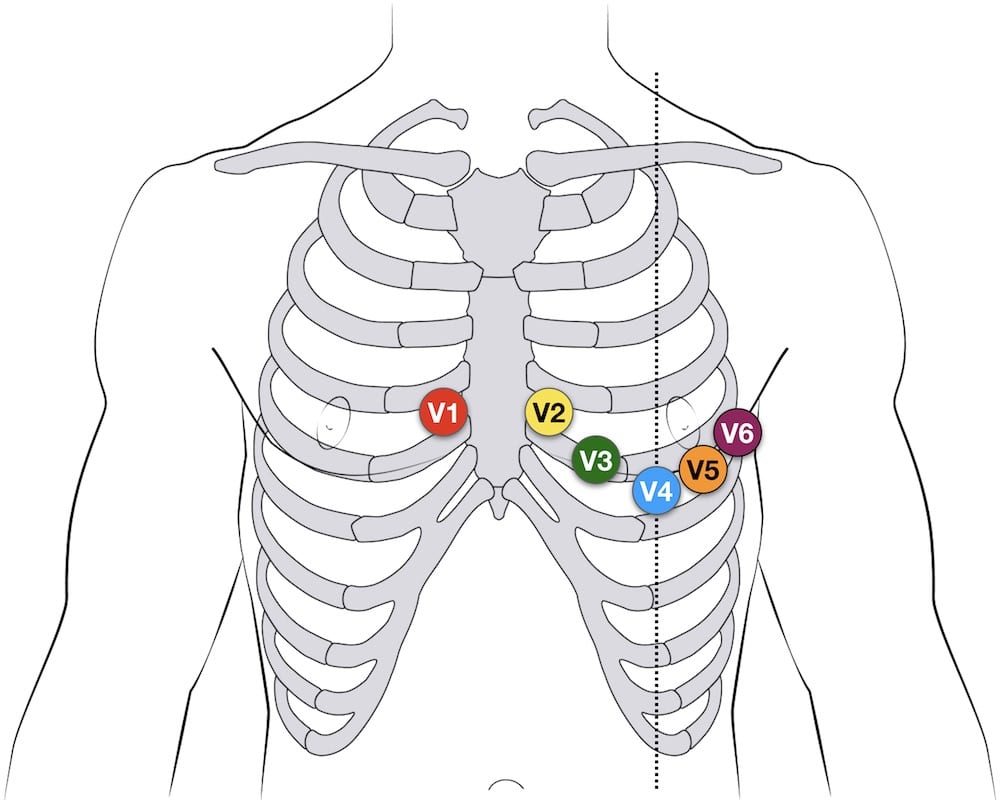
ECG Lead positioning • LITFL • ECG Library Basics
This applet lets you see the changes in blood pressure and volume curves, and explains them with popup windows, as you drag the mouse across the screen. Web an electrocardiogram (ecg) is a graphic record produced by an electrocardiograph that provides details about one’s heart rate and rhythm and any other related abnormalities; Web the electrocardiogram (ecg) is used to.
Web The Ecg Must Always Be Interpreted Systematically.
Um diese website zu betreiben, ist es für uns notwendig cookies zu verwenden. The book’s format is both innovative and captivating, ensuring that readers retain a wealth of practical knowledge for accurate ecg interpretation. Ecg changes generally do not manifest until there is a moderate degree of hyperkalaemia (≥ 6.0 mmol/l). The earliest manifestation of hyperkalaemia is an increase in t wave amplitude.
It Is Used To Record The Electrical Activity Of The Heart From Different Angles To Both Identify And Locate Pathology.
Cardiac axis represents the sum of depolarisation vectors generated by individual cardiac myocytes. Web learn how to draw a typical ecg by tracing out the shape of the electrocardiogram based on blood pressure and volume changes in the cardiac cycle. Uses 3 electrodes (ra, la and ll) monitor displays the bipolar leads (i, ii and iii) Web an ecg measures these changes in electrical signals (or, in fact, voltage) on different areas of skin and plots them as a graph.
Failure To Perform A Systematic Interpretation Of The Ecg May Be Detrimental.
Web unique animated representation of the interplay of ecg image and visualisation of cardiac conduction, atrial, chamber, valve and ventricular function in 29 ecg findings relevant for practice! Build your own ecgs rhythms, modify segments, and explore variations. It is used clinically to identify and locate pathology within the cardiac conducting system and within cardiac muscle. As with all investigations the most important things are your findings on history, examination and basic observations.
Web Electrocardiography Is The Process Of Producing An Electrocardiogram (Ecg Or Ekg), A Recording Of The Heart's Electrical Activity Through Repeated Cardiac Cycles.
This is the well labelled diagram of standard ecg. Clinically is is reflected by the ventricular axis, and interpretation relies on determining the relationship between the qrs axis and limb leads of the ecg (below diagram) Having a good system will avoid making errors. Web an ekg/ecg is a representation of the electrical activity of the heart muscle as it changes with time, usually printed on paper for easier analysis.