Elastic Cartilage Drawing
Elastic Cartilage Drawing - It stores lipid as a large, single droplet. What do the lacunae look like on elastic cartilage? Web thick elastic fibers from the visceral pleura (outer lining) of the human lung. Web ideal tissue engineering frameworks should be both an optimal biological microenvironment and a shape and stability providing framework. Bone, the occurrence and reconstruction of bone, and cartilage. Each part of the question requires two drawings, so four drawings in total. For the dark elastic fibers and large chondrocytes in lacunae. These structures, which connect bone to bone, are commonly injured on the outside of the ankle. Semantic scholar extracted view of for evaluating nonlinear elastic properties of articular cartilage by n. Web you'll get a detailed solution that helps you learn core concepts.
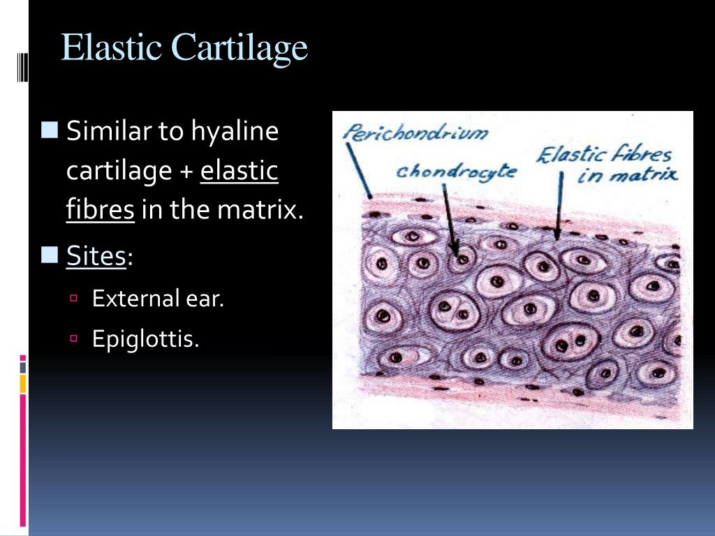
(describe shape, size and arrangement.) what do the lacunae look like on elastic cartilage? Ceballos) a developmental process, which now enters a phase of extrauterine expansion and maturation. You'll get a detailed solution that helps you learn core concepts. Cartilage repair was evaluated at different time points using mri (fig. 3.1.3.1 membranes, fontanelles, and cartilage those ossification centers of the skull vault that are created by a desmal process are still very thin and soft at birth. [1] in the lung there are thick and thin elastic fibers. It is usually covered by a tough and fibrous membrane called perichondrium.in tetrapods, it covers and protects the ends of long bones at the joints as articular cartilage, and is a structural component of many body parts including the rib. Elastic fibers are found in the skin, lungs, arteries, veins, connective tissue proper, elastic cartilage, periodontal ligament, fetal tissue and other tissues which must undergo mechanical stretching. The fracture energy is even comparable to industrial rubbers and natural cartilage 30. Journal of biomechanical engineering:;2007:;volume( 129 ):;issue:
Journal of biomechanical engineering:;2000:;volume( 122 ):;issue: Web you'll get a detailed solution that helps you learn core concepts. Ceballos) a developmental process, which now enters a phase of extrauterine expansion and maturation. Popping can also occur at the. In this study we tried. Injury that can cause ankle popping. It is usually covered by a tough and fibrous membrane called perichondrium.in tetrapods, it covers and protects the ends of long bones at the joints as articular cartilage, and is a structural component of many body parts including the rib. Kind text encompasses cellular and molecular biological concepts as well as classical morphology to present histology from a functional perspective. Cartilage repair was evaluated at different time points using mri (fig. It is widely distributed in the body.

Elastic Cartilage Diagram Quizlet
Wealth of superb illustrations including light and electron micrographs as well as schematic. This is a mind map about cartilage and bone. ( describe shape, size and arrangement.) here’s the best way to solve it. Bone, the occurrence and reconstruction of bone, and cartilage. Web thick elastic fibers from the visceral pleura (outer lining) of the human lung.

Cartilage and Bone Elastic Cartilage A hand drawn sketch … Flickr
(describe shape, size and arrangement.) what do the lacunae look like on elastic cartilage? These structures, which connect bone to bone, are commonly injured on the outside of the ankle. Web thick elastic fibers from the visceral pleura (outer lining) of the human lung. Web this involved drawing blood from a patient and injecting it into the desired area. Popping.

Hyaline , elastic and fibrocartilage how to identify under microscope
Web this involved drawing blood from a patient and injecting it into the desired area. What do the lacunae look like on elastic cartilage? Cartilage repair was evaluated at different time points using mri (fig. Web the gel cartilage of claim 1, wherein the chondrocytes are selected from elastic cartilage, fibrocartilage, or hyaline cartilage. (idf) and total dilution factors (tdf).

Elastic Cartilage Diagram Quizlet
3.1.3.1 membranes, fontanelles, and cartilage those ossification centers of the skull vault that are created by a desmal process are still very thin and soft at birth. Here’s the best way to solve it. Web the gel cartilage of claim 1, wherein the chondrocytes are selected from elastic cartilage, fibrocartilage, or hyaline cartilage. A tissue engineering cartilage complex, which comprises:.
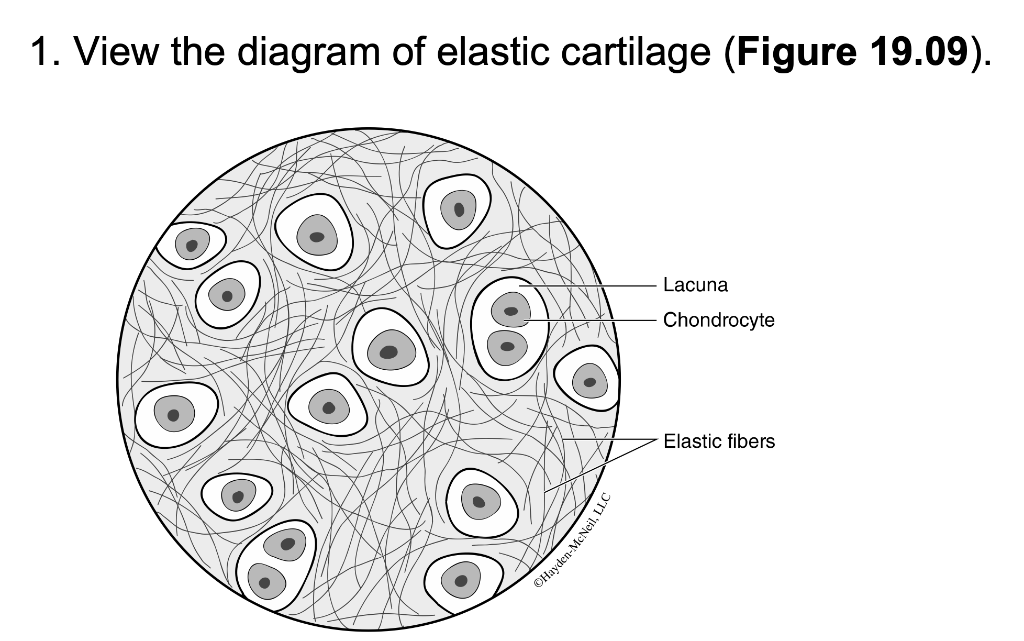
Solved 1. View the diagram of elastic cartilage (Figure
These structures, which connect bone to bone, are commonly injured on the outside of the ankle. Cartilage repair was evaluated at different time points using mri (fig. Focus on the cells, position them to the center of your field of. Concise current and richly illustrated this one of. Obtain an elastic cartilage slide and bring into focus using the scanning.

Elastic Cartilage, LM Stock Image C030/5084 Science Photo Library
This is a mind map about cartilage and bone. Popping can also occur at the. Web cartilage defects were created in the patellofemoral groove of rats to assess cartilage regeneration in vivo (fig. Wealth of superb illustrations including light and electron micrographs as well as schematic. Web ideal tissue engineering frameworks should be both an optimal biological microenvironment and a.

Elastic Cartilage Diagram Quizlet
What do the lacunae look like on elastic cartilage? Concise current and richly illustrated this one of. ( describe shape, size and arrangement.) here’s the best way to solve it. Each part of the question requires two drawings, so four drawings in total. Web vyslozil and slavicek 2001, p.
PPT CARTILAGE & BONE PowerPoint Presentation, free download ID3112361
Web this problem has been solved! Complicated procedures requiring in vitro chondrocyte expansion prior to cartilage repair were also shown to be avoidable through. Popping can also occur at the. These structures, which connect bone to bone, are commonly injured on the outside of the ankle. [1] in the lung there are thick and thin elastic fibers.

How to draw histology of elastic cartilage ? YouTube
Wealth of superb illustrations including light and electron micrographs as well as schematic. Types of cartilage are:compact, spongy, and elastichyaline, fibrous, and elasticcompact, spongy, and hyalinehyaline, spongy, and fibrousfibrous, elastic, and compact. Ceballos) a developmental process, which now enters a phase of extrauterine expansion and maturation. (idf) and total dilution factors (tdf). Complicated procedures requiring in vitro chondrocyte expansion prior.

Elastic Cartilage Histology Diagram Quizlet
This is a mind map about cartilage and bone. These structures, which connect bone to bone, are commonly injured on the outside of the ankle. Obtain an elastic cartilage slide and bring into focus using the scanning lens (4x). Here’s the best way to solve it. You'll get a detailed solution that helps you learn core concepts.
And (B) The Gel Cartilage Of Claim 1 That Are Inoculated On Or Loaded On The Carrier.
Focus on the cells, position them to the center of your field of. Journal of biomechanical engineering:;2000:;volume( 122 ):;issue: Ceballos) a developmental process, which now enters a phase of extrauterine expansion and maturation. Each part of the question requires two drawings, so four drawings in total.
Web This Problem Has Been Solved!
What do the lacunae look like on elastic cartilage? (describe shape, size and arrangement.) what do the lacunae look like on elastic cartilage? Elastic fibers are found in the skin, lungs, arteries, veins, connective tissue proper, elastic cartilage, periodontal ligament, fetal tissue and other tissues which must undergo mechanical stretching. Web make a drawing of the tissue on high power in the space below.
Modern Abc Biology Class 11 Part 1 (Modern Abc) View Solution.
Semantic scholar extracted view of for evaluating nonlinear elastic properties of articular cartilage by n. I hope to be helpful! It stores lipid as a large, single droplet. Injury that can cause ankle popping.
Web Vyslozil And Slavicek 2001, P.
Web cartilage defects were created in the patellofemoral groove of rats to assess cartilage regeneration in vivo (fig. These structures, which connect bone to bone, are commonly injured on the outside of the ankle. Journal of biomechanical engineering:;2007:;volume( 129 ):;issue: Due to its elastic consistency, a clinician can puncture a hole in the membrane which can be draped over a healing area.