Embryo Drawing
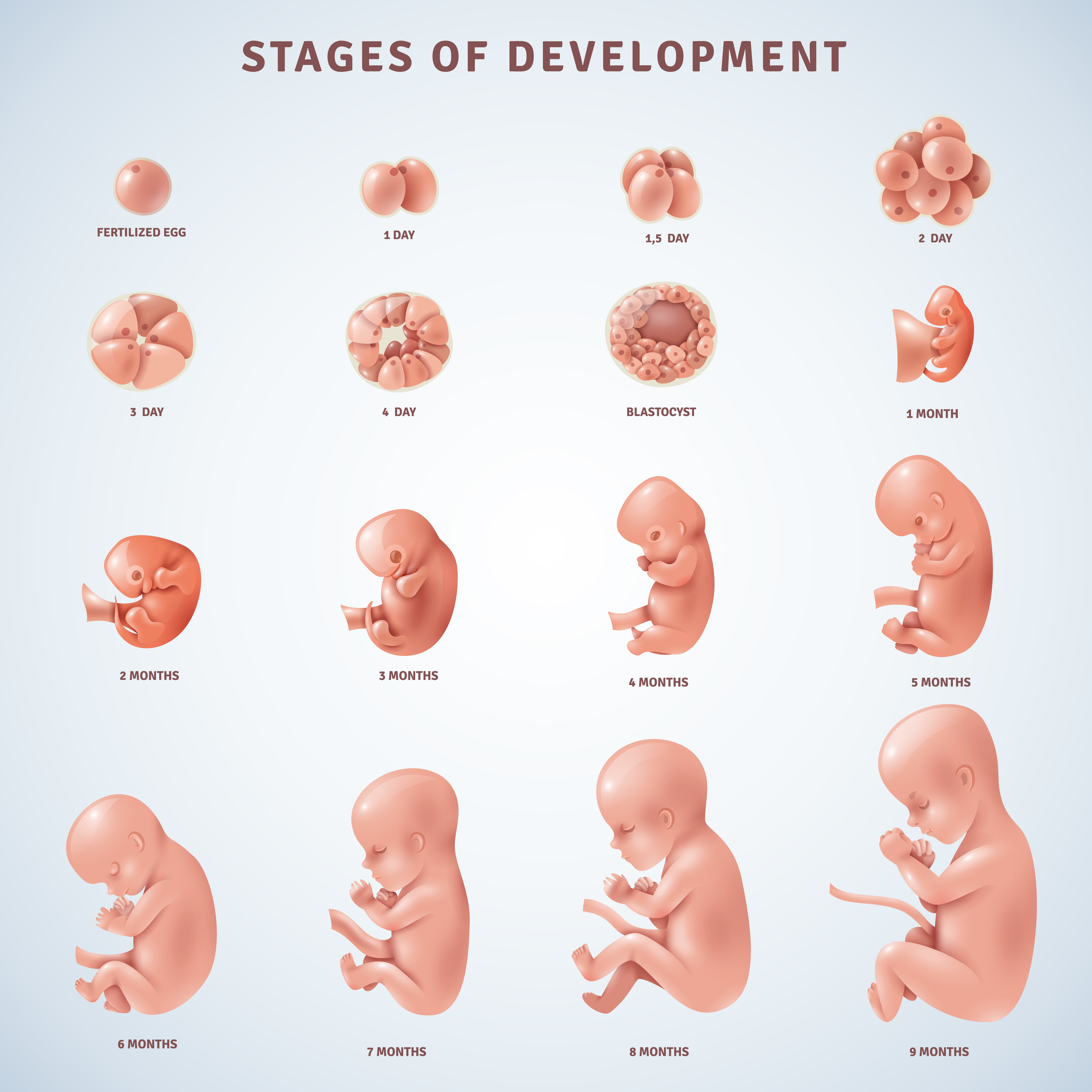
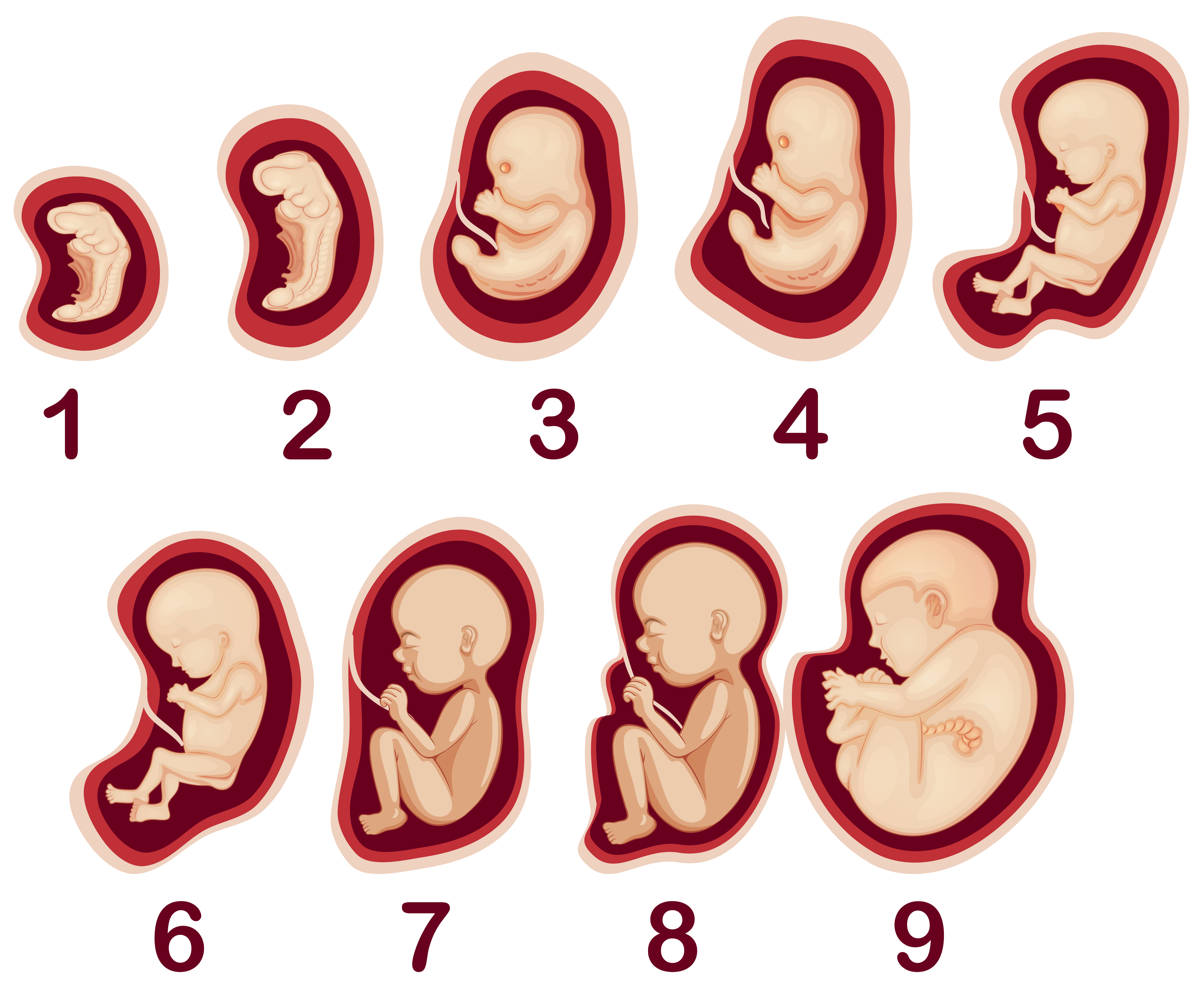
Embryo Drawing - Web embryo drawing is the illustration of embryos in their developmental sequence. Have the students label the right. Web studies of the fetus in the womb are two coloured annotated sketches by leonardo da vinci made in around 1511. In plants and animals, an embryo develops from a zygote, the single cell that results when an egg and sperm fuse during fertilization. All site content, unless otherwise specified, is licensed under a creative commons attribution license. Web embryo drawing refers to any representation of the illustration of embryos in their developmental sequence. Proceed through the 8 figures, discussing the changes and the points to look for. We haven’t always had ultrasounds to be able to check embryo growth inside the body. Previously, people had to study embryos in other ways. The studies correctly depict the human fetus in its proper position inside a dissected uterus.
In plants and animals, an embryo develops from a zygote, the single cell that results when an egg and sperm fuse during fertilization. This will give them a total of 8 areas in which to draw. Web embryo drawing is the illustration of embryos in their developmental sequence. In plants and animals, an embryo develops from a zygote, the single cell that results when an egg and sperm fuse during fertilization. The images that would not go away. Web scanning electron microscope image of an embryo : Web leonardo da vinci's embryological drawings of the fetus in the womb and his accompanying observational annotations are found in the third volume of his private notebooks. Have students fold a piece of white paper into quarters. In plants and animals, an embryo develops from a zygote, the single cell that results when an egg and sperm fuse during fertilization. In plants and animals, an embryo develops from a zygote, the single cell that results when an egg and sperm fuse during fertilization.
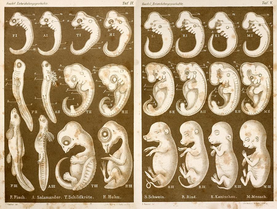
Proceed through the 8 figures, discussing the changes and the points to look for. In plants and animals, an embryo develops from a zygote, the single cell that results when an egg and sperm fuse during fertilization. All site content, unless otherwise specified, is licensed under a creative commons attribution license. I n the early stages of human embryonic development, a zygote divides into two identical totipotent. Have students fold a piece of white paper into quarters. In animals, the zygote divides repeatedly to form a ball of cells, which then forms a set of tissue layers that migrate and fold to. Explore evolution incorrectly asserts that haeckel s biogenetic law claims that the earliest stage of embryos are most similar. He produced a series of iconic drawings of embryos from a range of species, captured at various points through development, to. It has been widely noted that a number of the embryos in top row of the tables 6 and 7 from haeckel's anthropogenie (1874) are not realistic representations. This will give them a total of 8 areas in which to draw.
The Final Stages Of The Human Embryo Drawing by Mary Evans Picture Library
Web embryo drawing is the illustration of embryos in their developmental sequence. Web create a painting or drawing that depicts a mother cradling her pregnant belly, emphasizing the protective and nurturing bond between mother and child. This will give them a total of 8 areas in which to draw. Stewart/spl) some of the most iconic images in biology hold a.
Stages Human Embryonic Development 475937 Vector Art at Vecteezy
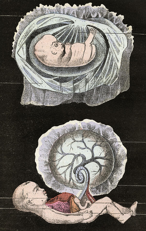
Stewart/spl) some of the most iconic images in biology hold a dark secret. Soon after their publication in 1868, a colleague alleged fraud, and haeckel’s many enemies have repeated the charge ever since. Web in one of his most famous drawings, leonardo depicts a human fetus lying inside a dissected uterus. Web create a painting or drawing that depicts a.

Human embryo Royalty Free Vector Image VectorStock
A smaller sketch of the same; In 1997, developmental biologist michael richardson compared his research team's embryo photographs to ernst haeckel's 1874 embryo drawings and called haeckel's work noncredible.science soon published <“>haeckel's embryos: Web embryo drawing is the illustration of embryos in their developmental sequence. Have students fold a piece of white paper into quarters. All site content, unless otherwise.
1874 Ernst Haeckel Embryo Drawings Photograph by Paul D Stewart Pixels
Soon after their publication in 1868, a colleague alleged fraud, and haeckel’s many enemies have repeated the charge ever since. This will give them a total of 8 areas in which to draw. Web create a painting or drawing that depicts a mother cradling her pregnant belly, emphasizing the protective and nurturing bond between mother and child. In animals, the.

Contour vector outline drawing of human embryo. Medical design editable
The studies correctly depict the human fetus in its proper position inside a dissected uterus. I n the early stages of human embryonic development, a zygote divides into two identical totipotent. Proceed through the 8 figures, discussing the changes and the points to look for. A large drawing of an embryo within a human uterus with a cow's placenta; Web.

Contour vector outline drawing of human embryo. Medical design editable
Web create a painting or drawing that depicts a mother cradling her pregnant belly, emphasizing the protective and nurturing bond between mother and child. Web studies of the fetus in the womb are two coloured annotated sketches by leonardo da vinci made in around 1511. He produced a series of iconic drawings of embryos from a range of species, captured.
Anatomical Human Embryo Drawing Etsy
Please refer to the terms of use. A large drawing of an embryo within a human uterus with a cow's placenta; In 1997, developmental biologist michael richardson compared his research team's embryo photographs to ernst haeckel's 1874 embryo drawings and called haeckel's work noncredible.science soon published <“>haeckel's embryos: Web among the most famous are drawings of embryos by the darwinist.
Embryo Sketch at Explore collection of Embryo Sketch
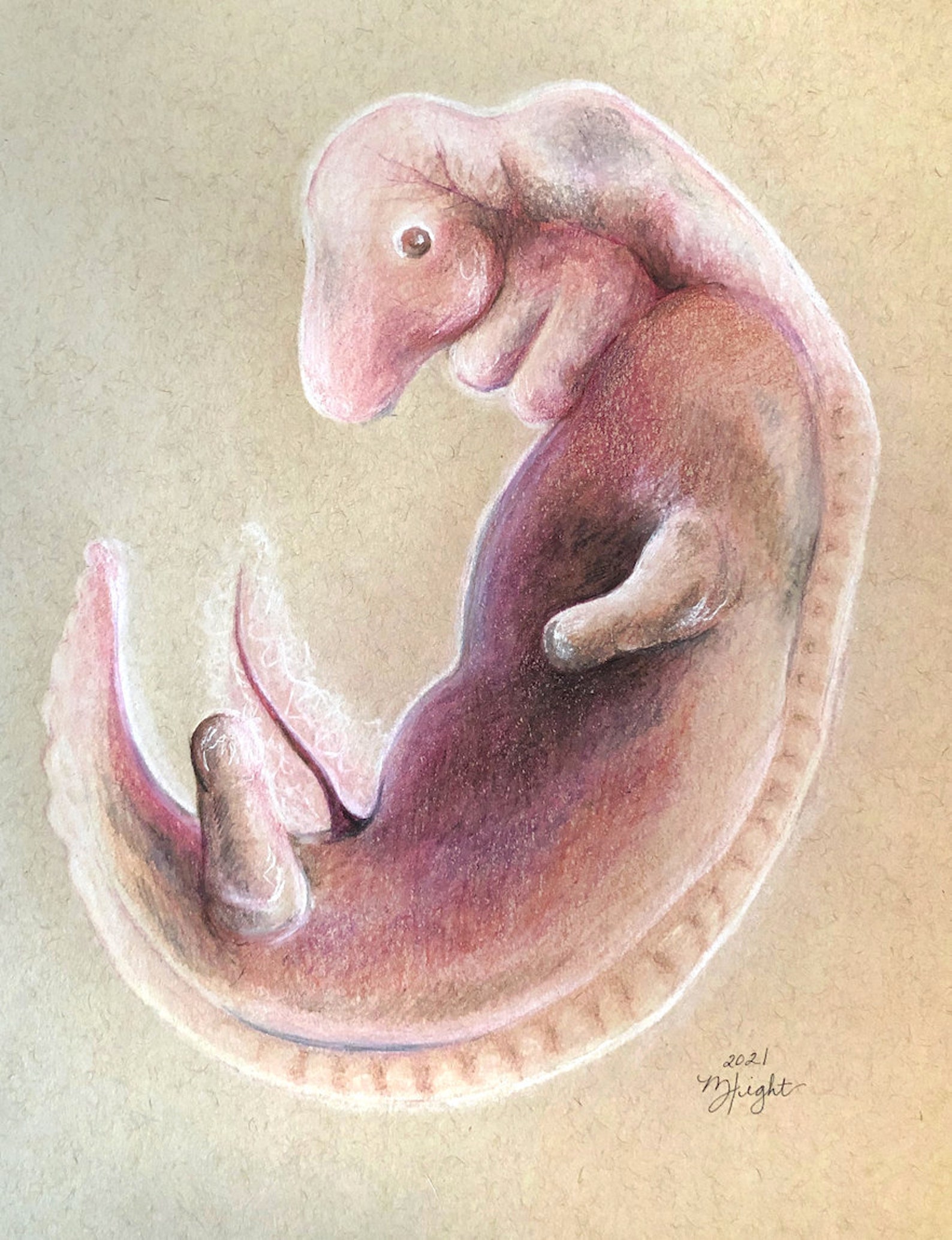
I n the early stages of human embryonic development, a zygote divides into two identical totipotent. He produced a series of iconic drawings of embryos from a range of species, captured at various points through development, to. A smaller sketch of the same; Illustrative drawings in detail of the placenta and. Web are you looking for the best images of.
A Vector of Human Embryo Development 294723 Vector Art at Vecteezy
In animals, the zygote divides repeatedly to form a ball of cells, which then forms a set of tissue layers that migrate and fold to. In plants and animals, an embryo develops from a zygote, the single cell that results when an egg and sperm fuse during fertilization. He was also the first to expertly draw the uterine artery and.
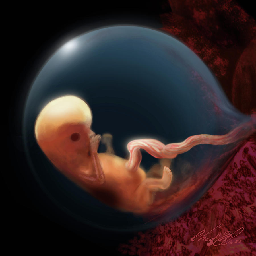
Embryo at 8 Weeks Drawing by Chad Glass Fine Art America
In plants and animals, an embryo develops from a zygote, the single cell that results when an egg and sperm fuse during fertilization. Web in one of his most famous drawings, leonardo depicts a human fetus lying inside a dissected uterus. A smaller sketch of the same; Web among the most famous are drawings of embryos by the darwinist ernst.
Web Embryo Drawing Is The Illustration Of Embryos In Their Developmental Sequence.
The studies correctly depict the human fetus in its proper position inside a dissected uterus. Web embryo drawing stock illustrations. Web in one of his most famous drawings, leonardo depicts a human fetus lying inside a dissected uterus. Soon after their publication in 1868, a colleague alleged fraud, and haeckel’s many enemies have repeated the charge ever since.
The Analysis Of External Morphological Characteristics And Gl Also Indicate An Average Difference Of 1.7 Weeks Between The Human Embryonic Ages Calculated By.
Web embryo drawing is the illustration of embryos in their developmental sequence. Web embryo images normal and abnormal mammalian development is a tutorial that uses scanning electron micrographs (sems) as the primary resource to teach mammalian embryology. Illustrative drawings in detail of the placenta and. Haeckel's concept of caenogenesis fully acknowledged that there can be signficant differences between embryos including at the earliest stages of development.
Web Among The Most Famous Are Drawings Of Embryos By The Darwinist Ernst Haeckel In Which Humans And Other Vertebrates Begin Identical, Then Diverge Toward Their Adult Forms.
In plants and animals, an embryo develops from a zygote, the single cell that results when an egg and sperm fuse during fertilization. He was also the first to expertly draw the uterine artery and the vascular system of the cervix and vagina. All site content, unless otherwise specified, is licensed under a creative commons attribution license. Stewart/spl) some of the most iconic images in biology hold a dark secret.
Web Without Being Able To Take Photographs Of Real Embryos To Support His Ideas, Haeckel Turned To His Sketchbook.
Have the students label the right. Web embryo drawing is the illustration of embryos in their developmental sequence. A large drawing of an embryo within a human uterus with a cow's placenta; A team of researchers labeled one of two cells in a developing embryo with gfp and used dna (blue) and actin (pink) labeling to track cell progeny to determine the contribution of each to developing structures.