Epidermis Drawing
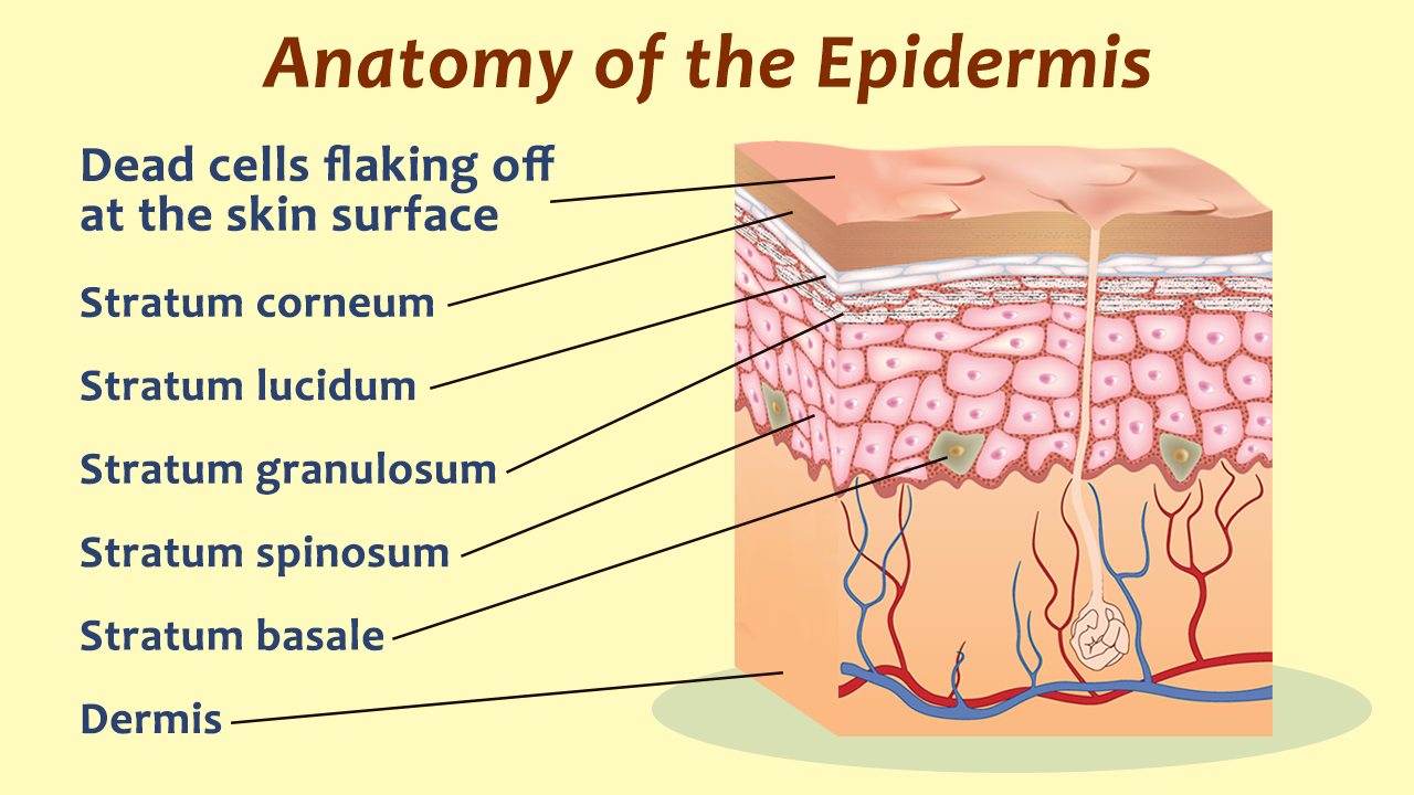
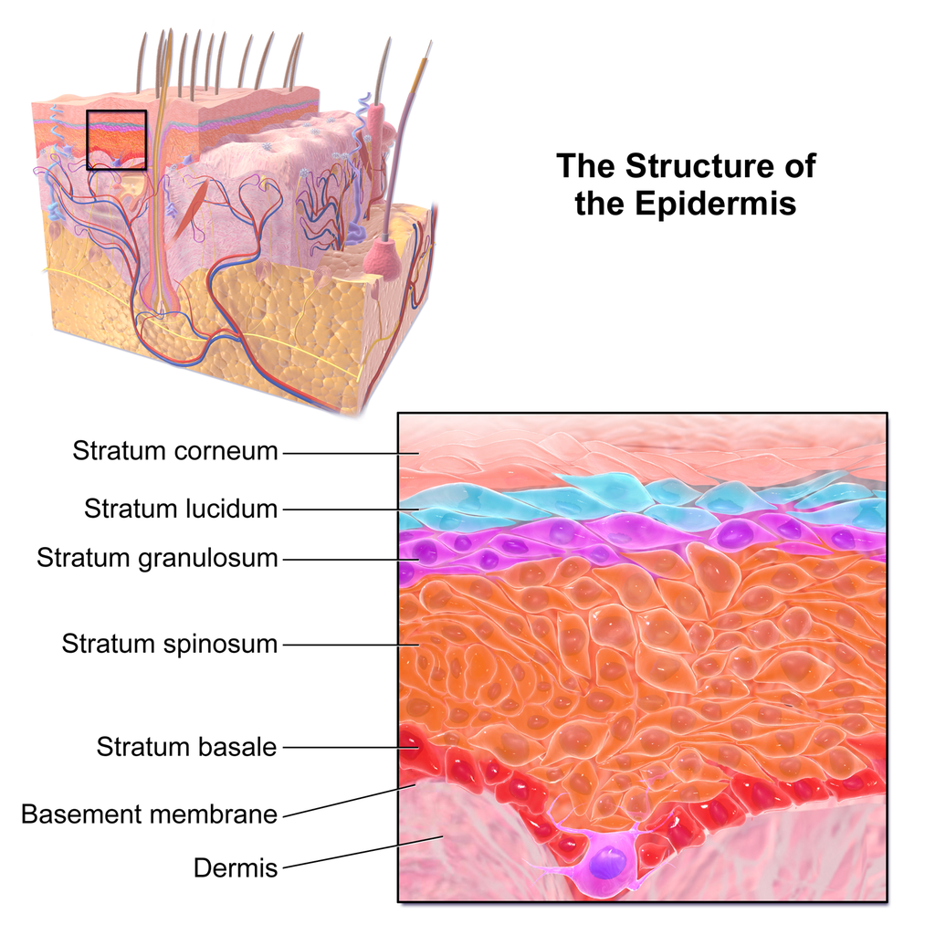
Epidermis Drawing - Web hello friends, this is my youtube channel and in this channel i used to share videos of different diagrams in easy way and step by step tutorials. Beneath the dermis lies the hypodermis, which is composed mainly of loose. Your skin has four layers of skin cells in the epidermis and an additional fifth layer in areas of thick skin. Web the main function of the epidermis is to protect the deeper tissues from water, microorganisms, mechanical and chemical trauma, and damage from uv light. Anatomy of the skin, showing the epidermis, dermis, and subcutaneous tissue. Web the epidermis is composed of multiple layers of flattened cells [4] that overlie a base layer ( stratum basale) composed of columnar cells arranged perpendicularly. Undoubtedly, the skin is the largest organ in the human body; The layers of cells develop from stem cells in the basal layer. So, five layers or strata, and each strata or. Sectional view of the skin.comparison illustration of protection effect between healthy skin and wounded skin.
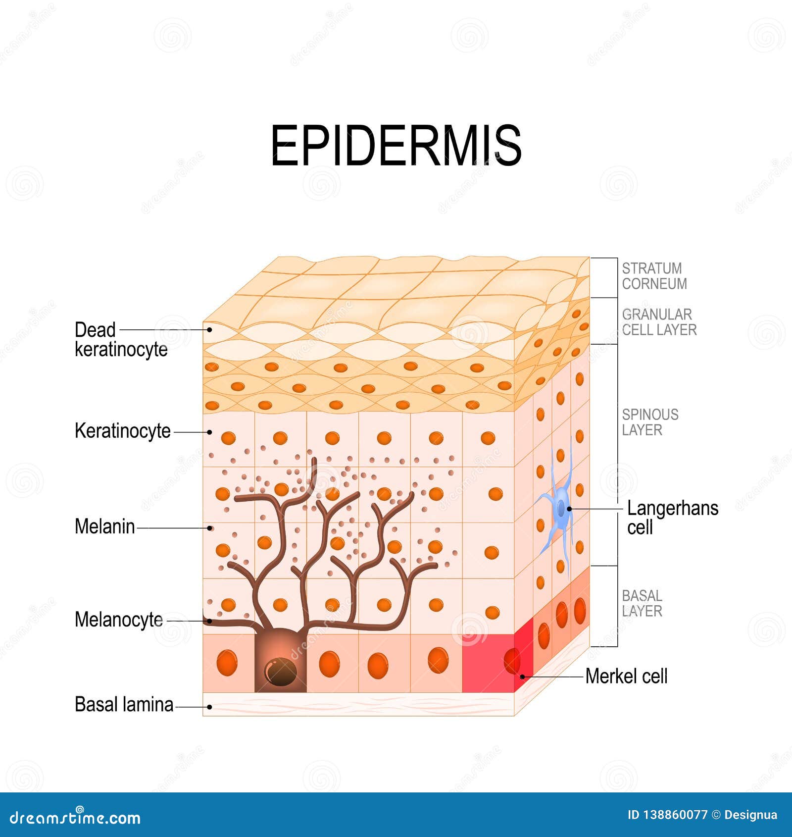
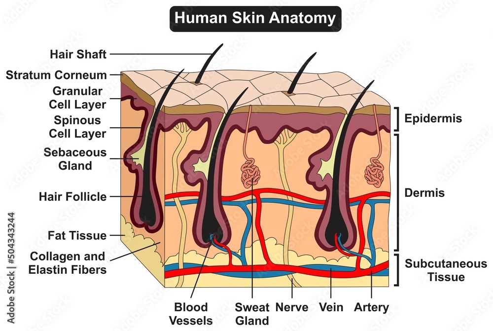
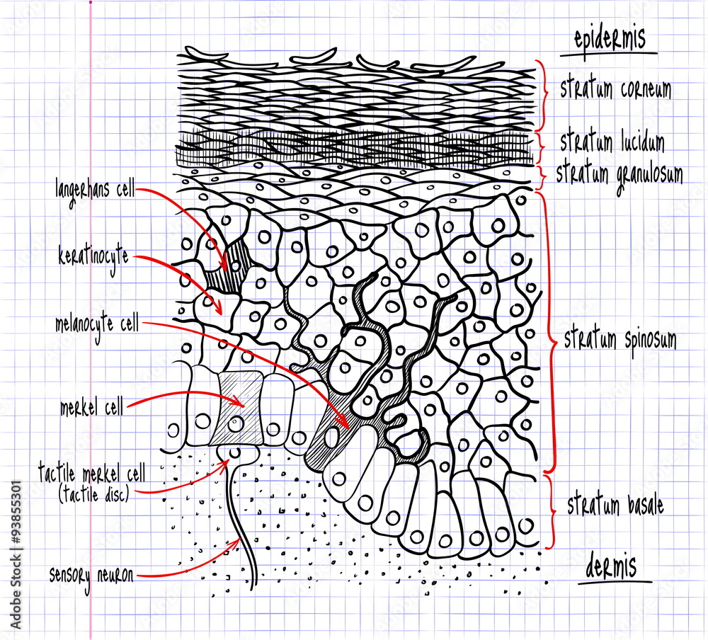
Web hello friends, this is my youtube channel and in this channel i used to share videos of different diagrams in easy way and step by step tutorials. Beneath the dermis lies the hypodermis, which is composed mainly of loose connective and fatty tissues. The epidermis includes five main layers: It is comprised of three major layers: They are far away from any blood supply, causing a lack of nutrients. Sectional view of the skin.comparison illustration of protection effect between healthy skin and wounded skin. Web your drawing with the epidermis, dermis (papillary layer), blood vessels, and dermis (reticular layer). Drawing shows normal skin anatomy, including the epidermis, dermis, hair follicles, sweat glands, hair shafts, veins, arteries, fatty tissue, nerves, lymph vessels, oil glands, and subcutaneous tissue. Anatomy of the skin with merkel cells; Web skin is the largest organ in the body and covers the body's entire external surface.
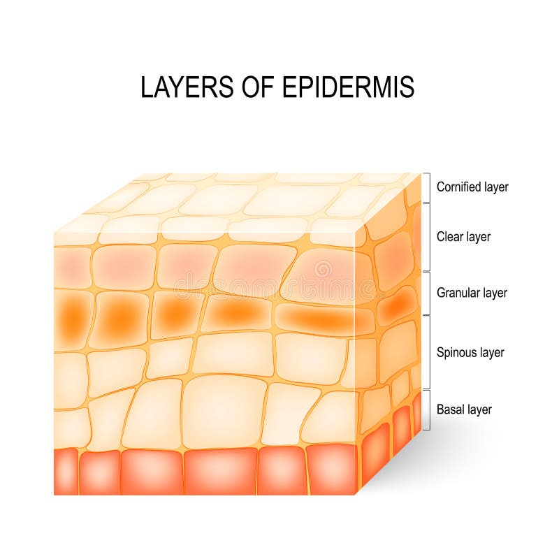
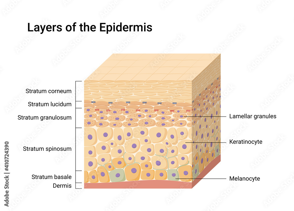
Using image a as a reference, label your drawing with the epidermis, dermis, hypodermis, hair follicle, and hair. The epidermis includes five main layers: Web hello friends, this is my youtube channel and in this channel i used to share videos of different diagrams in easy way and step by step tutorials. The four layers of cells, beginning at the bottom, are the stratum basale, stratum spinosum, stratum granulosum, and stratum corneum. Web this article will describe the anatomy and histology of the skin. Web diagram of human skin structure. The epidermis, made of closely packed epithelial cells, and the dermis, made of dense, irregular connective tissue that houses blood vessels, hair follicles, sweat glands, and other structures. Web the skin is composed of two main layers: It forms the outer covering for the entire body and protects the internal tissues from the external environment. Undoubtedly, the skin is the largest organ in the human body;
Do You Know Your Skin? Layers of the Epidermis and their Functions
The epidermis, made of closely packed epithelial cells, and the dermis, made of dense, irregular connective tissue that houses blood vessels, hair follicles, sweat glands, and other structures. Web the skin is by far the largest organ of the human body, weighing about 10 pounds (4.5 kg) and measuring about 20 square feet (2 square meters) in surface area. Web.
Epidermis Structure. Cell, And Layers Of A Human Skin. Illustration
Beneath the dermis lies the hypodermis, which is composed mainly of loose connective and fatty tissues. They're exposed to harsh chemicals contained in soaps, lotions, and other products. It is made up of three layers, the epidermis, dermis, and the hypodermis, all three of which vary significantly in their anatomy and function. In addition, the epidermis continuously makes new skin.
Epidermis Structure. Cell, And Layers Of A Human Skin. Stock Vector
Web your drawing with the epidermis, dermis (papillary layer), blood vessels, and dermis (reticular layer). Web skin is the largest organ in the body and covers the body's entire external surface. Your skin has four layers of skin cells in the epidermis and an additional fifth layer in areas of thick skin. In addition, the epidermis continuously makes new skin.
Vector illustration of Epidermis layers. Skin anatomy. Medical diagram
B&w, medical illustration (jpeg format) source: Beneath the dermis lies the hypodermis, which is composed mainly of loose connective and fatty tissues. Your skin has four layers of skin cells in the epidermis and an additional fifth layer in areas of thick skin. Web skin is the largest organ in the body and covers the body's entire external surface. It.

Layers of the Epidermis Sketchy Medicine
Web they're exposed to oxygen in the air, causing them to age faster. The skin's structure is made up of an intricate network which serves as the body’s initial barrier against pathogens, uv. Literally covering you from head to toe. B&w, medical illustration (jpeg format) source: The epidermis is composed of layers of skin cells called keratinocytes.

Epidermis Definition, Anatomy and Function
Beneath the dermis lies the hypodermis, which is composed mainly of loose connective and fatty tissues. The skin's structure is made up of an intricate network which serves as the body’s initial barrier against pathogens, uv. Web the main function of the epidermis is to protect the deeper tissues from water, microorganisms, mechanical and chemical trauma, and damage from uv.

Structure of the epidermis medical vector illustration, dermis anatomy
The skin consists of two distinct layers: Beneath the dermis lies the hypodermis, which is composed mainly of loose connective and fatty tissues. They are far away from any blood supply, causing a lack of nutrients. Web the skin is by far the largest organ of the human body, weighing about 10 pounds (4.5 kg) and measuring about 20 square.
10.3 Epidermis Human Biology
The skin's structure is made up of an intricate network which serves as the body’s initial barrier against pathogens, uv. The skin is composed of two main layers: Web ‘skin diagram || how to draw and label the parts of skin’ is demonstrated in this video tutorial step by step.the sense of touch had received supreme importa. The thickness of.
Human skin anatomy structure and parts infographic diagram epidermis
Web the epidermis is composed of multiple layers of flattened cells [4] that overlie a base layer ( stratum basale) composed of columnar cells arranged perpendicularly. It is made up of three layers, the epidermis, dermis, and the hypodermis, all three of which vary significantly in their anatomy and function. 100 kb referencing hub media. Sectional view of the skin.comparison..
drawing of the structure of the human epidermis Stock Vector Adobe Stock
Using image a as a reference, label your drawing with the epidermis, dermis, hypodermis, hair follicle, and hair. Web ‘skin diagram || how to draw and label the parts of skin’ is demonstrated in this video tutorial step by step.the sense of touch had received supreme importa. In addition, the epidermis continuously makes new skin that replaces the old skin.
Web Hello Friends, This Is My Youtube Channel And In This Channel I Used To Share Videos Of Different Diagrams In Easy Way And Step By Step Tutorials.
The stratum basale (also called the stratum germinativum) is the deepest epidermal layer and attaches the epidermis to the basal lamina, below which lie the layers of the dermis. The university of waikato te whare wānanga o waikato published 1 february 2011 size: Beneath the dermis lies the hypodermis, which is composed mainly of loose connective and fatty tissues. The four layers of cells, beginning at the bottom, are the stratum basale, stratum spinosum, stratum granulosum, and stratum corneum.
The Epidermis, Made Of Closely Packed Epithelial Cells, And The Dermis, Made Of Dense, Irregular Connective Tissue That Houses Blood Vessels, Hair Follicles, Sweat Glands, And Other Structures.
Web the skin is composed of two main layers: The skin's structure is made up of an intricate network which serves as the body’s initial barrier against pathogens, uv. It forms the outer covering for the entire body and protects the internal tissues from the external environment. Anatomy of the skin, showing the epidermis, dermis, and subcutaneous tissue.
It Is Made Up Of Three Layers, The Epidermis, Dermis, And The Hypodermis, All Three Of Which Vary Significantly In Their Anatomy And Function.
Drawing shows normal skin anatomy, including the epidermis, dermis, hair follicles, sweat glands, hair shafts, veins, arteries, fatty tissue, nerves, lymph vessels, oil glands, and subcutaneous tissue. In addition, the epidermis continuously makes new skin that replaces the old skin cells and produces melanin that provides skin color. The skin is composed of two main layers: The epidermis, made of closely packed epithelial cells, and the dermis, made of dense, irregular connective tissue that houses blood vessels, hair follicles, sweat glands, and other structures.
So, Five Layers Or Strata, And Each Strata Or.
Web the skin is by far the largest organ of the human body, weighing about 10 pounds (4.5 kg) and measuring about 20 square feet (2 square meters) in surface area. Literally covering you from head to toe. Your skin has four layers of skin cells in the epidermis and an additional fifth layer in areas of thick skin. Web ‘skin diagram || how to draw and label the parts of skin’ is demonstrated in this video tutorial step by step.the sense of touch had received supreme importa.