Fluid Mosaic Model Drawing
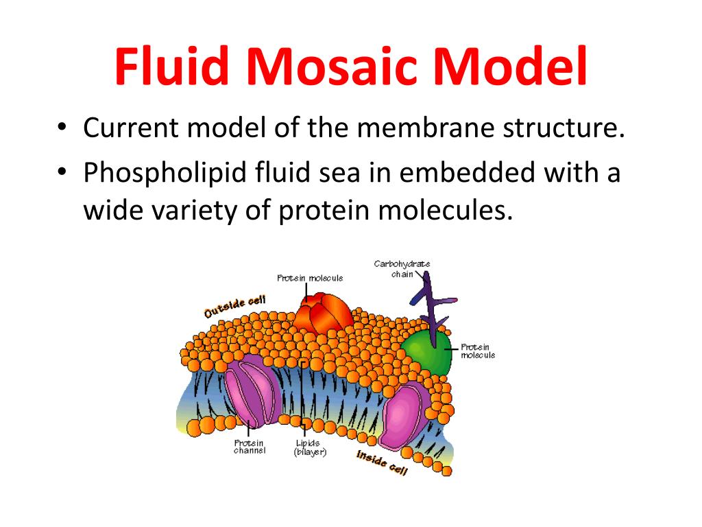
Fluid Mosaic Model Drawing - Web the fluid mosaic model was first proposed as a visual representation of research observations. Web according to the fluid mosaic model, the plasma membrane is a mosaic of components—primarily, phospholipids, cholesterol, and proteins—that move freely and fluidly in the plane of the membrane. Finished drawing sample for lesson 7. Web drawing the fluid mosaic model. Web the fluid mosaic model describes the plasma membrane structure as a mosaic of phospholipids, cholesterol, proteins, and carbohydrates. Web the fluid mosaic model is a way biologists use to describe the structure of biological membranes, such as the cell membrane. This model explains the structure of the plasma membrane of animal cells as a mosaic of components such as phospholipids, proteins, cholesterol, and carbohydrates. Passive and active movement between cells and their surroundings. Web the fluid mosaic model is one way of understanding biological membranes, consistent with most experimental observations. Model checklist, model diagramming pages, follow up activity questions for analysis and conclusions.
25k views 2 years ago science diagrams | explained and labelled science diagrams. Cell structure notes on the cell/plasma membrane. In other words, a diagram of the membrane (like the one below) is just a snapshot of a dynamic process in which phospholipids and proteins. Make a paper membrane model. Web fluid mosaic model of plasma membranehow to draw cell membranein this drawing,i will show you how to draw fluid mosaic model of plasma membrane or how to dra. The fluid mosaic model explains the structure of cell membranes. Explore the cell membrane's the three main components: This model explains the structure of the plasma membrane of animal cells as a mosaic of components such as phospholipids, proteins, cholesterol, and carbohydrates. It was first proposed by seymour jonathan singer and garth l. The fluid mosaic model also helps to explain:
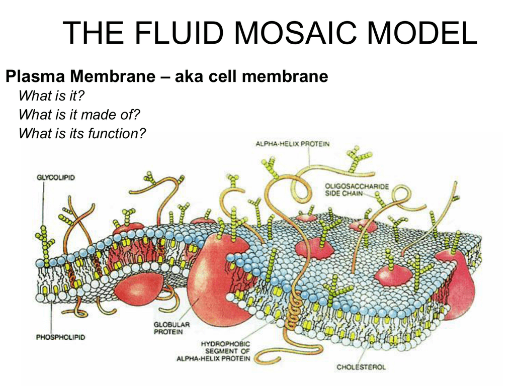
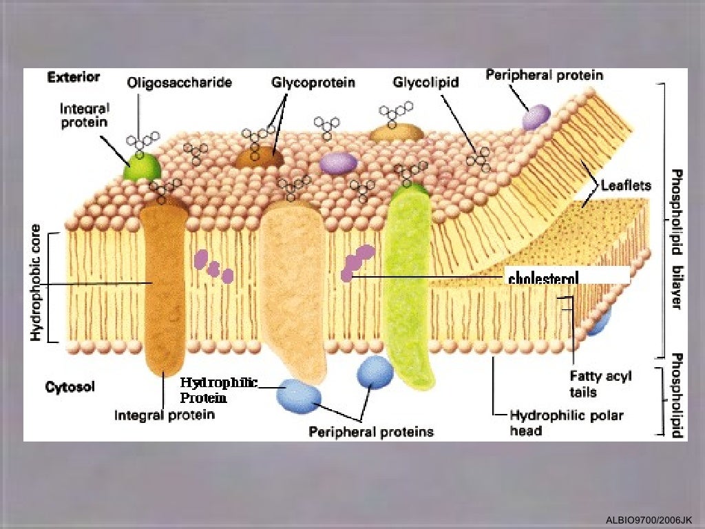
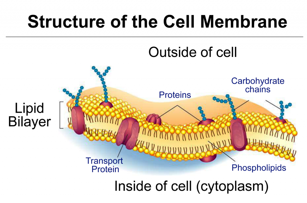
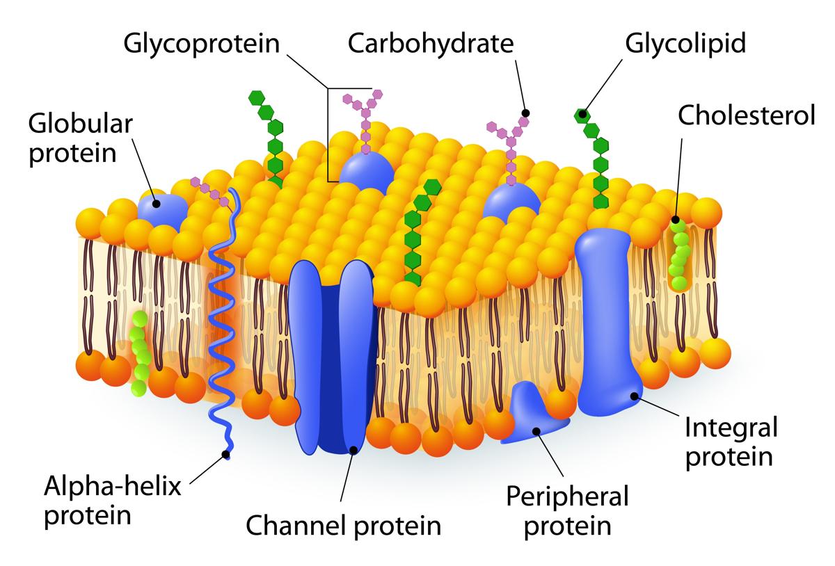
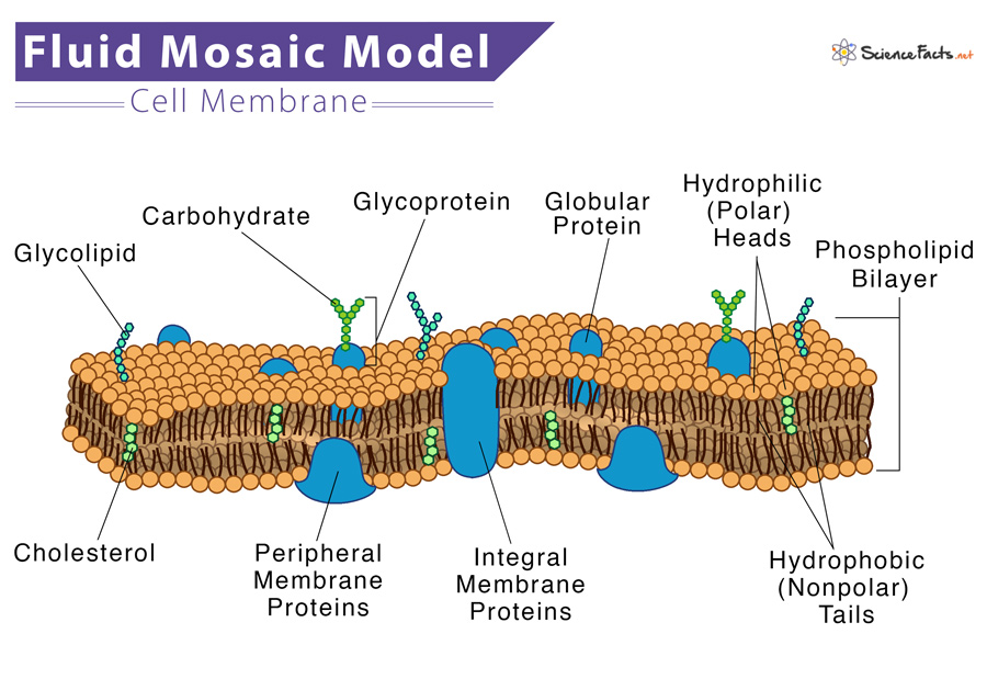
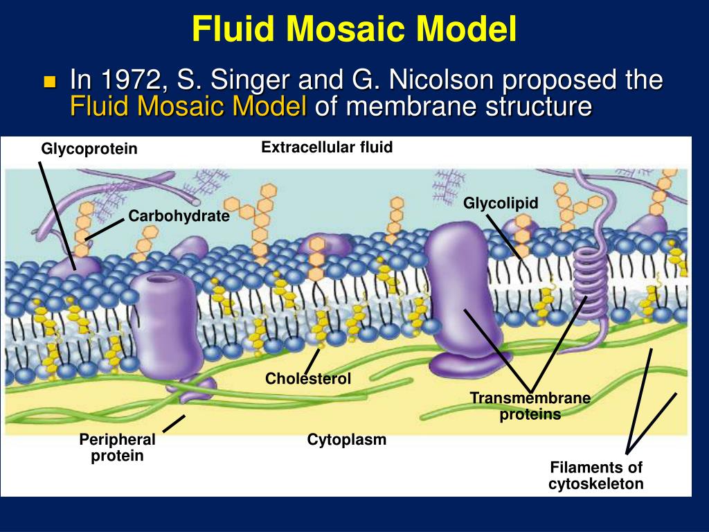
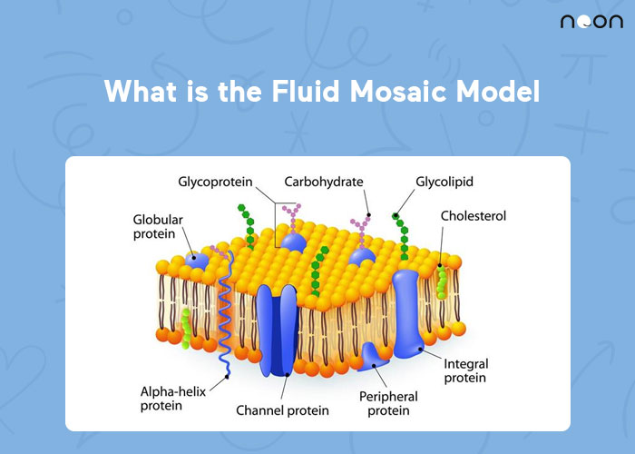
Describe the fluid mosaic model of cell membranes. Cell structure notes on the cell/plasma membrane. These membranes are composed of phospholipids, forming a bilayer with hydrophilic heads facing outwards and hydrophobic tails facing inwards. Plasma membranes range from 5 to 10 nm in thickness. Glycoproteins and glycolipids should be. Integral proteins are embedded in the phospholipid of the membrane, whereas peripheral proteins are attached to its surface. It was first proposed by seymour jonathan singer and garth l. Web the fluid mosaic model describes the structure of the plasma membrane as a mosaic of components —including phospholipids, cholesterol, proteins, and carbohydrates—that gives the membrane a fluid character. Model checklist, model diagramming pages, follow up activity questions for analysis and conclusions. The fluid mosaic model also helps to explain:
PPT Fluid Mosaic Model PowerPoint Presentation, free download ID
Plasma membranes range from 5 to 10 nm in thickness. In other words, a diagram of the membrane (like the one below) is just a snapshot of a dynamic process in which phospholipids and proteins. Proteins, glycolipids, glycoproteins, and cholesterol are embedded within this bilayer, creating a diverse mosaic. These membranes are composed of phospholipids, forming a bilayer with hydrophilic.
Fluid mosaic model of cell membrane coderbezy
This video will help you to explain about how to draw fluid mosaic model of plasma membrane easily step by step. 25k views 2 years ago science diagrams | explained and labelled science diagrams. The model has been modified in parts over time, keeping the basic concept the same. Web according to the fluid mosaic model, the plasma membrane is.
The fluid mosaic model of membrane structure
Web drawing the fluid mosaic model. Here is a great game about cell membranes (uses the parts we’ve been learning about!). 16k views 1 year ago. Model checklist, model diagramming pages, follow up activity questions for analysis and conclusions. These membranes are composed of phospholipids, forming a bilayer with hydrophilic heads facing outwards and hydrophobic tails facing inwards.
COMPONENTS OF THE CELL — Biology Notes
Web the fluid mosaic model describes the structure of the plasma membrane as a mosaic of components —including phospholipids, cholesterol, proteins, and carbohydrates—that gives the membrane a fluid character. The diagram of the plasma membrane must show the. Web the fluid mosaic model of the membrane was first outlined in 1972 and it explains how biological molecules are arranged to.
Fluid Mosaic Model Biology Wise
Web the fluid mosaic model is one way of understanding biological membranes, consistent with most experimental observations. Plasma membranes range from 5 to 10 nm in thickness. 332 views 1 year ago ib biology assessment drawings. Web the fluid mosaic model is the most acceptable model of the plasma membrane. Model checklist, model diagramming pages, follow up activity questions for.
Fluid Mosaic Model
Info page for drawing 7. Explore the cell membrane's the three main components: Web drawing the fluid mosaic model. Web the fluid mosaic model of the membrane was first outlined in 1972 and it explains how biological molecules are arranged to form cell membranes. Web the fluid mosaic model is the most acceptable model of the plasma membrane.
PPT Cell Membrane Structure and Function PowerPoint Presentation ID
Web the fluid mosaic model was first proposed as a visual representation of research observations. Web the fluid mosaic model is a way biologists use to describe the structure of biological membranes, such as the cell membrane. This video will help you to explain about how to draw fluid mosaic model of plasma membrane easily step by step. Passive and.
Fluid Mosaic Model
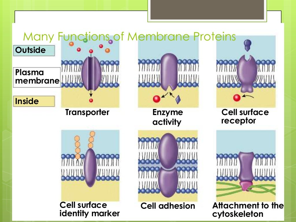
Learn about the cell membrane's vital role in regulating what enters and leaves a cell, and in helping a cell successfully maintain homeostasis, even when the environment around it is changing. In this definition of the cell membrane, its main function is to act as a barrier between the contents inside the cell and the extracellular environment. It was first.
What is the Fluid Mosaic Model? Learn At Noon
Web the fluid mosaic model was first proposed as a visual representation of research observations. 332 views 1 year ago ib biology assessment drawings. Model checklist, model diagramming pages, follow up activity questions for analysis and conclusions. The diagram of the plasma membrane must show the. The fluid mosaic model explains the structure of cell membranes.

Fluid Mosaic Model
Web drawing the fluid mosaic model. Info page for drawing 7. Cell structure notes on the cell/plasma membrane. Make a paper membrane model. The phospholipid bilayer, making it clear which part is the phosphate head and which parts are the hydrocarbon tails.
Learn About The Cell Membrane's Vital Role In Regulating What Enters And Leaves A Cell, And In Helping A Cell Successfully Maintain Homeostasis, Even When The Environment Around It Is Changing.
Web the fluid mosaic model is one way of understanding biological membranes, consistent with most experimental observations. This video will help you to explain about how to draw fluid mosaic model of plasma membrane easily step by step. It was first proposed by seymour jonathan singer and garth l. Finished drawing sample for lesson 7.
Integral Proteins Are Embedded In The Phospholipid Of The Membrane, Whereas Peripheral Proteins Are Attached To Its Surface.
Proteins, glycolipids, glycoproteins, and cholesterol are embedded within this bilayer, creating a diverse mosaic. You should show and label the following: Here is a great game about cell membranes (uses the parts we’ve been learning about!). The model has been modified in parts over time, keeping the basic concept the same.
This Model Explains The Structure Of The Plasma Membrane Of Animal Cells As A Mosaic Of Components Such As Phospholipids, Proteins, Cholesterol, And Carbohydrates.
Passive and active movement between cells and their surroundings. In this definition of the cell membrane, its main function is to act as a barrier between the contents inside the cell and the extracellular environment. Explore the cell membrane's the three main components: These membranes are composed of phospholipids, forming a bilayer with hydrophilic heads facing outwards and hydrophobic tails facing inwards.
The Fluid Mosaic Model Explains The Structure Of Cell Membranes.
Web the fluid mosaic model of the membrane was first outlined in 1972 and it explains how biological molecules are arranged to form cell membranes. 332 views 1 year ago ib biology assessment drawings. Info page for drawing 7. Web the fluid mosaic model was first proposed as a visual representation of research observations.