Jugular Blood Draw Dog Complications
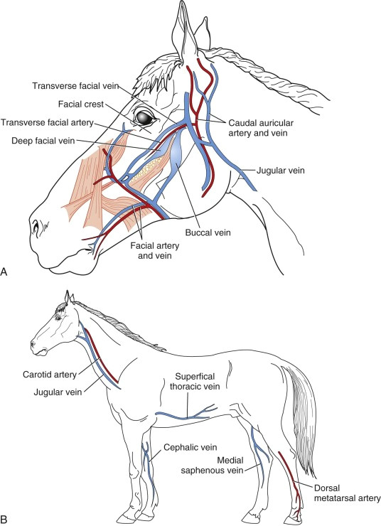
Jugular Blood Draw Dog Complications - Web the thrombus can be classified based on its location and the clinical signs it produces (eg, jugular venous thrombosis in large animals associated with prolonged venous catheterization, pulmonary arterial thromboembolism associated with heartworm disease in dogs). Web the incidence of mechanical and inflammatory complications associated with the jugular venous catheter was evaluated by adamantos et al., with complications observed in 39% of catheters, without differences between dogs and cats. Cats with cardiomyopathy, especially those with an enlarged left atrium, can develop large thromboemboli in the aorta or brachial artery. However, if performing tests requiring only a small amount of blood (e.g., glucose, pcv/tpp), peripheral veins may be used. Once blood collected, let off vein, before the needle removed, still hold onto leg, then cover puncture site when needle out. Facial veins that contribute to forming jugular vein. Peripheral veins, eg cephalic and saphenous, often detrimental → slow blood flow → sample artifacts (hemolysis and microclots). Web kelsey reinauer, cvt, demonstrates how to draw blood from a dog’s jugular vein. Secure/restrain patient, standing or sitting, or laying on their side! Lingofacial and maxillary veins of the dog.
Let off vein once iv catheter is in and move to hold off. Dissection and exploring jugular vein. High blood pressure (hypertension) |. The hematoma was about the size of two golf balls (it was oblong), and had gone down/been resorbed by saturday morning. Phlebotomy is an important and common technique that must be mastered by vet assistants, triage,. Peripheral veins, eg cephalic and saphenous, often detrimental → slow blood flow → sample artifacts (hemolysis and microclots). The volume of blood removed and the frequency of sampling should be based on the purpose of the scientific procedure and the total blood volume of the animal. Secure/restrain patient, standing or sitting, or laying on their side! Dog facial veins (upper part) lingual vein formation in dog. Web how veterinarians perform a routine jugular blood draw and why it is different than in humans.
Web ideally, blood should be collected from the jugular, as this generally allows for better sampling. The simple answer is from the vein. Venipuncture at this site could/canresult in hematoma formation or hemorrhage and upper airway compromise. Preferred sites for blood collection. Web jugular vein gives rapid, unobstructed flow of blood. In small animal practice the jugular vein is often the preferred site for collection. Dissection and exploring jugular vein. Web this study revealed a relatively high complication rate associated with central venous jugular catheterization in canine and feline patients, although few cases necessitated catheter removal. Secure/restrain patient, standing or sitting, or laying on their side! Web blood samples are commonly obtained from the jugular, cephalic or lateral saphenous veins;

Jugular Blood Draw, Canine YouTube
Let off vein once iv catheter is in and move to hold off. The hematoma was about the size of two golf balls (it was oblong), and had gone down/been resorbed by saturday morning. Web how veterinarians perform a routine jugular blood draw and why it is different than in humans. Dissection and exploring jugular vein. The volume of blood.

Jugular venipuncture canine YouTube
Web in this vetgirl online veterinary continuing education video, amy johnson, bs, lvt, rlatg, cvj, vetgirl’s manager of content development, and michaela witcher, ms, cvt, review how to draw blood from a jugular vein on a dog. Contributed by nichola gaither from animal hospital of statesville. This is the first time she has had blood drawn from the jugular to.
Jugular Blood Draw, Canine YouTube
Preferred sites for blood collection. Once blood collected, let off vein, before the needle removed, still hold onto leg, then cover puncture site when needle out. Lingofacial and maxillary veins of the dog. Thrombocytopenia, dic, vitamin k antagonism etc). Let off vein once iv catheter is in and move to hold off.

Dog Jugular Blood Draw Warehouse of Ideas
Once blood collected, let off vein, before the needle removed, still hold onto leg, then cover puncture site when needle out. If intubation is necessary, administer iv lidocaine ahead of time to reduce the associated cough reflex. Secure/restrain patient, standing or sitting, or laying on their side! She explains when the circumstances are appropriate for drawing from the jugular, needle.

dog jugular blood draw YouTube
Web in this vetgirl online veterinary continuing education video, amy johnson, bs, lvt, rlatg, cvj, vetgirl’s manager of content development, and michaela witcher, ms, cvt, review how to draw blood from a jugular vein on a dog. If a dog has neck pain after this procedure, there may be chances that he struggled a bit during the blood draw causing.
Drawing Blood From Jugular Dogs jfstudios
Dog facial veins (upper part) lingual vein formation in dog. Older and smaller patients required more catheterization attempts, on average, than did other patients. Aseptic preparation of the venipuncture site must be performed if blood is to be collected Contributed by nichola gaither from animal hospital of statesville. If a dog has neck pain after this procedure, there may be.

Jugular blood draw on canine YouTube
However, if performing tests requiring only a small amount of blood (e.g., glucose, pcv/tpp), peripheral veins may be used. Web generally jugular veins are not used if head injury! How maxillary vein is formed in a. Web avoid jugular blood collection and catheters, nasogastric tubes, and nasal oxygen catheters to decrease the risk for elevating intracranial pressure. Dog jugular blood.

Percutaneous external jugular vein catheterization in piglets using a
Contributed by nichola gaither from animal hospital of statesville. High blood pressure (hypertension) |. Web kelsey reinauer, cvt, demonstrates how to draw blood from a dog’s jugular vein. Web this study revealed a relatively high complication rate associated with central venous jugular catheterization in canine and feline patients, although few cases necessitated catheter removal. Web blood samples are commonly obtained.

Drawing Blood Dog's Jugular YouTube
Web our hound, jaina, developed a hematoma on friday after a blood draw for dhpp titer and heartworm tests. Web to cause minimal disturbance and discomfort to the animal. How maxillary vein is formed in a. High blood pressure (hypertension) |. She explains when the circumstances are appropriate for drawing from the jugular, needle size, and.

Jugular Blood Draw Dog Draw easy
Web blood samples are commonly obtained from the jugular, cephalic or lateral saphenous veins; Dissection and exploring jugular vein. Phlebotomy is an important and common technique that must be mastered by vet assistants, triage,. Web avoid jugular blood collection and catheters, nasogastric tubes, and nasal oxygen catheters to decrease the risk for elevating intracranial pressure. The rvn or svn should.
Preferred Sites For Blood Collection.
In small animal practice the jugular vein is often the preferred site for collection. This is the first time she has had blood drawn from the jugular to our knowledge. Read patient’s behavior cues and use foot/leg, wall, corner as restraint assistants. If intubation is necessary, administer iv lidocaine ahead of time to reduce the associated cough reflex.
That Might Include The Cephalic Vein.
Web how veterinarians perform a routine jugular blood draw and why it is different than in humans. Degenerative valve disease is the most common heart disease in dogs and accounts for about 75% of cardiovascular disease in this species. Thrombocytopenia, dic, vitamin k antagonism etc). The medial saphenous vein can also be used, and in general the use of the jugular vein is recommended.
If A Dog Has Neck Pain After This Procedure, There May Be Chances That He Struggled A Bit During The Blood Draw Causing The Muscles To Become Sore Or Perhaps An Intervertebral Disc In The Neck Was On The Verge Of Bulging.
Web kelsey reinauer, cvt, demonstrates how to draw blood from a dog’s jugular vein. Peripheral veins, eg cephalic and saphenous, often detrimental → slow blood flow → sample artifacts (hemolysis and microclots). Venipuncture at this site could/canresult in hematoma formation or hemorrhage and upper airway compromise. The most common ones are discussed below.
The Simple Answer Is From The Vein.
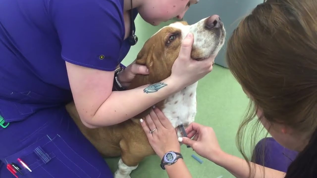
Web when blood is taken from the dog's jugular vein, the dog's head needs to be tilted in an upward position. Aseptic preparation of the venipuncture site must be performed if blood is to be collected Once blood collected, let off vein, before the needle removed, still hold onto leg, then cover puncture site when needle out. High blood pressure (hypertension) |.