Knee Anatomy Drawing
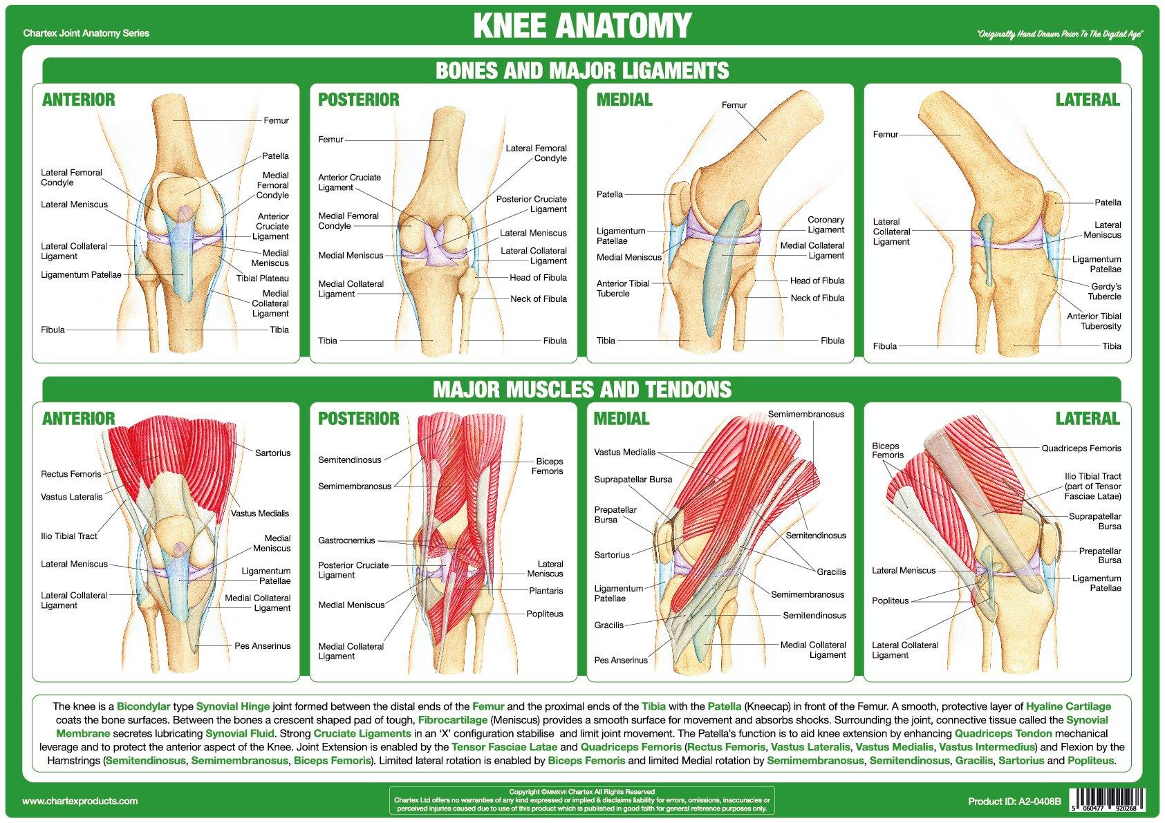
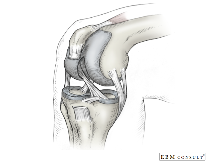
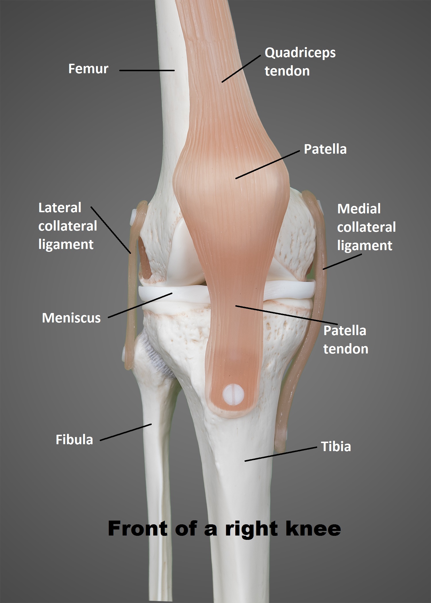
Knee Anatomy Drawing - The knee joints sit on mark 6, as that line corresponds to the bottom of the kneecaps. Web when drawing anatomy of the leg muscles, start at the top, where the leg connects to the pelvis. It is formed by articulations between the patella, femur and tibia. The most basic component of knee joint anatomy are the bones which provide the structure to the knee. From there, the legs follow the same general form and function of the arms, although the legs contain a few more muscle groups. The upper leg is facing the viewer and can be simplified as an elongated cuboid shape in perspective. The same geometry can be applied. When drawing a knee, we have two big volumes—the upper and lower leg. The tibiofemoral joint and patellofemoral joint. Web the knee joint is one of the strongest and most important joints in the human body.
Web the knee joint is a hinge type synovial joint, which mainly allows for flexion and extension (and a small degree of medial and lateral rotation). Hand drown anatomy illustration human. Web to draw the knee, begin by visualizing the bones and tendons underneath to help with the placement of landmarks. One between the femur and tibia, and one between the femur and patella. Anterior ligament of fibular head. Inside larynx nasal throttle anatomy. It is a complex hinge joint composed of two articulations; The knee joint is one of the largest and most complex joints in the body. Web let’s assume the figure in this human anatomy drawing is standing with the feet vertically aligned with the hip joints. Then draw the quadriceps muscles, and indicate the patella and its tendon down to the lower leg.
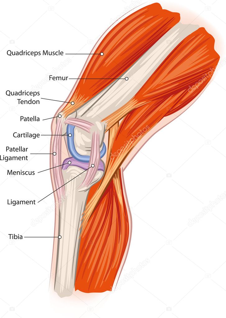
By understanding the anatomy of the knee we can infer about what is happening underneath when we. When drawing a knee, we have two big volumes—the upper and lower leg. When the leg is stretched out, the knee joint is placed on a straight line with the hip and ankle (left). The femur, tibia and patella. Web anatomy of human knee vector sketch of leg bones and joint, medicine design. Design your own anime and manga characters: The largest joint in the body, the knee is also one of the most easily injured. A pencil drawing of the muscles and the skeleton of a human leg. The knee joint is one of the largest and most complex joints in the body. The knee joint joins the thigh with the leg and consists of two articulations:
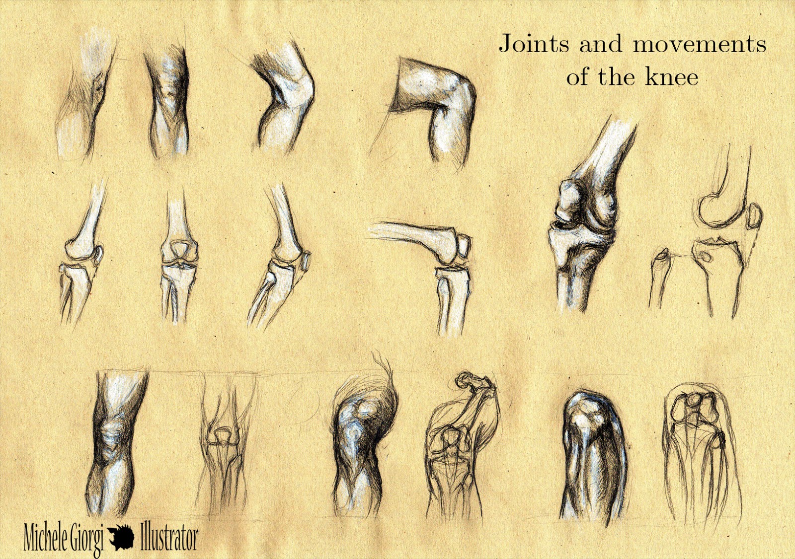
Michele Illustrator Anatomy Sketches Joints and movements of
Front 3/4 view of the pelvis (left) and back 3/4 view (right). A pencil drawing of the muscles and the skeleton of a human leg. The upper leg is facing the viewer and can be simplified as an elongated cuboid shape in perspective. Web knee anatomy involves more than just muscles and bones. It allows the lower leg to move.
Knee anatomy Stock Vector Image by ©Lukaves 18341225
The knee joints sit on mark 6, as that line corresponds to the bottom of the kneecaps. Web let’s assume the figure in this human anatomy drawing is standing with the feet vertically aligned with the hip joints. Then draw the quadriceps muscles, and indicate the patella and its tendon down to the lower leg. Movements at the knee joint.
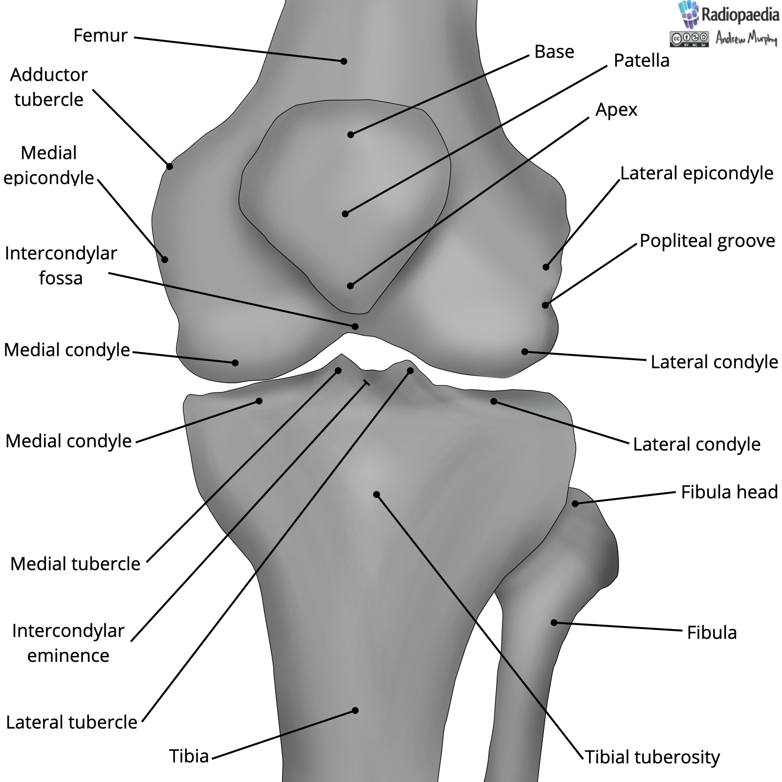
Radiopaedia Drawing Bones of the knee joint English labels
Anterior superior iliac spine at 1, and anterior inferior iliac spine at. It helps you stand, move and keep your balance. By understanding the anatomy of the knee we can infer about what is happening underneath when we. The snapshot icon at the top center will take a snapshot of your scene that can then be saved as a jpg.
Knee injuries causes, types, symptoms, knee injuries prevention & treatment
Inside larynx nasal throttle anatomy. Content is updated weekly and includes: Ligaments, tendons, and cartilage work together to connect the thigh bone, shin bone, and knee cap and allow the leg to bend back and forth like a hinge. A pencil drawing of the muscles and the skeleton of a human leg. The femur, tibia and patella.

Structure of the human knee Stock Vector Image by ©Silbervogel 72406683
Web cartoon illustration of the human knee joint anatomy illustration of the human knee joint anatomy knee joint icon gray illustration on white 3d vector of the human respiratory system, lungs, alveoli. Web let’s assume the figure in this human anatomy drawing is standing with the feet vertically aligned with the hip joints. Anatomy of the knee for drawing. Web.

Knee bones and joint sketch human anatomy Vector Image
The knee joints sit on mark 6, as that line corresponds to the bottom of the kneecaps. Shop best sellersread ratings & reviews It connects your thigh bone (femur) to your shin bone (tibia). Web to draw the knee, begin by visualizing the bones and tendons underneath to help with the placement of landmarks. It allows the lower leg to.

Anatomy of Knee
The knee is a complex joint that flexes, extends, and twists slightly from side to side. There are four knee bones that fit together to make two different knee joints: Knee joints, 19 century medical illustration. Web in this episode of simplified constructive anatomy, we cover the structure of the legs and knees. The knee joint joins the thigh with.
Knee Joint Anatomy Poster
While it's already relatively simple, we can still study th. Discover (and save!) your own pins on pinterest When the leg is stretched out, the knee joint is placed on a straight line with the hip and ankle (left). It allows the lower leg to move relative to the thigh while supporting the body’s weight. Ligaments, tendons, and cartilage work.
Anatomy Knee
Then draw the quadriceps muscles, and indicate the patella and its tendon down to the lower leg. It is a complex hinge joint composed of two articulations; Design your own anime and manga characters: Movements at the knee joint are essential to many everyday activities, including walking, running, sitting and standing. Web to draw the knee, begin by visualizing the.
The Knee UT Health San Antonio
Zygote body is a free online 3d anatomy atlas. The femur, tibia and patella. The knee joint is one of the largest and most complex joints in the body. When the leg is stretched out, the knee joint is placed on a straight line with the hip and ankle (left). Content is updated weekly and includes:
Discover (And Save!) Your Own Pins On Pinterest
Web anatomy of human knee vector sketch of leg bones and joint, medicine design. 60 day money backpremium canvas artmade in usa Shop best sellersread ratings & reviews When drawing a knee, we have two big volumes—the upper and lower leg.
Then Draw The Quadriceps Muscles, And Indicate The Patella And Its Tendon Down To The Lower Leg.
Problems with any part of the knee's anatomy can. Zygote body is a free online 3d anatomy atlas. The upper leg is facing the viewer and can be simplified as an elongated cuboid shape in perspective. The tibiofemoral joint is an articulation between the tibia and the femur, while the patellofemoral joint is an articulation between the patella.
The Largest Joint In The Body, The Knee Is Also One Of The Most Easily Injured.
The knee is the meeting point of the femur (thigh bone) in the upper leg and the tibia (shinbone) in. The front of the leg contains three main muscles that run about parallel from the hip down to the knee. Design your own anime and manga characters: I am sure you have seen paintings and drawings by the old masters which depict the knee area so realistically that you have no doubts about its shape.
Your Knees Also Contain Cartilage, Like Your Meniscus, And Ligaments, Including Your Lcl, Mcl, Acl And Pcl.
The most basic component of knee joint anatomy are the bones which provide the structure to the knee. From there, the legs follow the same general form and function of the arms, although the legs contain a few more muscle groups. When the leg is stretched out, the knee joint is placed on a straight line with the hip and ankle (left). The tibiofemoral joint and patellofemoral joint.