Lower Limb Drawing
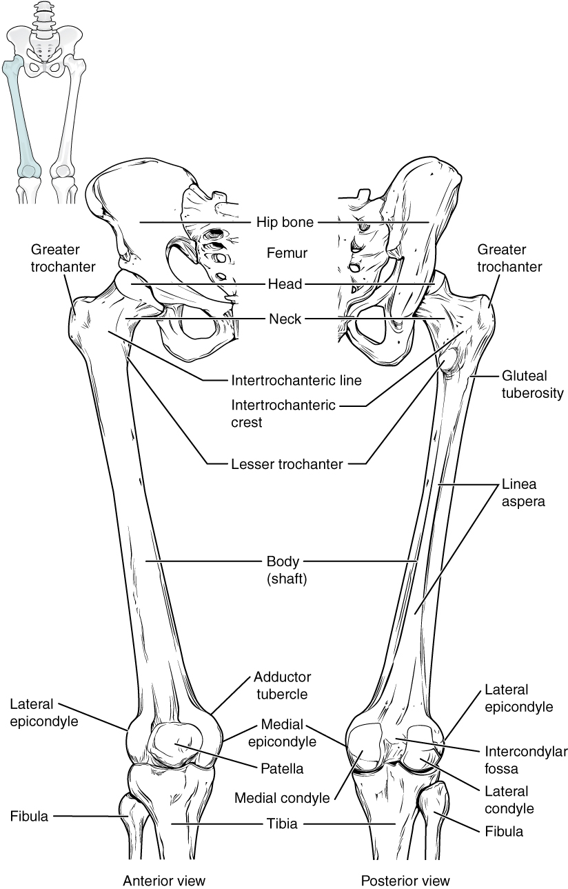
Lower Limb Drawing - 6 defendants and gun owners. Subtitle of part of web page, if appropriate. title: 16k views 1 year ago high yield videos. Web 3.9k views 4 years ago drawing and labelling: Lumbar plexus structure and branches. Each chart groups the muscles of that region into its component groups, making your revision a million times easier. Passes deep to the inguinal ligament and becomes the femoral artery. In this topic page, we will take a brief look at all of them and cover the basics of the entire lower limb. By the end of this section, you will be able to: Identify the divisions of the lower limb and describe the bones of each region.
The bones and muscles of the lower limb are larger. The adductors play an important role in normal gait as they help to draw the leg back towards the midline as we walk. In this comprehensive tutorial, we will guide you through the fundamentals of lower limb anatomy and demonstrate the simplest way to draw it. Subtitle of part of web page, if appropriate. title: Web 3d interactive models and tutorials on the anatomy of the lower limb, including the muscular compartments, osseus structures, blood supply and innervation. The superficial veins are located within the subcutaneous tissue whilst the deep veins are found deep to the deep fascia. Describe the bones and bony landmarks that articulate at each joint of the lower limb. This will help the students to draw venous drainage, lymphatic drainage. Superficial, deep and perforating veins. In this video the tricks of drawing the lower limbs are stated.
The adductors play an important role in normal gait as they help to draw the leg back towards the midline as we walk. In this topic page, we will take a brief look at all of them and cover the basics of the entire lower limb. The superficial veins are located within the subcutaneous tissue whilst the deep veins are found deep to the deep fascia. The deep veins accompany the major arteries and their branches and are usually paired. They can be divided into two groups; Web lower limb basics. Web the lower extremity can be divided into several parts or regions, as follows: Web the style of citing shown here is from the mla style citations (modern language association). The bones and muscles of the lower limb are larger. Web arteries of the lower extremity.

Bones of the Lower Limb Anatomy and Physiology I
Web star_border rate this article. In this comprehensive tutorial, we will guide you through the fundamentals of lower limb anatomy and demonstrate the simplest way to draw it. Web sign up here and try our free content: Bifurcates at approximately the fourth lumbar vertebra to form the left and right common iliac arteries. Superficial, deep and perforating veins.

How to Draw Legs, the Easy StepbyStep Guide with Simplified Anatomy
Subtitle of part of web page, if appropriate. title: 16k views 1 year ago high yield videos. Web before your first lower limb session, review the following movements of the lower limb and we will go over them in the first practical session. Web star_border rate this article. We can draw until it's done.
Lower Limb (Thigh, Leg and Foot)
Web anatomical drawing of limbs, hands and feet. Web the leg caput fibula the anterior surface of the tibie the medial and lateral maleolas achile tendon the calcaneus the base of the five metatarsal bone. Web in recent supreme court arguments, the former prosecutor has asked skeptical questions about criminal cases against former president donald trump, jan. Femuralis on the.

Lower Limb Skeletal Anatomy
Subtitle of part of web page, if appropriate. title: They can be divided into two groups; The muscles of the medial thigh are: Each chart groups the muscles of that region into its component groups, making your revision a million times easier. Web the lower extremity can be divided into several parts or regions, as follows:
8 THE LOWER LIMB Basicmedical Key
The superficial veins are located within the subcutaneous tissue whilst the deep veins are found deep to the deep fascia. Describe the bones and bony landmarks that articulate at each joint of the lower limb. Like the upper limb, the lower limb is divided into three regions. When citing a website the general format is as follows. Web in recent.
Pictures Of Bones Of The Lower Extremities
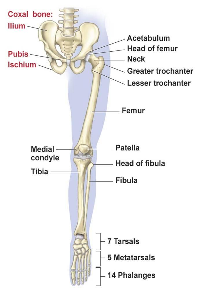
Fsc, biology practical copy, experiment 26 identification of the bones of the pelvic girdles, pectoral girdle,. The largest and most significant artery that brings oxygenated blood to the entire lower extremity is the femoral artery. Identify the divisions of the lower limb and describe the bones of each region. Subtitle of part of web page, if appropriate. title: Web arteries.
The lower limbs Human Anatomy and Physiology Lab (BSB 141)
Web sign up here and try our free content: Web the leg caput fibula the anterior surface of the tibie the medial and lateral maleolas achile tendon the calcaneus the base of the five metatarsal bone. Web 3 anatomical drawings of the posterior region of the leg allow the user to view the gastrocnemius muscle (medial and lateral head), the.
8.4 Bones of the Lower Limb Douglas College Human Anatomy and
Bifurcates at approximately the fourth lumbar vertebra to form the left and right common iliac arteries. The adductors play an important role in normal gait as they help to draw the leg back towards the midline as we walk. The superficial veins consist of great and small saphenous veins and their tributaries, which are situated beneath the skin in superficial.
Lower Limbs Skeleton Anatomy
The superficial veins are located within the subcutaneous tissue whilst the deep veins are found deep to the deep fascia. They can be divided into two groups; It gives off several branches throughout the thigh which supply the skin of the inguinal and the external genital areas, as well as some muscles of the thigh. Web in recent supreme court.
Lower Limb Bones, Muscles, Joints & Nerves » How To Relief
Each chart groups the muscles of that region into its component groups, making your revision a million times easier. Section of page if appropriate. The deep veins accompany the major arteries and their branches and are usually paired. Dermatomal map of the whole body Web the leg caput fibula the anterior surface of the tibie the medial and lateral maleolas.
By The End Of This Section, You Will Be Able To:
Temporary haemostasis by manual compression of the vessels of the lower limb: Web the lower extremity can be divided into several parts or regions, as follows: Web star_border rate this article. Subtitle of part of web page, if appropriate. title:
They Can Be Divided Into Two Groups;
Superficial, deep and perforating veins. Web a complete list of muscles. Web sign up here and try our free content: Section of page if appropriate.
The Superficial Veins Consist Of Great And Small Saphenous Veins And Their Tributaries, Which Are Situated Beneath The Skin In Superficial Fascia The Deep Veins Are The Venae Comitantes To The Anterior And Posterior Tibial Arteries, The
We’ve created muscle anatomy charts for every muscle containing region of the body: Learn to draw detailed bones, joints, and muscles to create your own portfolio of anatomical illustrations. The veins of the lower limb drain deoxygenated blood and return it to the heart. Web 3d interactive models and tutorials on the anatomy of the lower limb, including the muscular compartments, osseus structures, blood supply and innervation.
When Citing A Website The General Format Is As Follows.
The largest and most significant artery that brings oxygenated blood to the entire lower extremity is the femoral artery. This will help the students to draw venous drainage, lymphatic drainage. The deep veins accompany the major arteries and their branches and are usually paired. Dermatomal map of the whole body