Lysosome Drawing
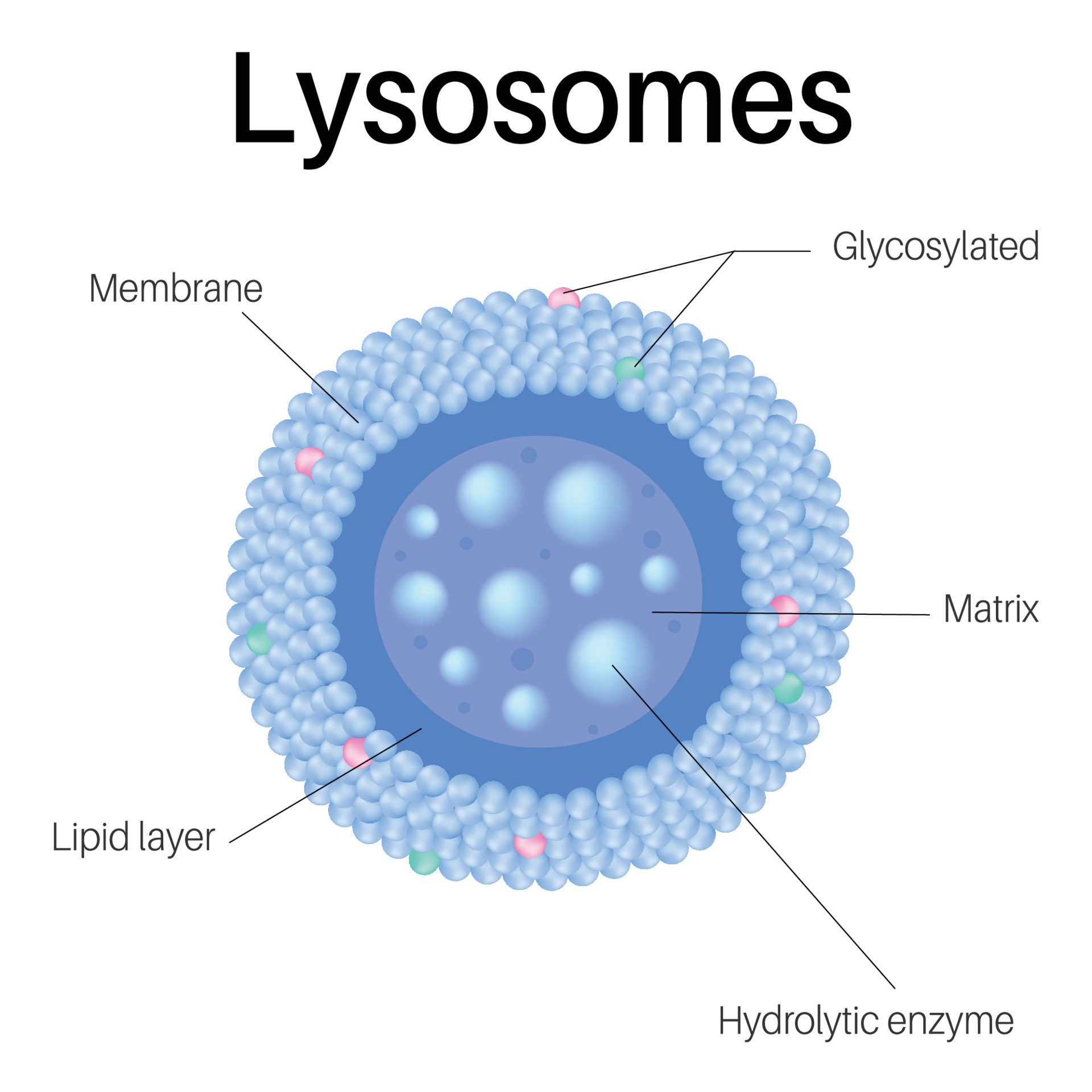
Lysosome Drawing - Web lysosome, subcellular organelle that is found in nearly all types of eukaryotic cells (cells with a clearly defined nucleus) and that is responsible for the digestion of macromolecules, old cell parts, and microorganisms. Web structure of lysosome. Illustration of human cell anatomy. Web lysosomes are formed by budding off of the golgi apparatus, and the hydrolytic enzymes within them are formed in the endoplasmic reticulum. It varies in shape and density. They are mostly globular or granular in appearance. There is variation in the shape and size from one cell to another and from time to time. Lysosomes are without any characteristic shape or structure i.e. The membrane surrounding the lysosome is vital to ensure these enzymes do not leak out into the cytoplasm and damage the cell. Lysosomes contain numerous hydrolytic enzymes which catalyse hydrolysis reactions.
There is variation in the shape and size from one cell to another and from time to time. Lysosome is round, vacuolar, and filled with dense material. Web lysosome, subcellular organelle that is found in nearly all types of eukaryotic cells (cells with a clearly defined nucleus) and that is responsible for the digestion of macromolecules, old cell parts, and microorganisms. Its size ranges from 0.2 to 0.5 μm. It varies in shape and density. Carcinoma cell, colored transmission electron micrograph (tem) cell. They maintain an interior acidity by using proton pumps from chemical reactions on their surface and in their interior. Lysosomes work by absorbing small pieces of cell debris and surrounding larger fragments. It is spherical and has the lipid bilayer, i.e., phospholipids. Web lysosomes are formed by budding off of the golgi apparatus, and the hydrolytic enzymes within them are formed in the endoplasmic reticulum.
Lysosomes are without any characteristic shape or structure i.e. Lysosome is round, vacuolar, and filled with dense material. Carcinoma cell, colored transmission electron micrograph (tem) cell. They are mostly globular or granular in appearance. It varies in shape and density. They maintain an interior acidity by using proton pumps from chemical reactions on their surface and in their interior. Web lysosome, subcellular organelle that is found in nearly all types of eukaryotic cells (cells with a clearly defined nucleus) and that is responsible for the digestion of macromolecules, old cell parts, and microorganisms. Each lysosome is surrounded by a membrane that maintains an acidic environment within the interior via a proton pump. It is spherical and has the lipid bilayer, i.e., phospholipids. Illustration of human cell anatomy.

How to draw structure of lysosome Lysosome drawing YouTube
Carcinoma cell, colored transmission electron micrograph (tem) cell. Each lysosome is surrounded by a membrane that maintains an acidic environment within the interior via a proton pump. They maintain an interior acidity by using proton pumps from chemical reactions on their surface and in their interior. It is spherical and has the lipid bilayer, i.e., phospholipids. They are mostly globular.
Lysosomes are membraneenclosed organelles. Lysosomes in cell. 8143455
It is spherical and has the lipid bilayer, i.e., phospholipids. Lysosomes work by absorbing small pieces of cell debris and surrounding larger fragments. Web how to draw lysosomes | structure of lysosome | step by step👉how to draw diagrams? Its size ranges from 0.2 to 0.5 μm. The diagram below shows the lysosome structure within a cell.

STRUCTURE AND FUNCTIONS OF LYSOSOMES
Web structure of lysosome. Its size ranges from 0.2 to 0.5 μm. Illustration of human cell anatomy. The membrane surrounding the lysosome is vital to ensure these enzymes do not leak out into the cytoplasm and damage the cell. The diagram below shows the lysosome structure within a cell.
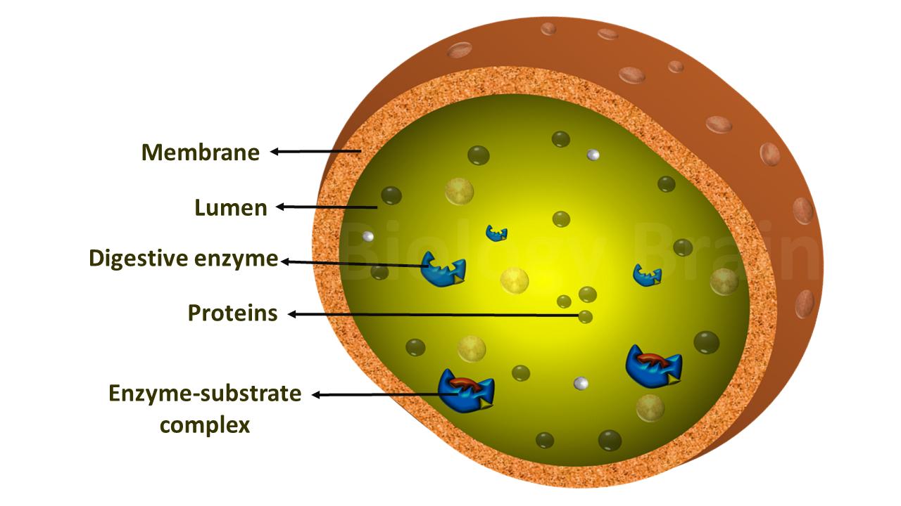
Diagram of Lysosomes and Types Biology Brain
It is spherical and has the lipid bilayer, i.e., phospholipids. Web lysosomes are formed by budding off of the golgi apparatus, and the hydrolytic enzymes within them are formed in the endoplasmic reticulum. Lysosomes work by absorbing small pieces of cell debris and surrounding larger fragments. The diagram below shows the lysosome structure within a cell. They maintain an interior.

How to draw structure of lysosomes step by step for beginners YouTube
They are mostly globular or granular in appearance. Each lysosome is surrounded by a membrane that maintains an acidic environment within the interior via a proton pump. Web lysosome, subcellular organelle that is found in nearly all types of eukaryotic cells (cells with a clearly defined nucleus) and that is responsible for the digestion of macromolecules, old cell parts, and.
Biological illustration of lysosome (Structure of cell lysosome) Stock
Web how to draw lysosomes | structure of lysosome | step by step👉how to draw diagrams? It varies in shape and density. Lysosomes are without any characteristic shape or structure i.e. They are mostly globular or granular in appearance. Illustration of human cell anatomy.

How to draw lysosomes Structure of Lysosome Step by step YouTube
The membrane surrounding the lysosome is vital to ensure these enzymes do not leak out into the cytoplasm and damage the cell. Web lysosomes are formed by budding off of the golgi apparatus, and the hydrolytic enzymes within them are formed in the endoplasmic reticulum. Web lysosome, subcellular organelle that is found in nearly all types of eukaryotic cells (cells.

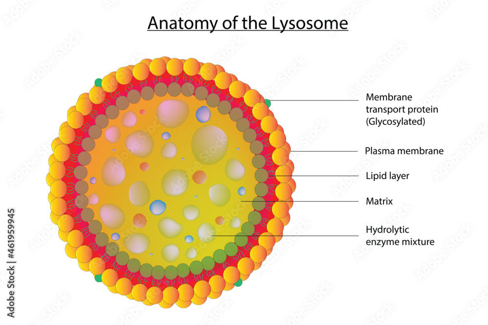
Anatomy of the Lysosome. Vector Diagram for Medical Use Stock Vector
Web lysosome, subcellular organelle that is found in nearly all types of eukaryotic cells (cells with a clearly defined nucleus) and that is responsible for the digestion of macromolecules, old cell parts, and microorganisms. The diagram below shows the lysosome structure within a cell. Lysosomes work by absorbing small pieces of cell debris and surrounding larger fragments. It is spherical.
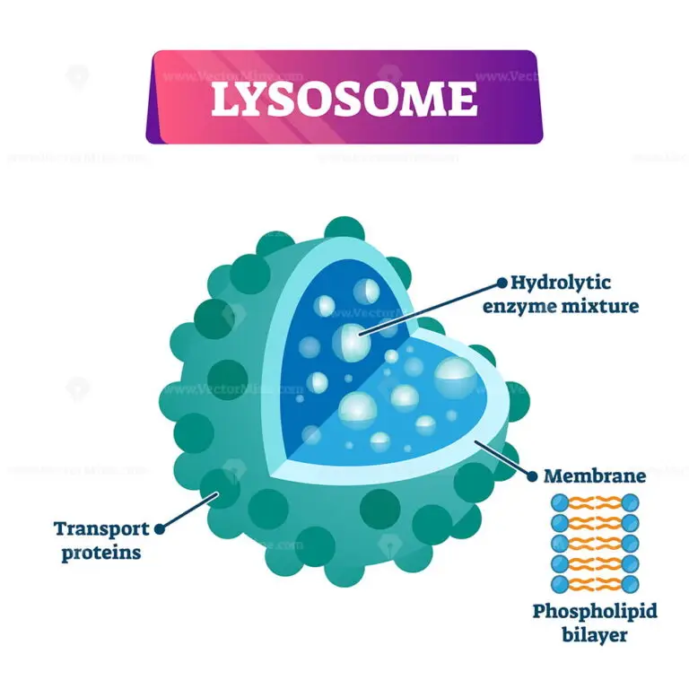
Lysosome cell organelle vector illustration labeled cross section
Web how to draw lysosomes | structure of lysosome | step by step👉how to draw diagrams? Web structure of lysosome. Lysosomes are without any characteristic shape or structure i.e. Each lysosome is surrounded by a membrane that maintains an acidic environment within the interior via a proton pump. Web lysosomes are formed by budding off of the golgi apparatus, and.

Structure of lysosomes infographics Royalty Free Vector
There is variation in the shape and size from one cell to another and from time to time. The membrane surrounding the lysosome is vital to ensure these enzymes do not leak out into the cytoplasm and damage the cell. It is spherical and has the lipid bilayer, i.e., phospholipids. Its size ranges from 0.2 to 0.5 μm. Web how.
Web Lysosome, Subcellular Organelle That Is Found In Nearly All Types Of Eukaryotic Cells (Cells With A Clearly Defined Nucleus) And That Is Responsible For The Digestion Of Macromolecules, Old Cell Parts, And Microorganisms.
Illustration of human cell anatomy. Lysosomes contain numerous hydrolytic enzymes which catalyse hydrolysis reactions. Carcinoma cell, colored transmission electron micrograph (tem) cell. Its size ranges from 0.2 to 0.5 μm.
Each Lysosome Is Surrounded By A Membrane That Maintains An Acidic Environment Within The Interior Via A Proton Pump.
Web lysosomes are formed by budding off of the golgi apparatus, and the hydrolytic enzymes within them are formed in the endoplasmic reticulum. They maintain an interior acidity by using proton pumps from chemical reactions on their surface and in their interior. There is variation in the shape and size from one cell to another and from time to time. It is spherical and has the lipid bilayer, i.e., phospholipids.
Lysosomes Are Without Any Characteristic Shape Or Structure I.e.
Lysosomes work by absorbing small pieces of cell debris and surrounding larger fragments. It varies in shape and density. Lysosome is round, vacuolar, and filled with dense material. Web structure of lysosome.
They Are Mostly Globular Or Granular In Appearance.
Web how to draw lysosomes | structure of lysosome | step by step👉how to draw diagrams? The diagram below shows the lysosome structure within a cell. The membrane surrounding the lysosome is vital to ensure these enzymes do not leak out into the cytoplasm and damage the cell.