Microscope Drawing With Parts
Microscope Drawing With Parts - Can be rotated to change magnification. Web microscope parts and functions with labeled diagram and functions how does a compound microscope work?. Use this with the microscope parts activity to help students identify and label the main parts of a microscope and then describe their functions. Web parts of the optical parts are as follows: The part that is looked through at the top of the compound microscope. Below this, draw another curved line, leaving the shape open on one side. Parts of a powell and leland microscope diagram Download the label the parts of the microscope: As a side note, the microscope used in this post is a great entry level or beginner microscope if you are trying to get someone interested in microscopes, microbiology, or science in general. Web all microscopes share features in common.
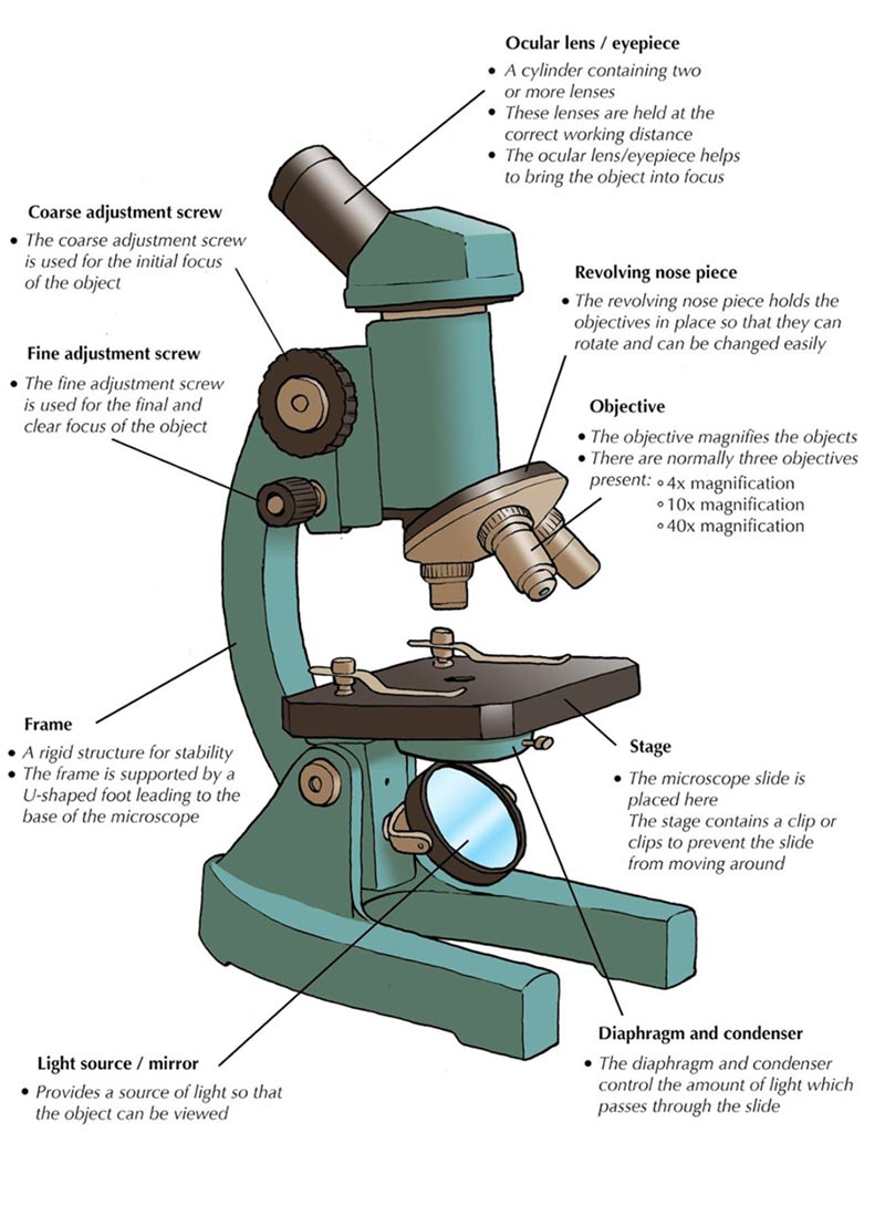
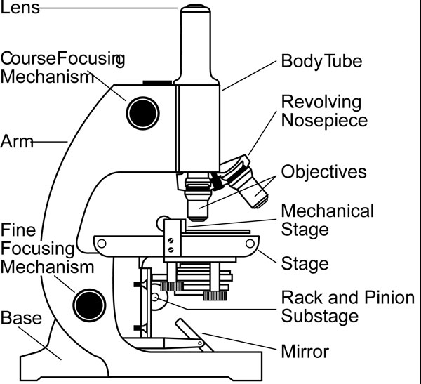
Notice the bend in the middle of each line. Connect them at the bottom using curved lines. Knobs (fine and coarse) by adjusting the knob, you can adjust the focus of the microscope. Use this with the microscope parts activity to help students identify and label the main parts of a microscope and then describe their functions. The eyepiece (or ocular lens) is the lens part at the top of a microscope that the viewer looks through. Web optical components of a compound microscope. Let us discuss the different parts of a compound microscope. It is also called a body tube or eyepiece tube. Web the most familiar type of microscope is the optical, or light, microscope, in which glass lenses are used to form the image. Then, draw three straight, parallel lines.
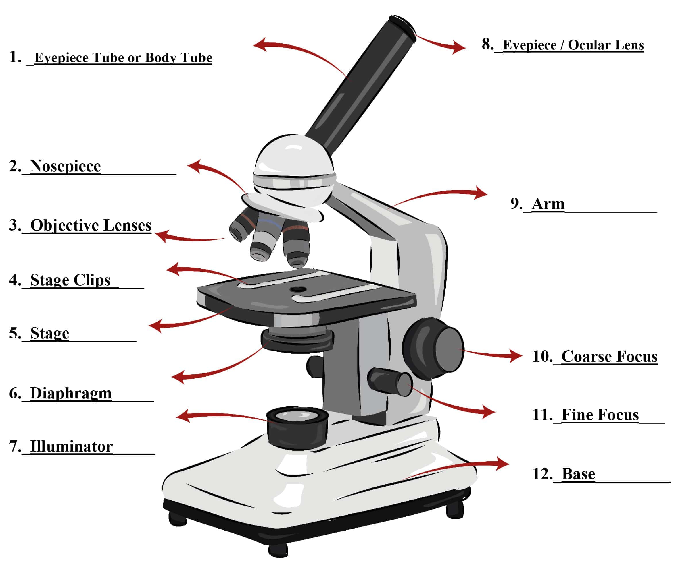
Web parts of a microscope include the following: The circled parts of the microscope are the fine and coarse adjustment knobs. So, something that was 1 mm wide in real life would be 400 mm wide in the microscope image. Answers pdf printable version here. A steady light source (110 volts) used in place of a mirror. The eyepiece (or ocular lens) is the lens part at the top of a microscope that the viewer looks through. Web the common light microscope used in the laboratory is called a compound microscope. 🔍 the microscope is an essential tool for scientists, researchers, and medical professionals. Supports the tube and connects it to the base. F = focal length of the convex lens.

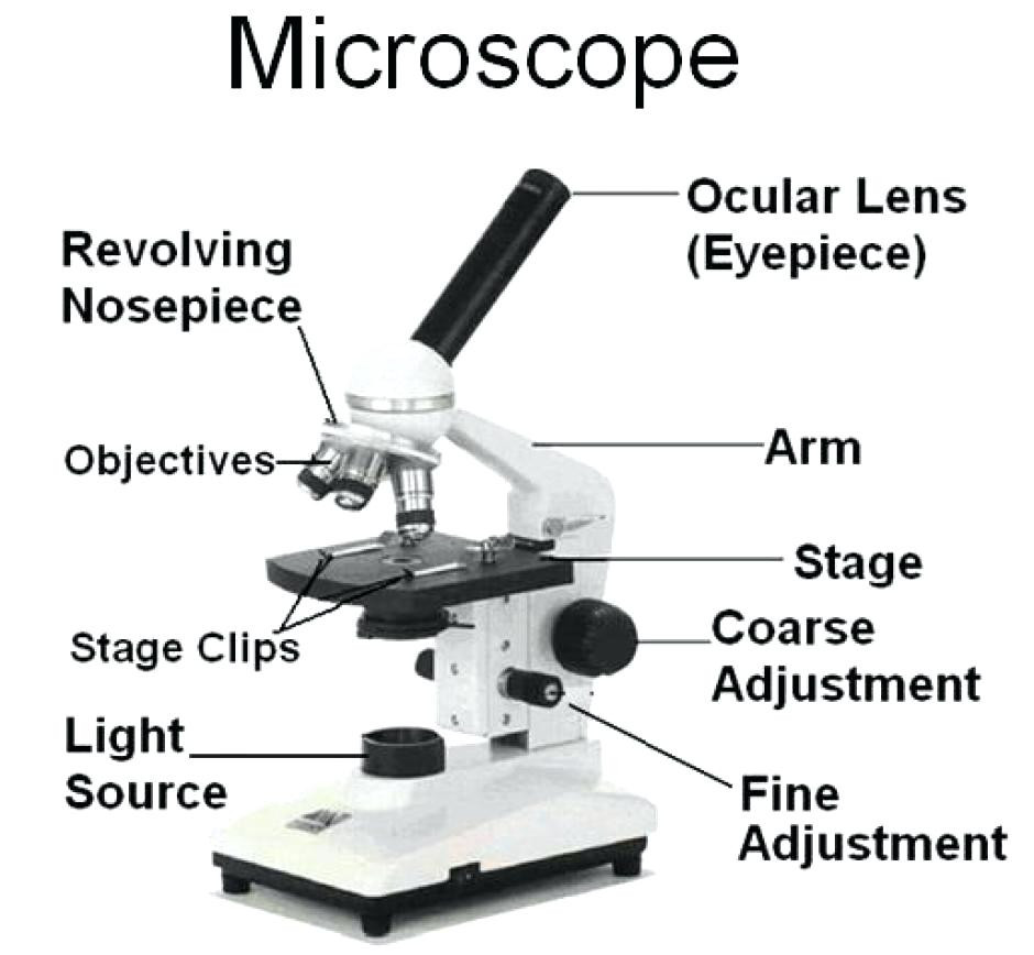
Parts of a microscope with functions and labeled diagram
Web the field diaphragm control is located around the lens located in the base. 🧬 the main function of a microscope is to provide a magnified view of small objects or organisms, such as bacteria, cells, or. Download the label the parts of the microscope: It is also called a body tube or eyepiece tube. The hand magnifying glass can.

Simple Microscope Drawing With Parts Micropedia
Use this with the microscope parts activity to help students identify and label the main parts of a microscope and then describe their functions. Web microscope parts and functions with labeled diagram and functions how does a compound microscope work?. Platform where the specimen is placed for observation. Notice the bend in the middle of each line. Supports the tube.

Simple Microscope Definition, Principle, Magnification, Parts
Web the most familiar type of microscope is the optical, or light, microscope, in which glass lenses are used to form the image. Connects the eyepiece to the objective lenses. Let us discuss the different parts of a compound microscope. Gathers light from the specimen and magnifies the image. Knobs (fine and coarse) by adjusting the knob, you can adjust.
Parts Of A Microscope With Functions And Labeled Diagram Images
It is to be noted that. The majority of the microscope models today have the knobs mounted on the same part of the device. Can be rotated to change magnification. Knobs (fine and coarse) by adjusting the knob, you can adjust the focus of the microscope. 🔍 the microscope is an essential tool for scientists, researchers, and medical professionals.
Parts of a Microscope SmartSchool Systems
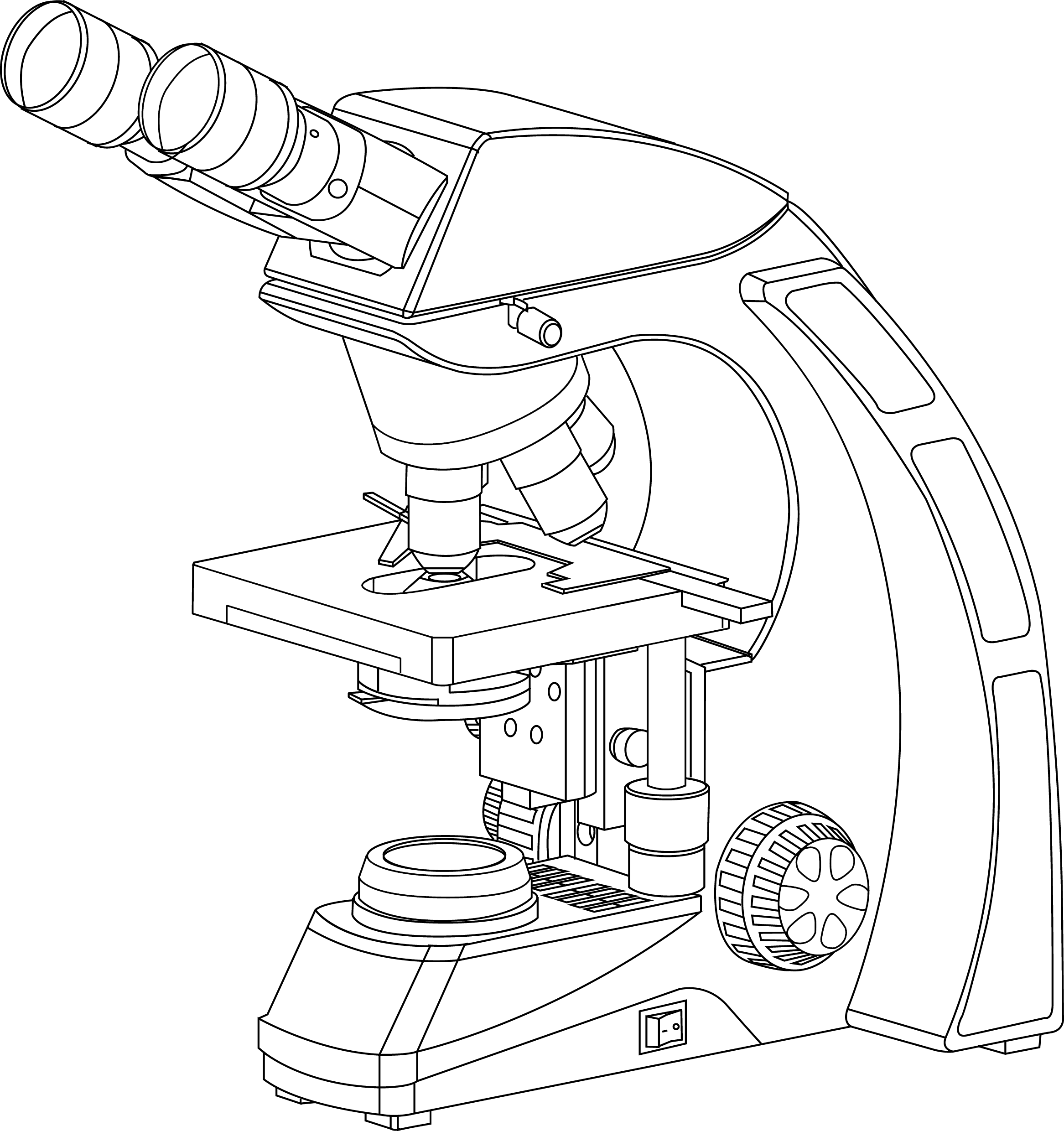
It’s actually not a toy microscope, it’s a functional microscope that produces great. It is because it contains two types of lenses; Web download the label the parts of the microscope pdf printable version here. Web the microscope illustrated in figure 5 below was manufactured by hugh powell and peter lealand around 1850. Parts of a powell and leland microscope.
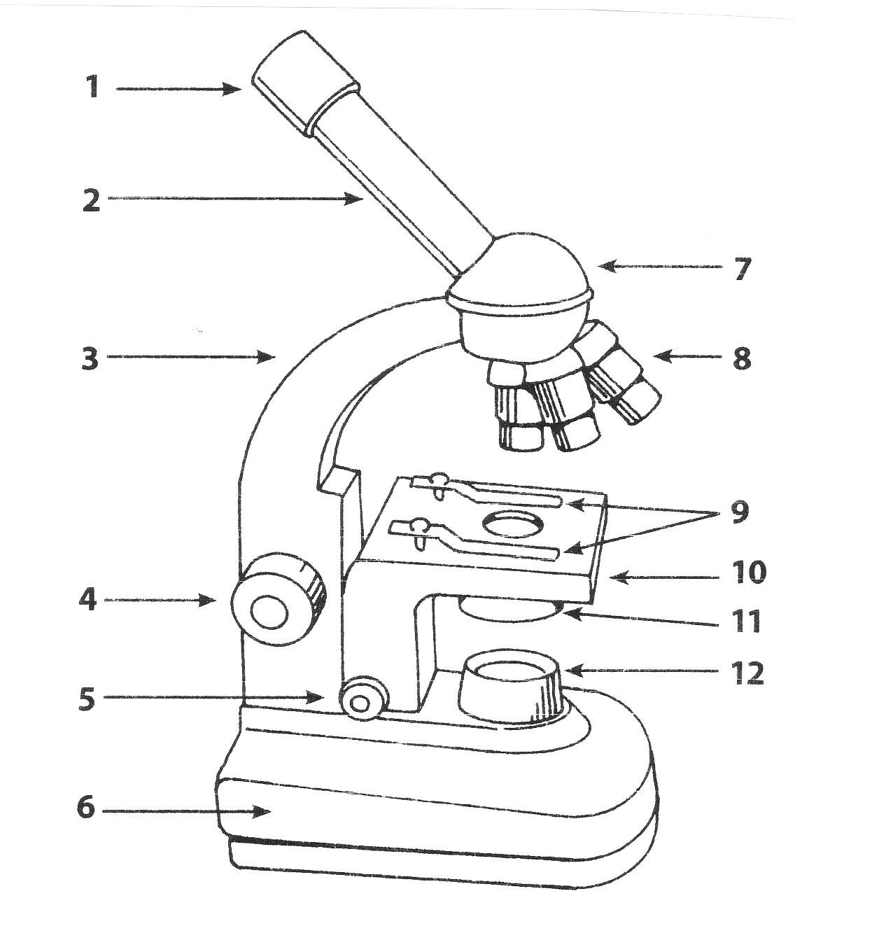
Parts Of A Microscope Drawing at GetDrawings Free download
If you want to redo an answer,. The eyepiece (or ocular lens) is the lens part at the top of a microscope that the viewer looks through. It is to be noted that. The standard eyepiece has a magnification of 10x. Many optical parts of a microscope work together to magnify and produce an image of the specimen placed on.
Microscope Drawing Easy at Explore collection of
The eyepiece (or ocular lens) is the lens part at the top of a microscope that the viewer looks through. Web microscope parts and functions with labeled diagram and functions how does a compound microscope work?. Mechanical parts of a compound microscope foot or base. Web the magnification power of a simple microscope is expressed as: The part that is.
Parts of a Microscope Microscope Parts and Functions Labkafe
Many optical parts of a microscope work together to magnify and produce an image of the specimen placed on a slide. Web parts of a microscope. The part that is looked through at the top of the compound microscope. Let us discuss the different parts of a compound microscope. Can be rotated to change magnification.

Labeled Microscope Diagram Tim's Printables
F = focal length of the convex lens. Web all microscopes share features in common. First, the purpose of a microscope is to. Download the label the parts of the microscope: Eyepieces typically have a magnification between 5x & 30x.
Microscope Types + Parts + History + Facts Science4Fun
F = focal length of the convex lens. The ocular lens is the lens close to the eye, and the objective lens is the lens close to the object. It’s actually not a toy microscope, it’s a functional microscope that produces great. Diagram of parts of a microscope. First, the purpose of a microscope is to.
The Standard Eyepiece Has A Magnification Of 10X.
Web parts of a microscope include the following: Web magnification is a measure of how much larger a microscope (or set of lenses within a microscope) causes an object to appear. Web the web page titled “parts of a microscope with labeled diagram and functions” has the following key takeaways: These lenses work together to magnify the image of an object.
Supports The Tube And Connects It To The Base.
🧬 the main function of a microscope is to provide a magnified view of small objects or organisms, such as bacteria, cells, or. Before exploring microscope parts and functions, you should probably understand that the compound light microscope is more complicated than just a microscope with more than one lens. Web the microscope illustrated in figure 5 below was manufactured by hugh powell and peter lealand around 1850. Many optical parts of a microscope work together to magnify and produce an image of the specimen placed on a slide.
Optical Microscopes Can Be Simple, Consisting Of A Single Lens, Or Compound, Consisting Of Several Optical Components In Line.
It is to be noted that. Web microscope parts and functions with labeled diagram and functions how does a compound microscope work?. Can be rotated to change magnification. In this interactive, you can label the different parts of a microscope.
Then, Draw Three Straight, Parallel Lines.
D = the lease possible distance of distinct vision of eye, typically 25cm. Supports the microscope head and attaches it to the base. Web the field diaphragm control is located around the lens located in the base. Focuses light onto the specimen.