Motor Neuron Drawing
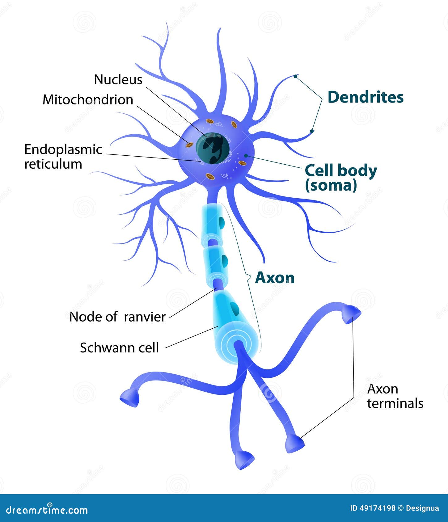
Motor Neuron Drawing - Those diagnosed with mnd eventually lose the ability to move and therefore to perform physical gestures and facial expressions ( motor neurone disease. Dendrites, a cell body, and an axon. Web cajal’s graceful drawings of neurons show them as separate, individual cells. Read this article to find out a diagram, types and related diseases of the motor neurons, only at byju’s. The soma, the axon, and the dendrites. Multiple sclerosis failure myelin cells. When these cells are damaged in some way, motor neuron disease can arise. Axon is myelinated by oligodendrocytes in the central nervous system (cns) and schwann cells in the peripheral nervous system (pns). For instance, if you picked up a hot coal, it motor neurons innervating the muscles in your fingers would cause your hand to let go. For more science drawing lessons visit www.ellenjmchenry.com, click on videos.
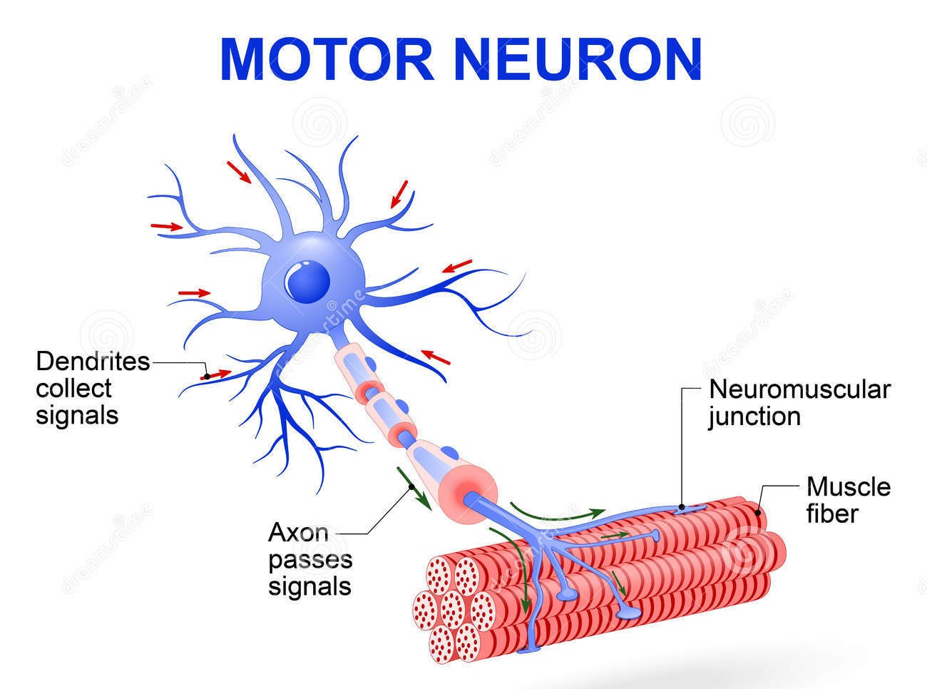
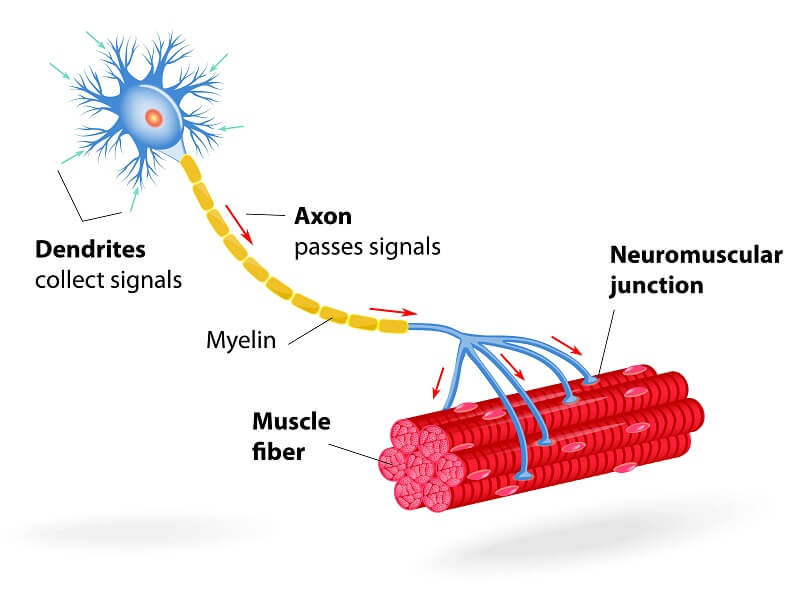
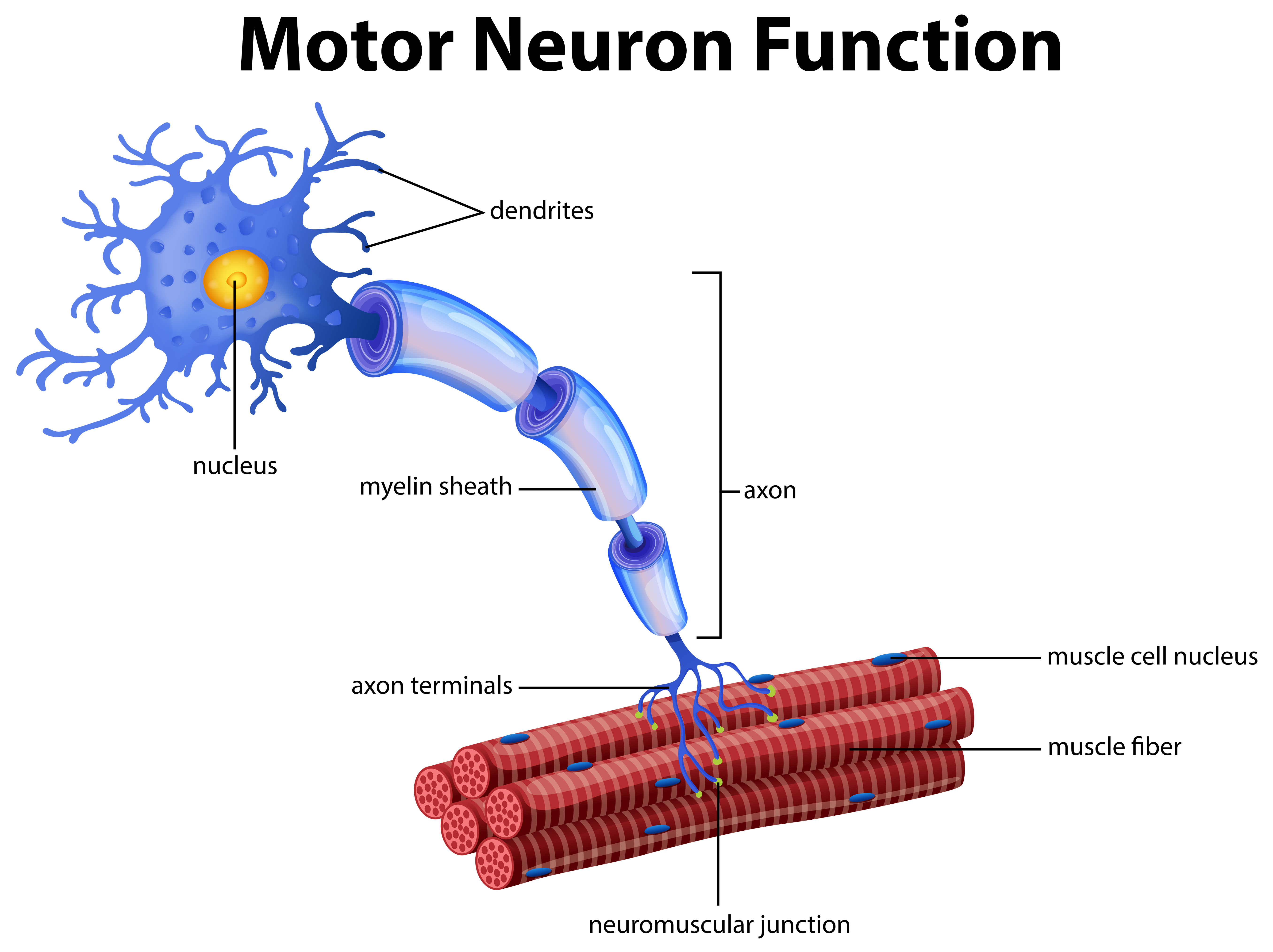
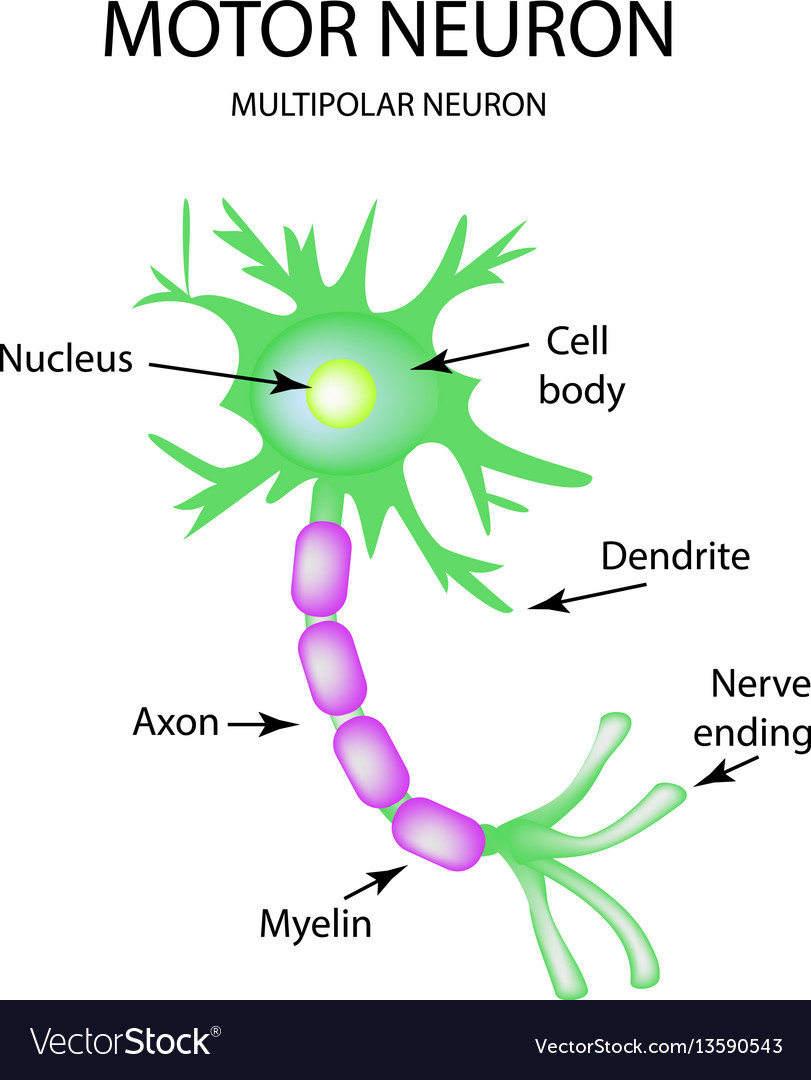
Motor neurons are located in the spinal cord, and their axon protrudes outside to the muscle fibers. (fig.1) the upper motor neurons originate in the cerebral cortex and travel down to the brain stem or spinal cord; It is also one among the few topics having the highest weightage of marks. Web in the exam you may be asked to label a diagram of a motor, relay or sensory neuron. Web the structure of a motor neuron is characterized by three components: He was the first to realize that the nervous system is not a network of continuous fibers, as was widely believed at the time. We find that the degree of m1 engram neuron reactivation correlates with motor performance. For more science drawing lessons visit www.ellenjmchenry.com, click on videos. Web motor neurons get information from other neurons and convey commands to your muscles, organs and glands. Courtesy of the cajal institute and the spanish national research council.
Web a motor neuron is basically a nerve cell whose function is to respond to sensory stimulation by producing the required muscular movement. Interneurons carry nerve impulses back and forth often between sensory and motor neurons within the spinal cord or brain. The soma, the axon, and the dendrites. Dendrites, a cell body, and an axon. This is characterized by muscle wasting (atrophy) and loss of motor function. Web a motor neuron is a cell of the central nervous system. Read this article to find out a diagram, types and related diseases of the motor neurons, only at byju’s. Motor neurons have the structure of a typical neuron. (fig.1) the upper motor neurons originate in the cerebral cortex and travel down to the brain stem or spinal cord; Web the structure of a motor neuron is characterized by three components:

How TO Draw neuron step by step easy/diagram of neuron/neuron drawing
For more science drawing lessons visit www.ellenjmchenry.com, click on videos. Web motor neurons get information from other neurons and convey commands to your muscles, organs and glands. Multiple sclerosis failure myelin cells. Web motor neurons, also known as efferent neurons, are nerve cells that run throughout the body and allow both voluntary and involuntary movements by innervating effector muscles and.

Neuron Diagram Straight from a Scientist
Dendrites, a cell body, and an axon. Web cajal’s graceful drawings of neurons show them as separate, individual cells. Cajal’s two brilliant insights — that every neuron in the. Difference between sensory and motor neuron. Web motor neurons (also referred to as efferent neurons) are the nerve cells responsible for carrying signals away from the central nervous system towards muscles.
Myelinated Motor Neurons Function, Location & Types
Specific regions of the cns coordinate different somatic processes using sensory inputs and motor outputs of peripheral nerves. He was the first to realize that the nervous system is not a network of continuous fibers, as was widely believed at the time. Those diagnosed with mnd eventually lose the ability to move and therefore to perform physical gestures and facial.
Motor Neuron The Definitive Guide Biology Dictionary
A simple case is a reflex caused by a synapse between a dorsal sensory neuron axon. Courtesy of the cajal institute and the spanish national research council. Those diagnosed with mnd eventually lose the ability to move and therefore to perform physical gestures and facial expressions ( motor neurone disease. Web motor neurons, also known as efferent neurons, are nerve.
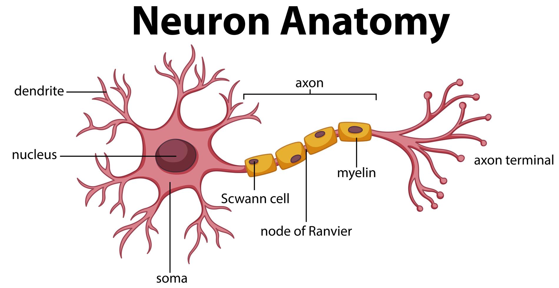
Diagram of Neuron Anatomy 358962 Vector Art at Vecteezy
A simple case is a reflex caused by a synapse between a dorsal sensory neuron axon. Web cajal’s graceful drawings of neurons show them as separate, individual cells. Motor neurons transmit signals to muscle cells or glands to control their functional output. He was the first to realize that the nervous system is not a network of continuous fibers, as.
Structure of a Motor Neuron Stock Vector Illustration of care, body
Web these are spanish neuroanatomist santiago ramón y cajal's drawings of neurons. Web in the exam you may be asked to label a diagram of a motor, relay or sensory neuron. For more science drawing lessons visit www.ellenjmchenry.com, click on videos. This is characterized by muscle wasting (atrophy) and loss of motor function. We find that the degree of m1.
A Vector of Motor Neuron Function 296405 Vector Art at Vecteezy
We find that the degree of m1 engram neuron reactivation correlates with motor performance. The diagram or the structure of the neuron is useful for both class 11 and 12 board exams as it has been repetitively asked in the board examinations. Courtesy of the cajal institute and the spanish national research council. Lower motor neurons, located in the spinal.

Motor neuron Alila Medical Images
Cajal’s two brilliant insights — that every neuron in the. Neurons [11:24] neurons are the basic building blocks of the nervous system. Diagram of neuron with labels. Neurons (or nerve cells) are specialized cells that transmit and receive electrical signals in the body. Dendrites, a cell body, and an axon.
The structure of the motor neuron infographics on Vector Image
Motor neurons transmit signals to muscle cells or glands to control their functional output. Motor neurons have a large cell body, or soma, and long projections used in transmitting information away from the soma. The lower motor neurons begin in the spinal cord and go on to innervate muscles and glands throughout the body. Web motor neurons, also known as.

Neuroanatomy, Motor Neuron StatPearls NCBI Bookshelf
Web motor neurons get information from other neurons and convey commands to your muscles, organs and glands. Motor neurons are located in the spinal cord, and their axon protrudes outside to the muscle fibers. Web motor (also called efferent) neurons, like the one in figure \(\pageindex{2}\), carry nerve impulses from the central nervous system to muscles and glands. Web a.
Diagram Of Neuron With Labels.
Web a motor neuron is a cell of the central nervous system. Hello friends in this video i will show you how to draw a neuron. Motor neurons transmit signals to muscle cells or glands to control their functional output. For instance, if you picked up a hot coal, it motor neurons innervating the muscles in your fingers would cause your hand to let go.
Neurons Are Composed Of Three Main Parts:
Multiple sclerosis failure myelin cells. A simple case is a reflex caused by a synapse between a dorsal sensory neuron axon. Neurons (or nerve cells) are specialized cells that transmit and receive electrical signals in the body. Lower motor neurons, located in the spinal cord and brain stem, send signals to skeletal muscles via neuromuscular junctions.
Axon Is Myelinated By Oligodendrocytes In The Central Nervous System (Cns) And Schwann Cells In The Peripheral Nervous System (Pns).
Dendrites, a cell body, and an axon. 91k views 6 years ago science diagrams | explained and labelled science diagrams. The lower motor neurons begin in the spinal cord and go on to innervate muscles and glands throughout the body. Neurons are the information processing units of the brain responsible for sending, receiving, and transmitting electrochemical signals throughout the body.
It Is Also One Among The Few Topics Having The Highest Weightage Of Marks.
Courtesy of the cajal institute and the spanish national research council. Motor neurons have a large cell body, or soma, and long projections used in transmitting information away from the soma. He was the first to realize that the nervous system is not a network of continuous fibers, as was widely believed at the time. Web in the exam you may be asked to label a diagram of a motor, relay or sensory neuron.