Muscle Cell Drawing
Muscle Cell Drawing - This article is about skeletal myocytes. While several associated proteins help, actin and myosin form thick and thin filaments which slide past each other to contract small units of a. View muscle cell drawing videos. Skeletal muscle fibers can be quite large compared to other cells, with diameters up to 100 μ m and lengths up to. Web it illustrates the distinctive structure of muscle cells, including striated myofibrils (components of muscle cells only). Muscle fibers and connective tissue layers make up the skeletal muscle. Web interactive guide to the muscular system | innerbody. Web human body maps. How the structure of a neuron allows it to receive and transmit information. I thought smooth muscle did not have the striations described in the video and only had one nucleus per cell instead of many.
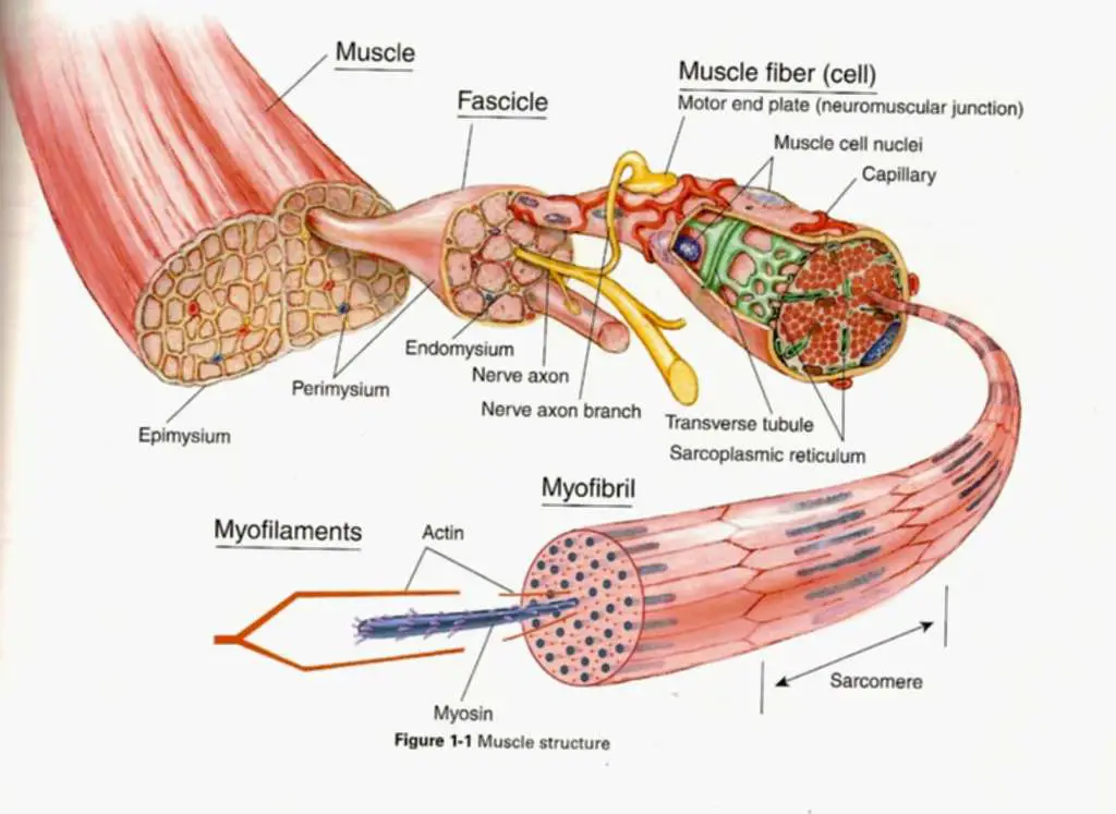
Web i'll draw one of these dudes coming out right here. 133 views 1 year ago little lectures for 1st semester a&p. Cardiac striated muscle, myocardium , show more. Web the actin and myosin proteins are arranged very regularly in the cytoplasm of individual muscle cells (referred to as fibers) in both skeletal muscle and cardiac muscle, which creates a pattern, or stripes, called striations. This is an individual muscle cell that's covered by the endomysium. For more about the contents of cells generally see animal cells. Muscle tissue is characterized by properties that allow movement. Muscle cell drawing pictures, images and stock photos. They are bound together by perimysium, a sheath of connective tissue, into bundles called fascicles, which are in. Structure of the skeletal muscle.
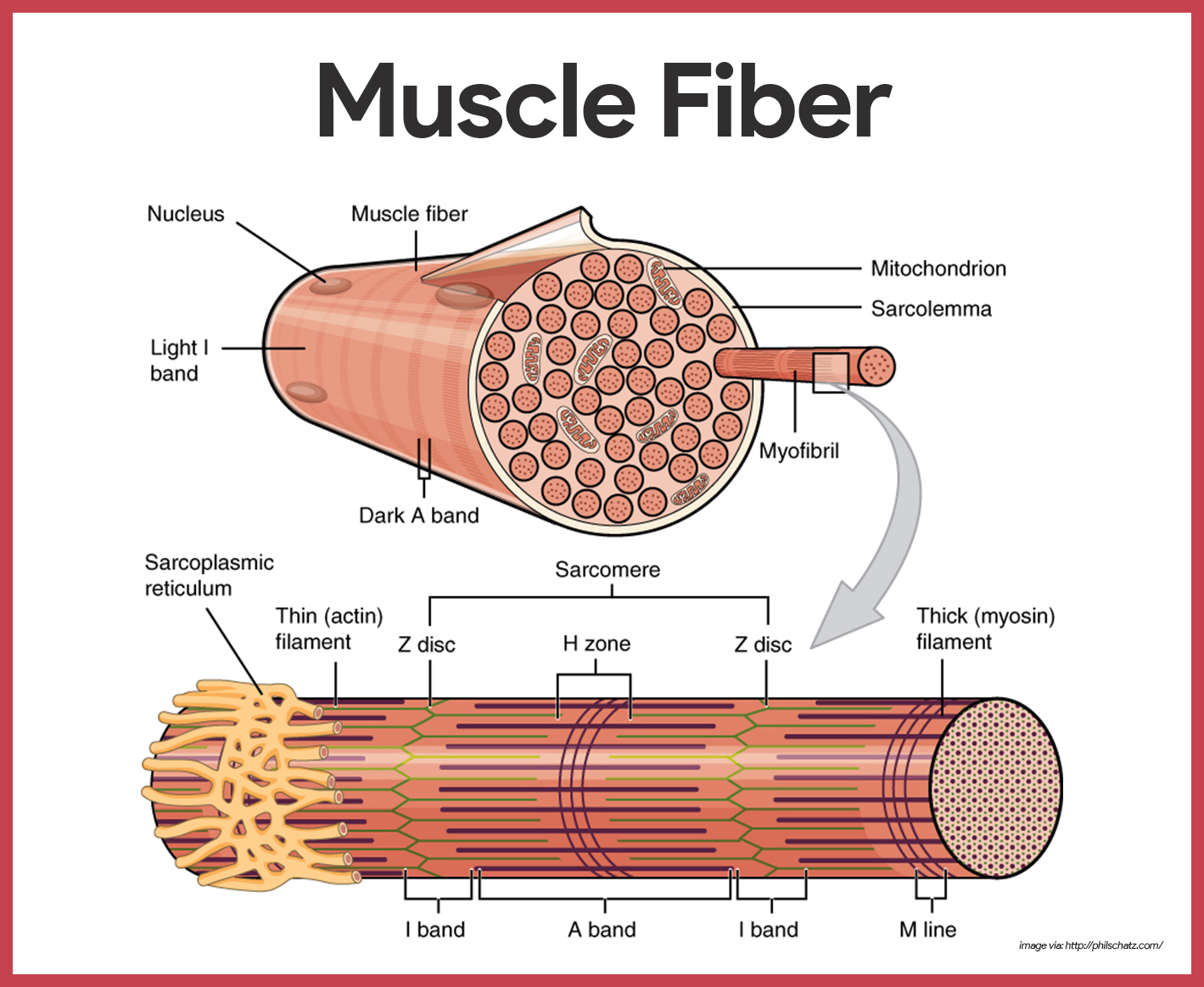
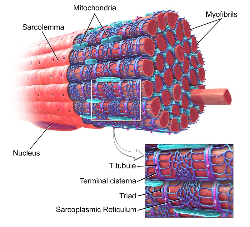
Identify the three types of muscle tissue. This article is about skeletal myocytes. They are bound together by perimysium, a sheath of connective tissue, into bundles called fascicles, which are in. Specialised animal cells have components that allow them to complete a specific purpose. Web figure 10.4 muscle fiber a skeletal muscle fiber is surrounded by a plasma membrane called the sarcolemma, which contains sarcoplasm, the cytoplasm of muscle cells. The primary job of muscles is to move the bones of the skeleton, but muscles also enable the heart to beat and constitute the walls of other vital. Compare and contrast the functions of each muscle tissue type. Web i'll draw one of these dudes coming out right here. The striations are visible with a light microscope under high magnification (see figure 10.2 ). Web remove from my bitesize.

Types of muscle cells vector illustration in 2022 Types of muscles
How different types of animal cell are adapted to carry out their function. It attaches to bones and the orbits through tendons. Web let's draw a skeletal muscle cell: Web it illustrates the distinctive structure of muscle cells, including striated myofibrils (components of muscle cells only). Web human body maps.
Muscular System Anatomy and Physiology Nurseslabs
Muscle cell drawing pictures, images and stock photos. Contractile tissue is able to generate tension of force. Muscle fibers and connective tissue layers make up the skeletal muscle. Web the actin and myosin proteins are arranged very regularly in the cytoplasm of individual muscle cells (referred to as fibers) in both skeletal muscle and cardiac muscle, which creates a pattern,.

Diagram showing types of muscle cells illustration Stock Vector Image
Myocytes, sometimes called muscle fibers, form the bulk of muscle tissue. Web remove from my bitesize. Learn about the three types of muscle as you use our 3d models to explore the anatomical structure and physiology of human muscles. Skeletal (striated or voluntary) muscle, smooth muscle (that we don't control, like the muscles in our bowel), and cardiac muscle? Cardiac.

Types of muscle cell diagram 1783902 Vector Art at Vecteezy
Web introduction to neurons and glia. Cardiac muscle cells or cardiomyocytes (also known as cardiac myocytes) are the muscle cells (myocytes) that make up the heart muscle. Muscle contraction in striated muscle. Compare and contrast the functions of each muscle tissue type. Muscle cells, commonly known as myocytes, are the cells that make up muscle tissue.

Skeletal Muscle Cell Structure
This is an individual muscle cell that's covered by the endomysium. Cardiac and skeletal myocytes are sometimes referred to as muscle fibers due to their long and. Web introduction to neurons and glia. Web interactive guide to the muscular system | innerbody. Web because skeletal muscle cells are long and cylindrical, they are commonly referred to as muscle fibers (or.

How To Draw Muscle Cell Step by Step YouTube
Skeletal muscle fibers can be quite large compared to other cells, with diameters up to 100 μ m and lengths up to. How do you know where you are right now? Web isn't it true that there are 3 kinds of muscle cells: Web by the end of this section, you will be able to: For more about the contents.
Muscle cell diagram
Web human body maps. Muscle cells, commonly known as myocytes, are the cells that make up muscle tissue. For more about the contents of cells generally see animal cells. Web it illustrates the distinctive structure of muscle cells, including striated myofibrils (components of muscle cells only). Hand drawn seamless pattern vector.
Muscle Cell (Myocyte) Definition, Function & Structure Biology
A muscle fiber is composed of many fibrils, which give the cell its striated appearance. The primary job of muscles is to move the bones of the skeleton, but muscles also enable the heart to beat and constitute the walls of other vital. Myocytes, sometimes called muscle fibers, form the bulk of muscle tissue. What is your request drawing? Web.

Types of muscle cell diagram 1762350 Vector Art at Vecteezy
Hand drawn seamless pattern vector. :) thanks for watching our channel. Web muscle cell definition. What is your request drawing? This article is about skeletal myocytes.
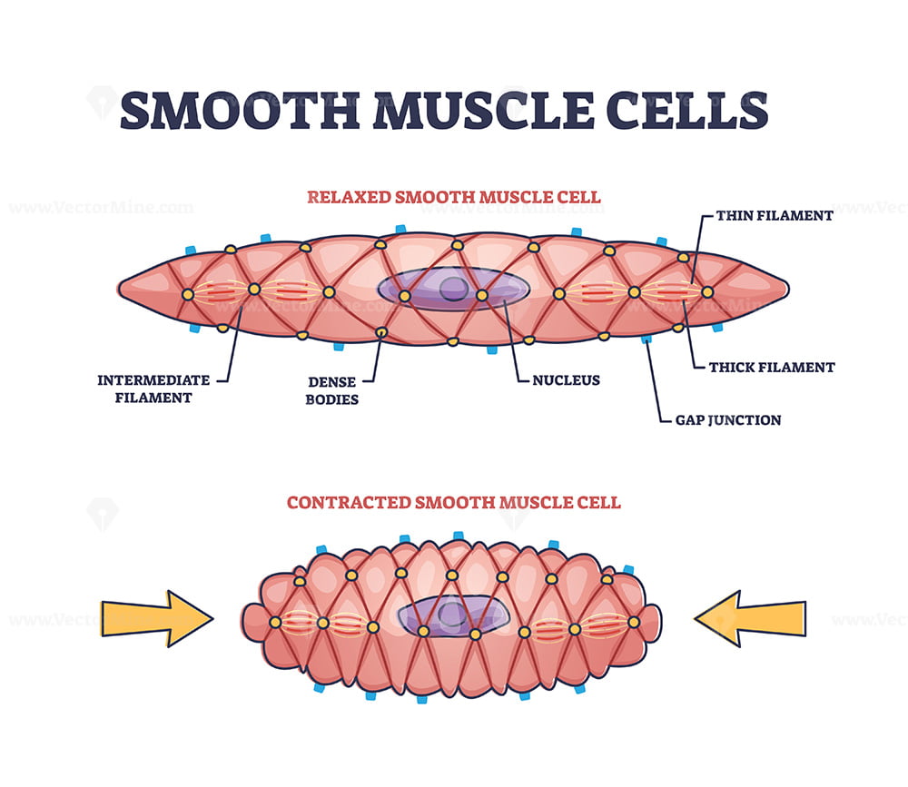
Smooth Muscle Cell Structure
Web let's draw a skeletal muscle cell: There are 3 types of muscle cells in the human body; Web it illustrates the distinctive structure of muscle cells, including striated myofibrils (components of muscle cells only). Web interactive guide to the muscular system | innerbody. Specialised animal cells include red blood cells, sperm, eggs, nerve cells, muscle.
What Is Your Request Drawing?
The striations are visible with a light microscope under high magnification (see figure 10.2 ). As it is broken down, atp must therefore be regenerated and replaced quickly to allow for sustained contraction. How different types of animal cell are adapted to carry out their function. Muscle cell drawing pictures, images and stock photos.
Specialised Animal Cells Have Components That Allow Them To Complete A Specific Purpose.
Web because skeletal muscle cells are long and cylindrical, they are commonly referred to as muscle fibers (or myofibers). Muscle cell diagram, how to draw smooth muscle cell, how to draw. Cardiac striated muscle, myocardium , show more. Web muscle cell definition.
A Muscle Cell, Also Known As A Myocyte, Is A Mature Contractile Cell In The Muscle Of An Animal.
There are 3 types of muscle cells in the human body; Cardiac and skeletal myocytes are sometimes referred to as muscle fibers due to their long and. It is also important to remember that organelles essential for all cells (not all of which are illustrated in the diagram above) are also present in muscle cells. Specialised animal cells include red blood cells, sperm, eggs, nerve cells, muscle.
Identify The Three Types Of Muscle Tissue.
Web how to draw muscle cell step by step. This article is about skeletal myocytes. View muscle cell drawing videos. It attaches to bones and the orbits through tendons.