Onion Root Tip Mitosis Drawing
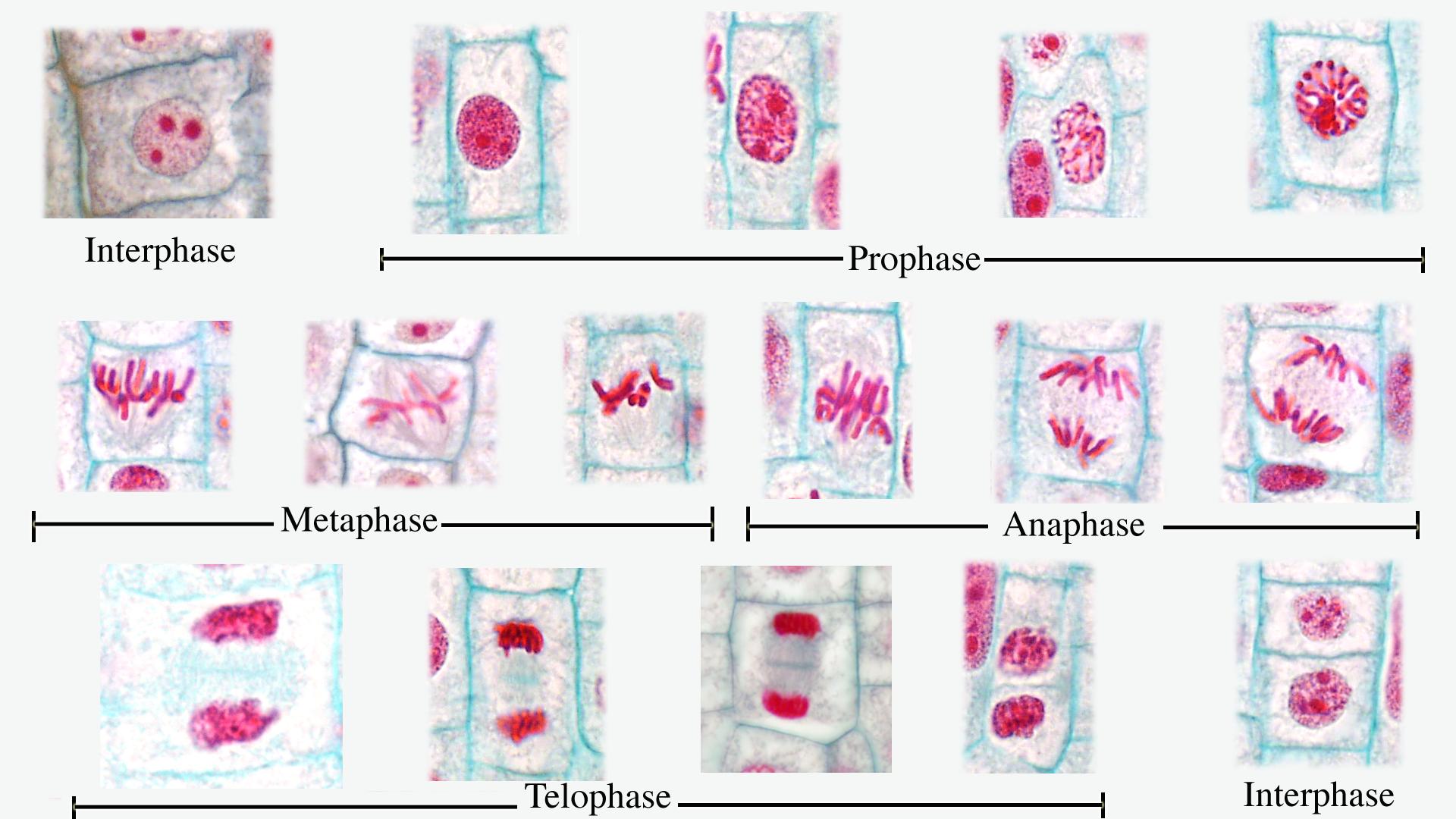
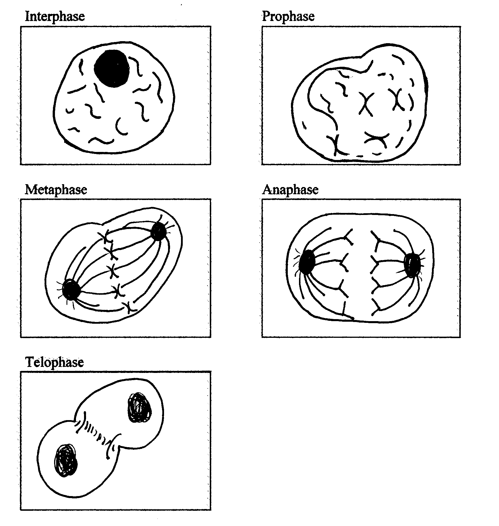
Onion Root Tip Mitosis Drawing - Because growth in roots occurs at the tips, this is where cells will most actively undergo mitosis. Nuclear membrane breaks down, chromatin condenses, mitotic spindle forms and attaches to kinetochores. To study and demonstrate mitosis by preparing the mount of an onion root tip cells. 2.1k views 1 year ago. Web onion root tip cell mitosis. Students count the number of cells they see in interphase, prophase,. Number of cells in each stage. In plants, the roots continue to grow as they search for water and nutrients. Web diagrammatic representation of a cell during telophase (late telophase), onion root tip mitosis. Focus in on low power and then switch to medium or high power.
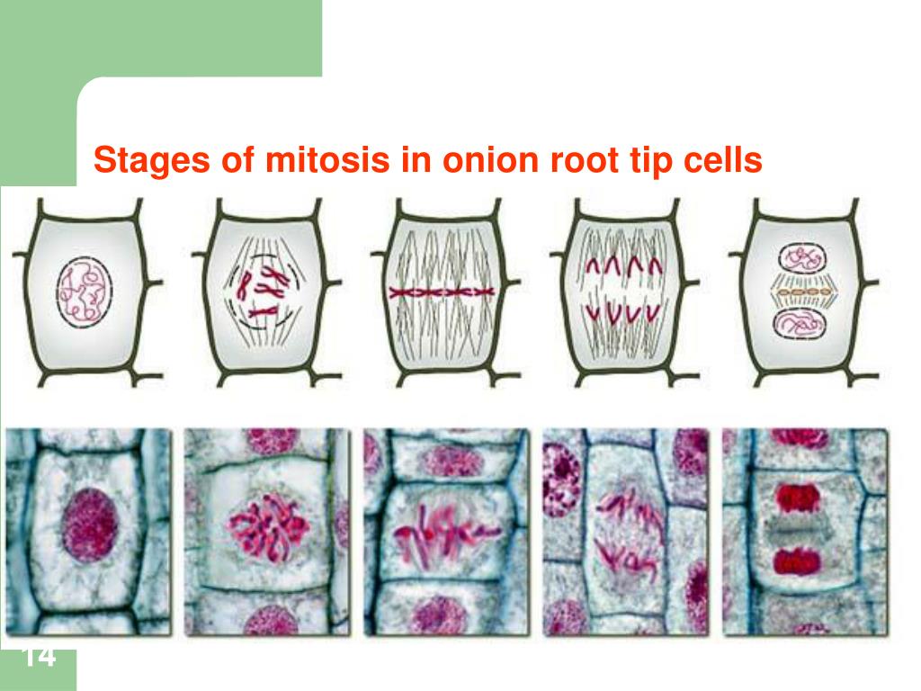
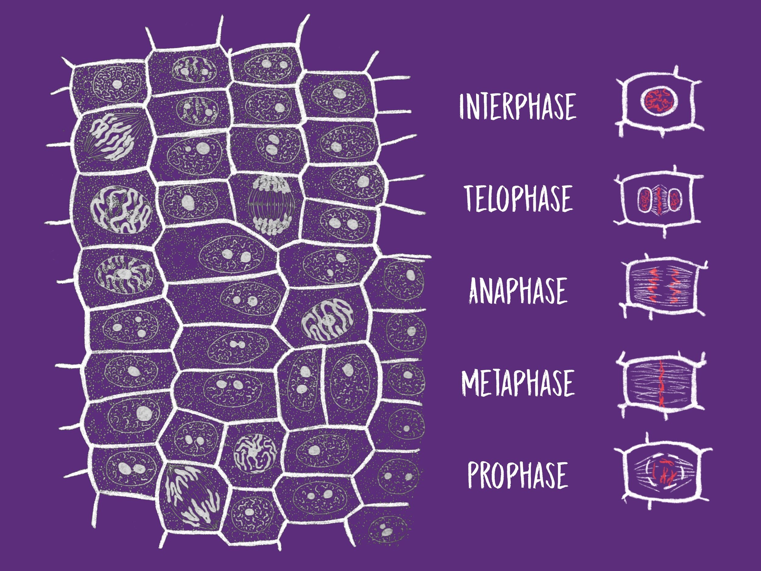
The simulation “mitosis in onion root tips'” aims to investigate the different stages of mitotic cell division in onion root tip cells. Web online onion root tips. The white arrow indicates the location of the root apical meristem. Identify and draw a cell in each of the four stages of mitosis in the onion slide. Web introduction to mitosis in onion root tips. Web mitosis in onion root tips (assignment) : Web onion root tips are often used in lessons on mitosis because they contain actively dividing cells in the root meristem, making it a great resource to observe different stages of the cell cycle, including mitosis. Set up a compound light microscope and turn on the. Repeat for prophase, metaphase, anaphase, and telophase cells, and cells in cytokinesis. Label, in each drawing, the defining features that you will look for when identifying each stage under the microscope.
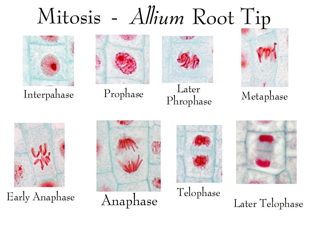
Use the image slider below to learn how to use a microscope to identify cells dividing by mitosis on an onion root tip slide. Focus in on low power and then switch to medium or high power. Flemming's solution (chromic acid, osmic acid, and acetic acid) image size. Web using correct microscope procedure, observe an onion root tip under high power (400x). To understand the process and different stages of mitosis and to. Project an image of onion tip cells on a whiteboard. Label, in each drawing, the defining features that you will look for when identifying each stage under the microscope. Web figure \ (\pageindex {3}\): Chalk (can be used directly on lab bench to draw cellular structures and then washed off) • apply an analytical technique by which the relative length of each stage of mitosis can be estimated.

Oniion Root Tip Microscope Images Flowering Plants
These regions of growth are good for studying the cell cycle because at any given time, you can find cells that are undergoing mitosis. Find, identify, and draw the phases of mitosis in the onion root tip and whitefish blastula. Web mitosis in onion root tips (assignment) : A cell undergoes mitotic cell division, a process of cell duplication in.
Mitosis Cell in the Root Tip of Onion Under a Microscope. Stock Photo
Flemming's solution (chromic acid, osmic acid, and acetic acid) image size. Biotechnology and biomedical engineering : Region of cell elongation region of cell division protective root cap. Label, in each drawing, the defining features that you will look for when identifying each stage under the microscope. Place a slide containing a stained preparation of the onion root tip.

Biology Encore — The image above shows a stained slide of an onion...
Place a slide containing a stained preparation of the onion root tip. Project an image of onion tip cells on a whiteboard. This lab requires students to use a microscope and preserved cells of an onion root that show dividing cells. In your notebook, make a drawing of each phase of mitosis, as well as interphase, in a plant cell..
of all stages of mitosis in onion root tip labeled UWDC
Web • prepare your own specimens of onion root in which you can visualize all of the stages of mitosis. Region of cell elongation region of cell division protective root cap. Web in this chapter, you can use pictures of onion root tip cells to learn how to identify the different phases of mitosis and better understand what events occur.
Onion Root Tip Mitosis Diagram
Number of cells in each stage. Web • prepare your own specimens of onion root in which you can visualize all of the stages of mitosis. Amrita vishwa vidyapeetham virtual lab. Web onion root tip mitosis. Region of cell elongation region of cell division protective root cap.

Second Onion Root Tip View with Stages of Mitosis Labeled … Flickr
Label, in each drawing, the defining features that you will look for when identifying each stage under the microscope. Use the image slider below to learn how to use a microscope to identify cells dividing by mitosis on an onion root tip slide. Nuclear membrane breaks down, chromatin condenses, mitotic spindle forms and attaches to kinetochores. Microtubules align chromosomes along.
of all stages of mitosis in onion root tip labeled UWDC
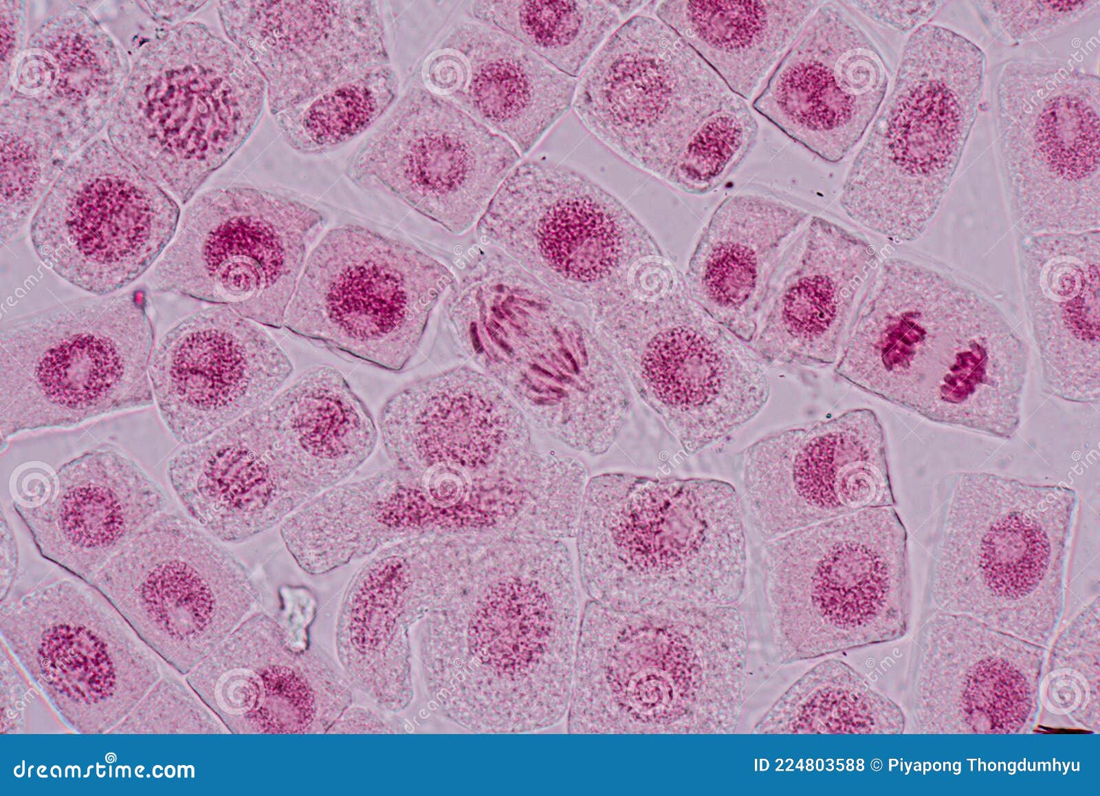
• apply an analytical technique by which the relative length of each stage of mitosis can be estimated. In plants, the roots continue to grow as they search for water and nutrients. Are the predictions you made in step 1 supported by your observations? Consulting with your team, identify and mark all the cells you feel are in interphase, and.
Mitosis Phases in Onion Root Tip Cells Tuva
Consulting with your team, identify and mark all the cells you feel are in interphase, and mark them with a particular color of marker pen. The images shown below were taken using a regular light microscope with an oil immersion lens at 1000x. Use the image slider below to learn how to use a microscope to identify cells dividing by.
ap lab 3 sample 3 mitosis
Cell division is of two types: Web the student will correctly identify and draw four stages of mitosis using microscope slide images of onion root tips and whitefish blastulae. Chalk (can be used directly on lab bench to draw cellular structures and then washed off) In plants, the roots continue to grow as they search for water and nutrients. Biotechnology.
Mitosis in Onion Root Tips — DataClassroom
Web onion root tip cell mitosis. In plants, the roots continue to grow as they search for water and nutrients. Consulting with your team, identify and mark all the cells you feel are in interphase, and mark them with a particular color of marker pen. These regions of growth are good for studying the cell cycle because at any given.
Are The Predictions You Made In Step 1 Supported By Your Observations?
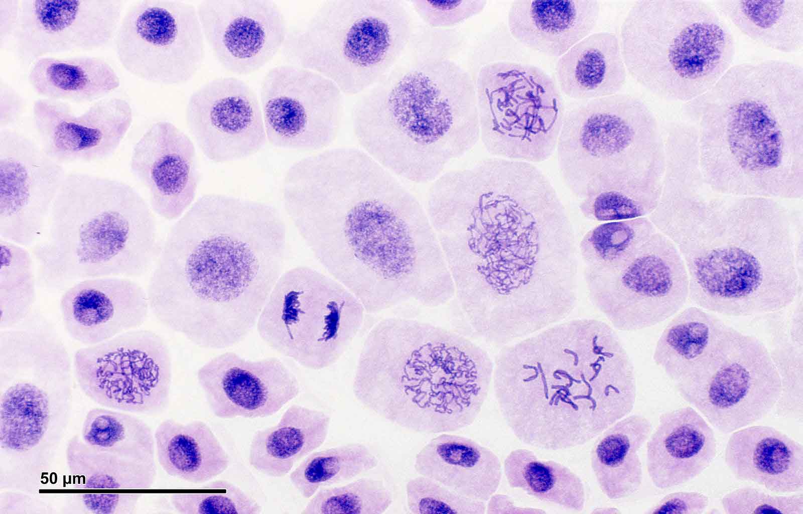
Students count the number of cells they see in interphase, prophase,. Viewing mitosis in onion root tips. Web the student will correctly identify and draw four stages of mitosis using microscope slide images of onion root tips and whitefish blastulae. Consulting with your team, identify and mark all the cells you feel are in interphase, and mark them with a particular color of marker pen.
As Nuclear Envelopes Develop Around Each Set Of Chromosomes Located At The Opposite Poles Of The Cell, Two Nuclei Are Formed In The Cell.
Onion root tip whitefish blastula; 48,000 x 36,000 5.2 gb. Below find micrographs of the four stages of mitosis. Web onion root tips are often used in lessons on mitosis because they contain actively dividing cells in the root meristem, making it a great resource to observe different stages of the cell cycle, including mitosis.
Cells In This Onion Root Tip Were Caught In Various Stages Of The Cell Cycle.
Web onion root tip graphic. Use the image slider below to learn how to use a microscope to identify cells dividing by mitosis on an onion root tip slide. Web mitosis in onion root tips (assignment) : To study and demonstrate mitosis by preparing the mount of an onion root tip cells.
In Your Notebook, Make A Drawing Of Each Phase Of Mitosis, As Well As Interphase, In A Plant Cell.
2.1k views 1 year ago. Web figure \ (\pageindex {3}\): For entities to mature, grow, maintain tissues, repair and synthesize new cells, cell division is required. Label, in each drawing, the defining features that you will look for when identifying each stage under the microscope.