Paramecium Drawing
Paramecium Drawing - Does a paramecium make a poo? Slipper animalcule, illustrated by louis joblot, 1718. Cv contractile vacuoles, fv food vacuoles, manu macronucleus, mino micronucleus, pe peristome, tr trichocysts and ve vestibulum. Original file (svg file, nominally 1,142 × 1,007 pixels, file size: Paramecium were among the first ciliates to be observed. Web at the low paramecium figure 5.11: Web the structure of pellicle and cilia. They are also found attached to the surface. A paramecium is a microscopic organism that lives in ponds and streams. Paramecia, illustrated by otto müller, 1773.
How fast can a paramecium move? Paramecia, illustrated by otto müller, 1773. They are also found attached to the surface. What is inside the cell. Members of the genus paramecium are microscopic but visible to the naked eye. Simulation of helical trajectories of paramecium. A paramecium is a microscopic organism that lives in ponds and streams. Original file (svg file, nominally 1,142 × 1,007 pixels, file size: Web panel 1 (a,b): See how cilia do the wave.
Earliest known illustration of paramecium. Web biological drawing showing the characteristics of paramecium, biology teaching resources by d g mackean. Members of the genus paramecium are microscopic but visible to the naked eye. Cytostome, cytopharynx, and food vacuole. Slipper animalcule, illustrated by louis joblot, 1718. Web this video explains how to draw paramecium : Download 77 paramecium diagram stock illustrations, vectors & clipart for free or amazingly low rates! How fast can a paramecium move? Fresh water, free living, omnipresent and is found in stagnant water. This video helps you to draw science diagrams with great ease and.

How TO Draw paramecium easy with pencil YouTube
151k views 3 years ago science diagrams | explained and labelled science diagrams. Web this will also help you to draw the structure and diagram of paramecium. 272 × 240 pixels | 544 × 480 pixels | 871 × 768 pixels | 1,161 × 1,024 pixels | 2,323 × 2,048 pixels | 1,142 × 1,007 pixels. Paramecium is found in.

Paramecium stock illustration. Image of outline, anatomy 33357022
Paramecium were among the first ciliates to be observed. Cytostome, cytopharynx, and food vacuole. #paramecium #howtodraw #biology this is a diagram of the paramecium. Earliest known illustration of paramecium. In this video i have shown the simplest way of drawing paramecium.

Paramecium diagram by lucidhysteria on DeviantArt
25k views 2 years ago #howtodraw #biology #paramecium. Web panel 1 (a,b): Web the structure of pellicle and cilia. Web this video explains how to draw paramecium : Members of the genus paramecium are microscopic but visible to the naked eye.

Simplest Way Of Drawing Paramecium Diagram How To Draw Paramecium in
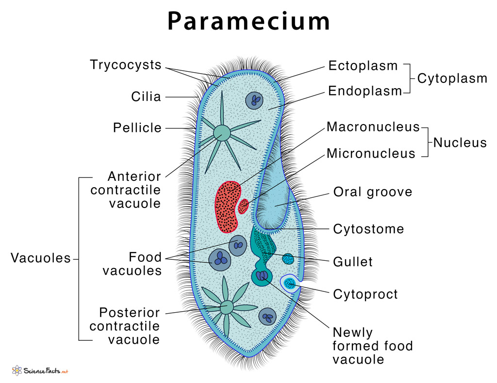
How does a paramecium eat? It is the pencil diagram of. Paramecium structure consists of trichocysts, contractile vacuoles, and cilia among other specialized organelles. Simulation of helical trajectories of paramecium. The specialized “skin” of paramecium cell body.

How To Draw Paramecium Diagram YouTube
You can learn to draw and colour the parts. 2.2k views 10 months ago easy science drawing. Web published 21 february 2022. In this video i have shown the simplest way of drawing paramecium. Paramecium were among the first ciliates to be observed.
ILLUSTRATED DIAGRAM OF PARAMECIUM (PARAMECIUM SP.) 1000X Stock Photo
Web this video explains how to draw paramecium : Length ranges between 80 to 350 µ. Paramecium is found in freshwater habitats and is used as a model organism in scientific studies. A paramecium is a microscopic organism that lives in ponds and streams. Web size of this png preview of this svg file:
Paramecium Definition, Structure, Characteristics and Diagram
Drawing of paramecium illustrating light microscopic features: #paramecium #howtodraw #biology this is a diagram of the paramecium. How fast can a paramecium move? They are found in freshwater, marine and brackish water. Slipper animalcule, illustrated by louis joblot, 1718.

DRAW IT NEAT How to draw Paramecium
Their size differs from species to species. A paramecium is a microscopic organism that lives in ponds and streams. Slipper animalcule, illustrated by louis joblot, 1718. Paramecium drawing pictures, images and stock photos. Web paramecium or paramoecium is a genus of unicellular ciliated protozoa.

How to draw paramecium step by step easy paramecium diagram YouTube
#paramecium #howtodraw #biology this is a diagram of the paramecium. Browse 50+ paramecium drawing stock photos and images available, or start a new search to explore more stock photos and images. How does a paramecium eat? Paramecia were among the first ciliates to be seen by. It is very easy drawing detailed method to help you.

DRAW IT NEAT How to draw Paramecium
Does a paramecium make a poo? Earliest known illustration of paramecium. 272 × 240 pixels | 544 × 480 pixels | 871 × 768 pixels | 1,161 × 1,024 pixels | 2,323 × 2,048 pixels | 1,142 × 1,007 pixels. Paramecium drawing pictures, images and stock photos. Web © 2023 google llc.
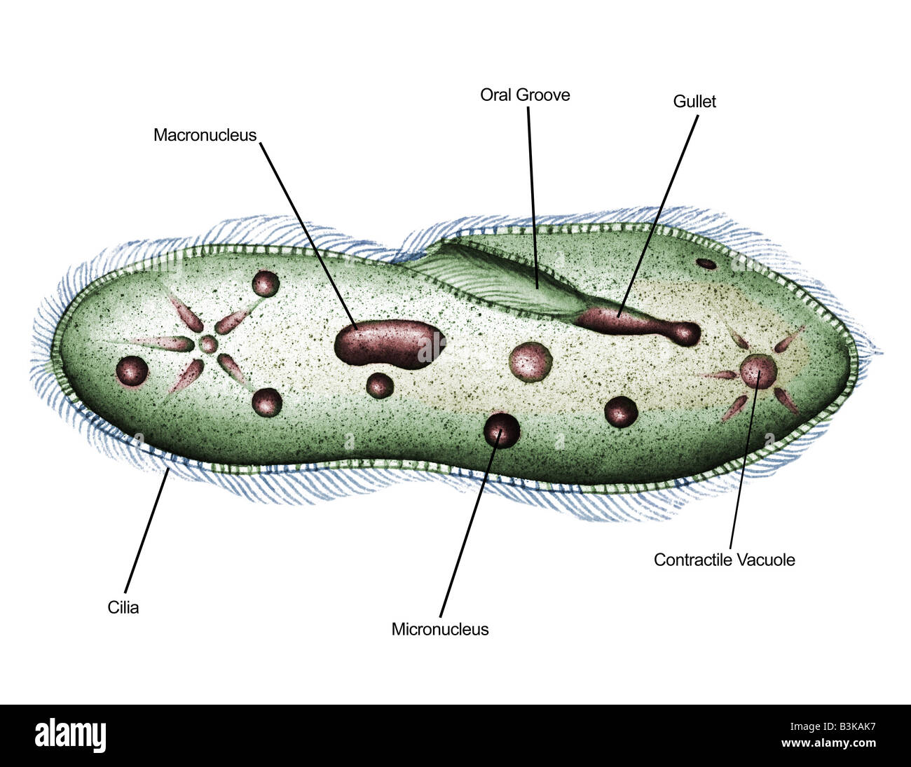
The Basic Anatomy Of Paramecium Shows The Following Distinct And Specialized.
Web panel 1 (a,b): Length ranges between 80 to 350 µ. Web paramecium or paramoecium is a genus of unicellular ciliated protozoa. Does a paramecium make a poo?
Web Hello Friends In This Video I Will Tell You About How To Draw Labelled Diagram Of Paramecium Step By Step For Beginners In Easy Wayso Friends If You Have Pro.
Download 77 paramecium diagram stock illustrations, vectors & clipart for free or amazingly low rates! Web © 2023 google llc. It is very easy drawing detailed method to help you. 151k views 3 years ago science diagrams | explained and labelled science diagrams.
Paramecium Structure Consists Of Trichocysts, Contractile Vacuoles, And Cilia Among Other Specialized Organelles.
Earliest known illustration of paramecium. #paramecium #howtodraw #biology this is a diagram of the paramecium. Original file (svg file, nominally 1,142 × 1,007 pixels, file size: (a) depending on the initial orientation of paramecium, the 2d projection will vary.
Cv Contractile Vacuoles, Fv Food Vacuoles, Manu Macronucleus, Mino Micronucleus, Pe Peristome, Tr Trichocysts And Ve Vestibulum.
Paramecia were among the first ciliates to be seen by. Light microscopic appearance of paramecium caudatum. Web published 21 february 2022. Paramecia, illustrated by otto müller, 1773.