Reticular Tissue Drawing
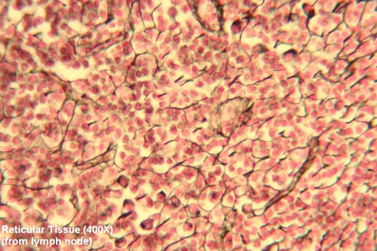
Reticular Tissue Drawing - Reticular fibers are composed of thin and delicately woven strands of type iii collagen. These tissues have a peculiar feature; Fine reticular fibers stain faintly; They are not visible with hematoxylin & eosin (h&e), but are specifically stained by silver. Web reticular connective tissue is located in the bone marrow, peyer’s patches, lymph nodes, kidney, liver, and spleen. Reticular fibers are abundant in lymphoid organs (lymph nodes, spleen), bone marrow and liver. Use the hotspot image below to learn more about the characteristics of reticular tissue. Web reticular connective tissue is a type of connective tissue [1] with a network of reticular fibers, made of type iii collagen [2] ( reticulum = net or network). Rather, you will always find reticular cells and fibers in association with other cells. Web reticular connective tissues are arranged along with different cells in various organs like bone marrow, lymph nodes, spleen, liver, kidneys, and even under the skin.
Web reticular tissue is a special subtype of connective tissue that is indistinguishable during routine histological staining. Reticular fibers form the stroma Web reticular 1 | digital histology. Web dense irregular connective tissue is a type of connective tissue proper with a matrix containing densely packed interwoven collagen fibers that fill most of the extracellular space and a thick jellylike ground substance comprising the remainder of the matrix. Reticular fibers are not unique to reticular connective tissue, but only in this tissue type are they dominant. Reticular tissue, a type of loose connective tissue in which reticular fibers are the most prominent fibrous component, forms the supporting framework of the lymphoid organs (lymph nodes, spleen, tonsils), bone marrow and liver. Reticular connective tissue forms a scaffolding for other cells in several organs, such as lymph nodes and bone marrow. Appearance and features of the reticular connective tissue. Use the image slider below to learn how to use a microscope to identify and study dense regular connective tissue on a. Reticular fibers are abundant in lymphoid organs (lymph nodes, spleen), bone marrow and liver.
The cells that make the reticular fibers are fibroblasts called reticular cells. Web reticular tissue is a special subtype of connective tissue that is indistinguishable during routine histological staining. Draw and label reticular tissue: Reticular cells produce the reticular fibers that form the network onto which other cells attach. Use the image slider below to learn how to use a microscope to identify and study reticular tissue on a microscope slide of a lymph node. Reticular fibers form the stroma Web dense irregular connective tissue is a type of connective tissue proper with a matrix containing densely packed interwoven collagen fibers that fill most of the extracellular space and a thick jellylike ground substance comprising the remainder of the matrix. Main menu » tissues » connective » special » reticular » reticular connective tissue is composed of a meshwork of reticular fibers (type iii collagen) in an open arrangement. Reticular fibers are not unique to reticular connective tissue, but only in this tissue type are they dominant. White (unilocular) and brown (multilocular) fat.

Reticular connective Tissue Diagram Quizlet
Use the hotspot image below to learn more about the characteristics of reticular tissue. Differentiate among the subclasses of connective tissue discussed in this chapter, including: The reticular fibers form the network onto which other cells attach. May anchor to collagenous septa, which divide organs into lobes. Web obtain a slide of a spleen or lymph node with reticular connective.

Reticular Connective Tissue, 40X Histology
These tissues have a peculiar feature; Web reticular tissue is a special subtype of connective tissue that is indistinguishable during routine histological staining. Reticular fibers form the stroma The cells that make the reticular fibers are fibroblasts called reticular cells. Web reticular connective tissue is a type of connective tissue [1] with a network of reticular fibers, made of type.
[Solved] RETICULAR TISSUE Draw and label. Include function and location
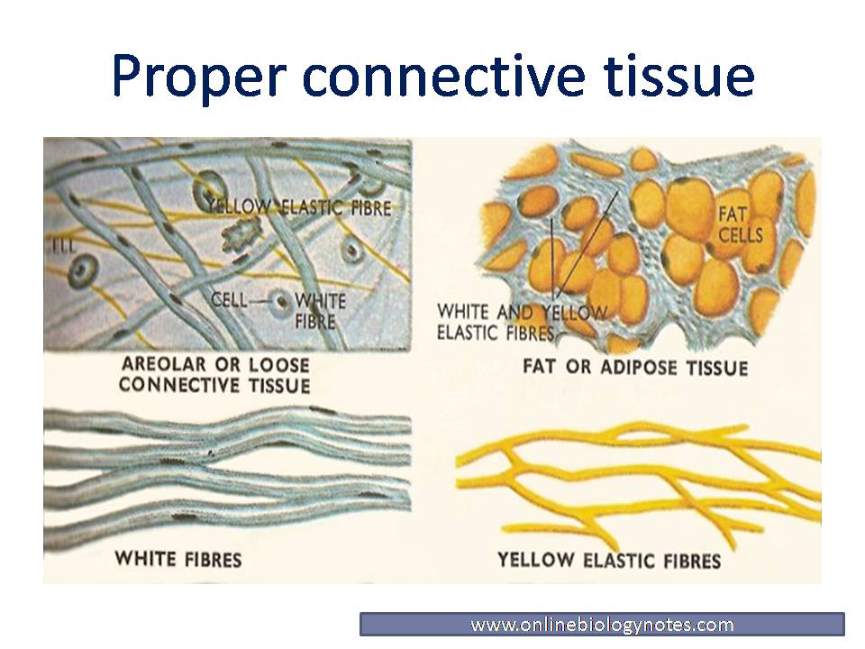
It derives its name from the latin reticulus, which means “little net.” dense connective tissue They are not visible with hematoxylin & eosin (h&e), but are specifically stained by silver. Differentiate among the subclasses of connective tissue discussed in this chapter, including: Watch the video tutorial now. Loose, irregular (areolar) connective tissue.

Reticular connective tissue cells and structure (preview) Human
Web reticular connective tissue 10x. Web reticular tissue is a special subtype of connective tissue that is indistinguishable during routine histological staining. Rather, you will always find reticular cells and fibers in association with other cells. Reticular fibers form the stroma Web reticular fibers provide most of the support for the liver and bone marrow as well.

4.3 Connective Tissue Supports and Protects Anatomy and Physiology
Reticular fibers are not unique to reticular connective tissue, but only in this tissue type are they dominant. They are not visible with hematoxylin & eosin (h&e), but are specifically stained by silver. Fine reticular fibers stain faintly; Draw and label reticular tissue: Watch the video tutorial now.
Reticular Tutorial Histology Atlas for Anatomy and Physiology
Loose, irregular (areolar) connective tissue. Shop our huge selectionread ratings & reviewsfast shippingdeals of the day Web reticular connective tissue is located in the bone marrow, peyer’s patches, lymph nodes, kidney, liver, and spleen. Web o correlate the histological compositions and organizations of ct proper, reticular ct, and adipose ct and their locations and functions. Fill out the blanks next.

Reticular Connective Tissue 20x Histology
In the circle below, draw a representative sample of key features you identified, taking care to correctly and clearly draw their true shapes and directions. Web dense irregular connective tissue is a type of connective tissue proper with a matrix containing densely packed interwoven collagen fibers that fill most of the extracellular space and a thick jellylike ground substance comprising.
Loose Connective Tissue Reticular
Web reticular 1 | digital histology. Learn everything about it in the full version of this video:. It derives its name from the latin reticulus, which means “little net.” dense connective tissue Use the image slider below to learn more about the characteristics of dense regular connective tissue. The cells that make the reticular fibers are fibroblasts called reticular cells.

Reticular Connective Tissue Diagram Quizlet
Web reticular connective tissues are arranged along with different cells in various organs like bone marrow, lymph nodes, spleen, liver, kidneys, and even under the skin. Web reticular connective tissue 10x. They are not visible with hematoxylin & eosin (h&e), but are specifically stained by silver. Reticular fibers are abundant in lymphoid organs (lymph nodes, spleen), bone marrow and liver..
Reticular tissue histology Kenhub
Use the image slider below to learn more about the characteristics of dense regular connective tissue. Web reticular connective tissue, 40x. Web reticular connective tissue 10x. Use the hotspot image below to learn more about the characteristics of reticular tissue. Its subunits, the reticular fibers, are predominant structures in the human body, but they are mainly scattered and mixed with.
Comprises An Abundance Of Reticular Fibers That Form Complicated Branching And Interweaving Patterns.
This chapter will enable you to: Reticular cells produce the reticular fibers that form the network onto which other cells attach. Web dense irregular connective tissue is a type of connective tissue proper with a matrix containing densely packed interwoven collagen fibers that fill most of the extracellular space and a thick jellylike ground substance comprising the remainder of the matrix. Web reticular 1 | digital histology.
Reticular Connective Tissue Forms A Scaffolding For Other Cells In Several Organs, Such As Lymph Nodes And Bone Marrow.
Web o correlate the histological compositions and organizations of ct proper, reticular ct, and adipose ct and their locations and functions. White (unilocular) and brown (multilocular) fat. Loose, irregular (areolar) connective tissue. Main menu » tissues » connective » special » reticular » reticular connective tissue is composed of a meshwork of reticular fibers (type iii collagen) in an open arrangement.
Rather, You Will Always Find Reticular Cells And Fibers In Association With Other Cells.
Use the image slider below to learn how to use a microscope to identify and study reticular tissue on a microscope slide of a lymph node. The cells that make the reticular fibers are fibroblasts called reticular cells. Reticular tissue, a type of loose connective tissue in which reticular fibers are the most prominent fibrous component, forms the supporting framework of the lymphoid organs (lymph nodes, spleen, tonsils), bone marrow and liver. Web reticular connective tissue is a type of connective tissue [1] with a network of reticular fibers, made of type iii collagen [2] ( reticulum = net or network).
Web Reticular Fibers Provide Most Of The Support For The Liver And Bone Marrow As Well.
Its subunits, the reticular fibers, are predominant structures in the human body, but they are mainly scattered and mixed with other types of fibers. Web reticular connective tissue, 40x. View the slide on an appropriate objective. May anchor to collagenous septa, which divide organs into lobes.