Retina Drawing Template
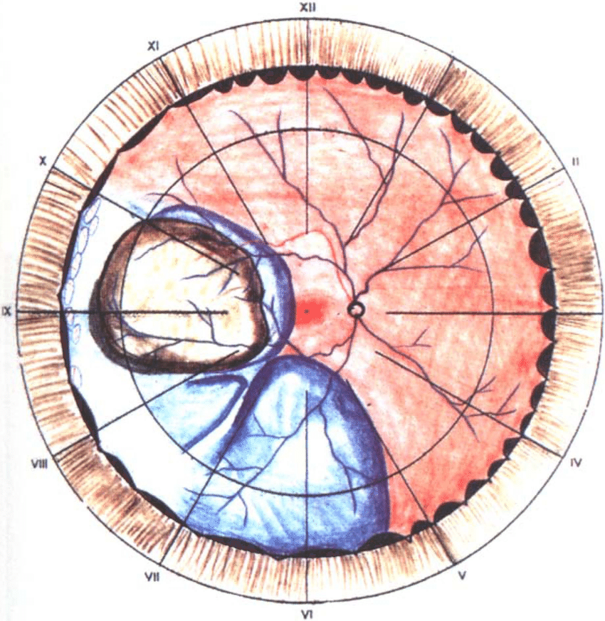
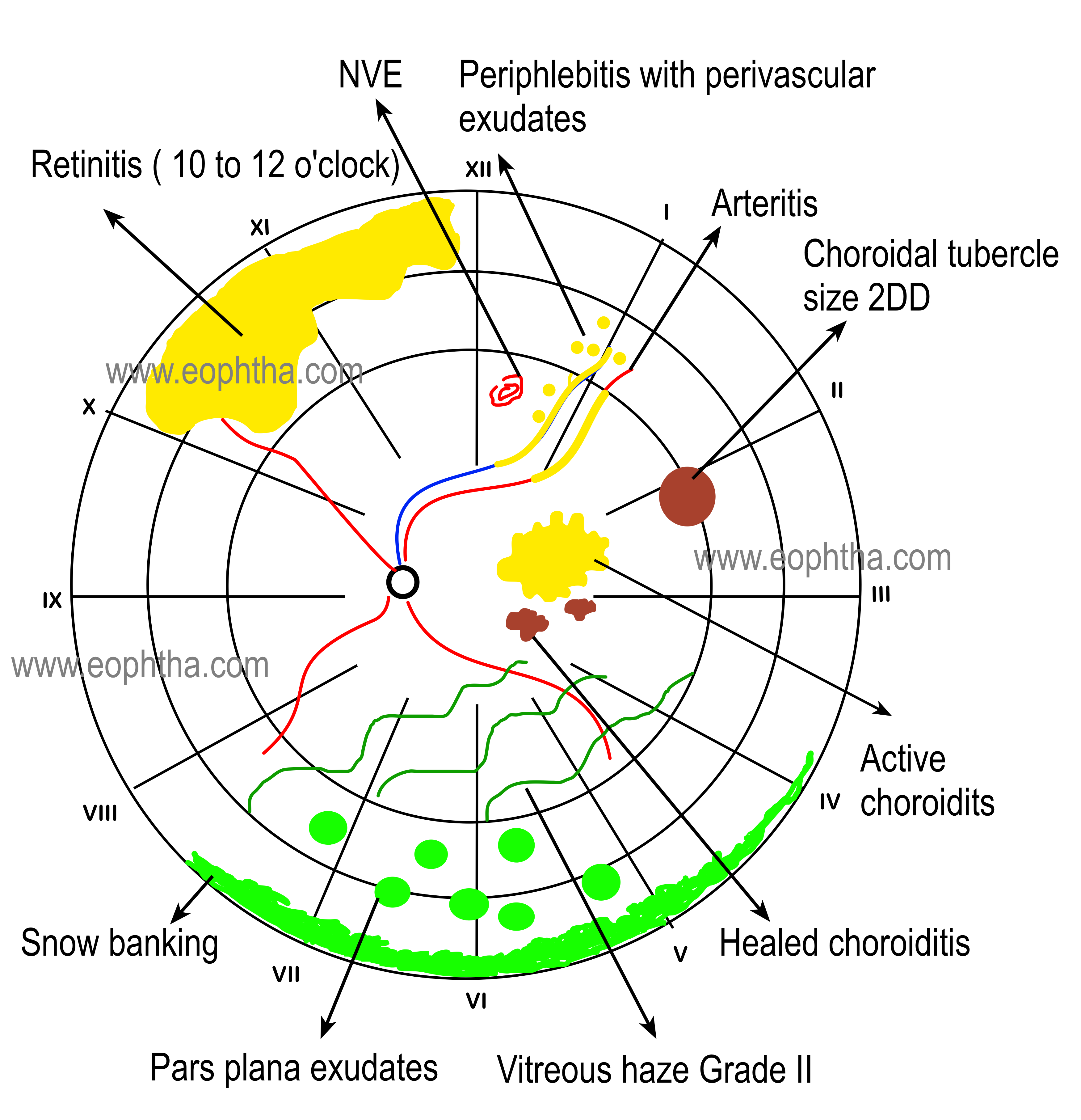
Retina Drawing Template - Web retinal drawing this is an open access journal, and articles are distributed under the terms of the creative commons attribution‑noncommercial‑sharealike 4.0 license, which allows others to remix, Web if they’re documenting central findings, such as arterial occlusion or haemorrhage in the case of retinal vein thrombosis, beginners should remember to simply turn the drawing template upside down, then they’ll be viewing and copying the fundus in. Web a true retina drawing will contain three concentric circles. Crystalgraphics creates templates designed to make even average presentations look incredible. Look no further—in this blog post, we have seven different free printables that are sure to provide clarity and help take your artwork up a notch! Existing color coding addresses most of the common retinal pathologies including preretinal, intraretinal, and subretinal lesions. Web the retina specialist confirms the diagnosis of macular hole in the right eye and documents it with a retinal drawing with labels and an interpretation and report. It lists the requisites needed which include an examination table, indirect ophthalmoscope, 20d lens, scleral depressor, and colored pencils. Web 91 views 1 year ago. The correct code for this eo would be 92225 ophthalmoscopy, extended, initial.
Web 91 views 1 year ago. In this video, i discussed about how to draw fundus picture and colour coding in normal and various retinal pathological conditions….more. Web ophthalmoscopy, extended, with retinal drawing and scleral depression of peripheral retinal disease (eg, for retinal tear, retinal detachment, retinal tumor). Illustrating emotive and captivating eyes. Web if they’re documenting central findings, such as arterial occlusion or haemorrhage in the case of retinal vein thrombosis, beginners should remember to simply turn the drawing template upside down, then they’ll be viewing and copying the fundus in. We'll help you figure it out. Existing color coding addresses most of the common retinal pathologies including preretinal, intraretinal, and subretinal lesions. An examination table, indirect ophthalmoscope, 20 d lens, a scleral depressor (or paper clips, etc), colored pencils. Not sure which apps are best for you? Crystalgraphics creates templates designed to make even average presentations look incredible.
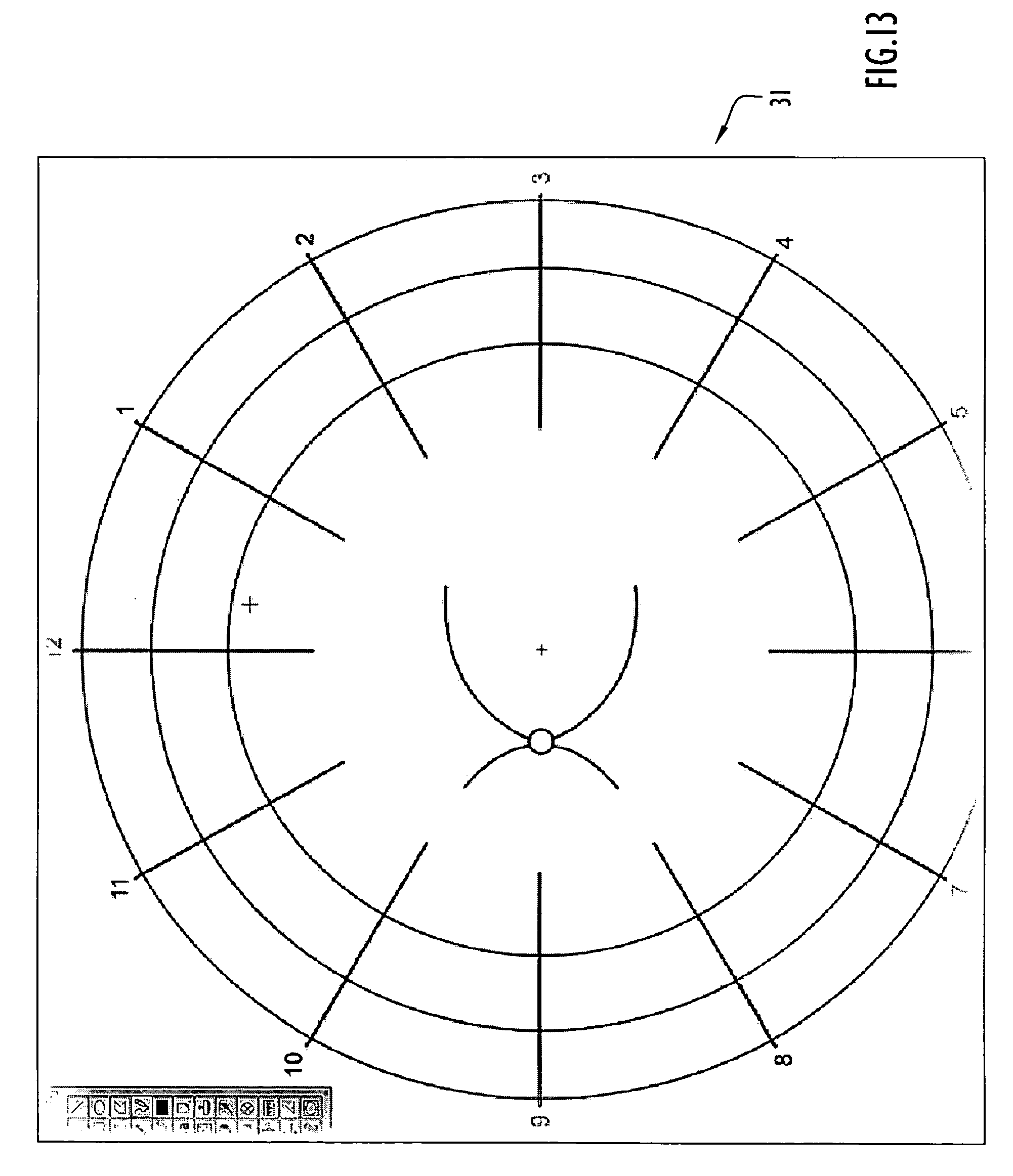
If you want to get good at drawing portraits, one of the best things you can do is practice individually drawing different parts of the face. 8.2k views 4 years ago. There are some stencils available commercially which aid in the quick reproduction of cornea and retina diagrams, front of eye, and slit shape outlines. Web february 13, 2023 | dee. Examination table, indirect ophthalmoscope, 20 d lens, scleral depressor, colored pencils (red, blue, green, yellow, brown, and black), fundus drawing chart, and eraser. Aug 13, 2019 • download as pptx, pdf •. Web the retina specialist confirms the diagnosis of macular hole in the right eye and documents it with a retinal drawing with labels and an interpretation and report. Web skip to one of these steps: The first represents the equator, the second represents the ora serrata, and the third represents the pars plana. We'll help you figure it out.
Retinal Drawing at Explore collection of Retinal
In a rrd with an intact fovea, the distance of a detachment from the fovea indicates the distance that ensures central visual acuity, as fovea‐off rrd are at risk of a worse visual outcome (salicone et. Web retinal drawing this is an open access journal, and articles are distributed under the terms of the creative commons attribution‑noncommercial‑sharealike 4.0 license, which.

My efforts as an artist learning to draw the retina — Matt Weed, MD
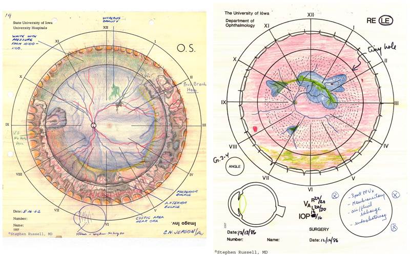
Web for rhegmatogenous retinal detachments (rrd), drawings of retinal breaks and degree of detachment can aid patient management. Web the requisites for drawing include six colored pencils or pens such as black, blue, brown, red, green, and yellow and an eraser to allow modifications of drawing. How to draw an iris. Web if they’re documenting central findings, such as arterial.
Retina Drawing Template
Not sure which apps are best for you? In this video, i discussed about how to draw fundus picture and colour coding in normal and various retinal pathological conditions….more. The correct code for this eo would be 92225 ophthalmoscopy, extended, initial. Ophthalmologists needs for a better practice and skills, as well as for documentation in electronic. Crystalgraphics creates templates designed.
Retina Drawing Template
Pdf | on jul 1, 2011, luann dvorak and others published retinal drawing: This document provides guidance on how to draw fundus diagrams. A lost art of medicine | find, read and cite all the research you need on researchgate. Are you looking to find an eye template printable to use as inspiration in your art? It is a useful.
Retina Drawing Template
Web retinal drawing this is an open access journal, and articles are distributed under the terms of the creative commons attribution‑noncommercial‑sharealike 4.0 license, which allows others to remix, I’ve rounded up 25 eye drawing sketch ideas for you to use for inspiration. Web skip to one of these steps: Existing color coding addresses most of the common retinal pathologies including.

Pin by Ansari sharique on optometry Drawing templates, Templates
An examination table, indirect ophthalmoscope, 20 d lens, a scleral depressor (or paper clips, etc), colored pencils. There should also be 12 tick marks indicating each clock hour of the retina. Web for rhegmatogenous retinal detachments (rrd), drawings of retinal breaks and degree of detachment can aid patient management. Web retina drwaing | ppt. Web how to draw eyes in.
Retina Drawing Template
Fundus drawing is universally acceptable records of the retinal disease process. Web the requisites for drawing include six colored pencils or pens such as black, blue, brown, red, green, and yellow and an eraser to allow modifications of drawing. The correct code for this eo would be 92225 ophthalmoscopy, extended, initial. There should also be 12 tick marks indicating each.

Documentation & Drawing in Ophthalmology
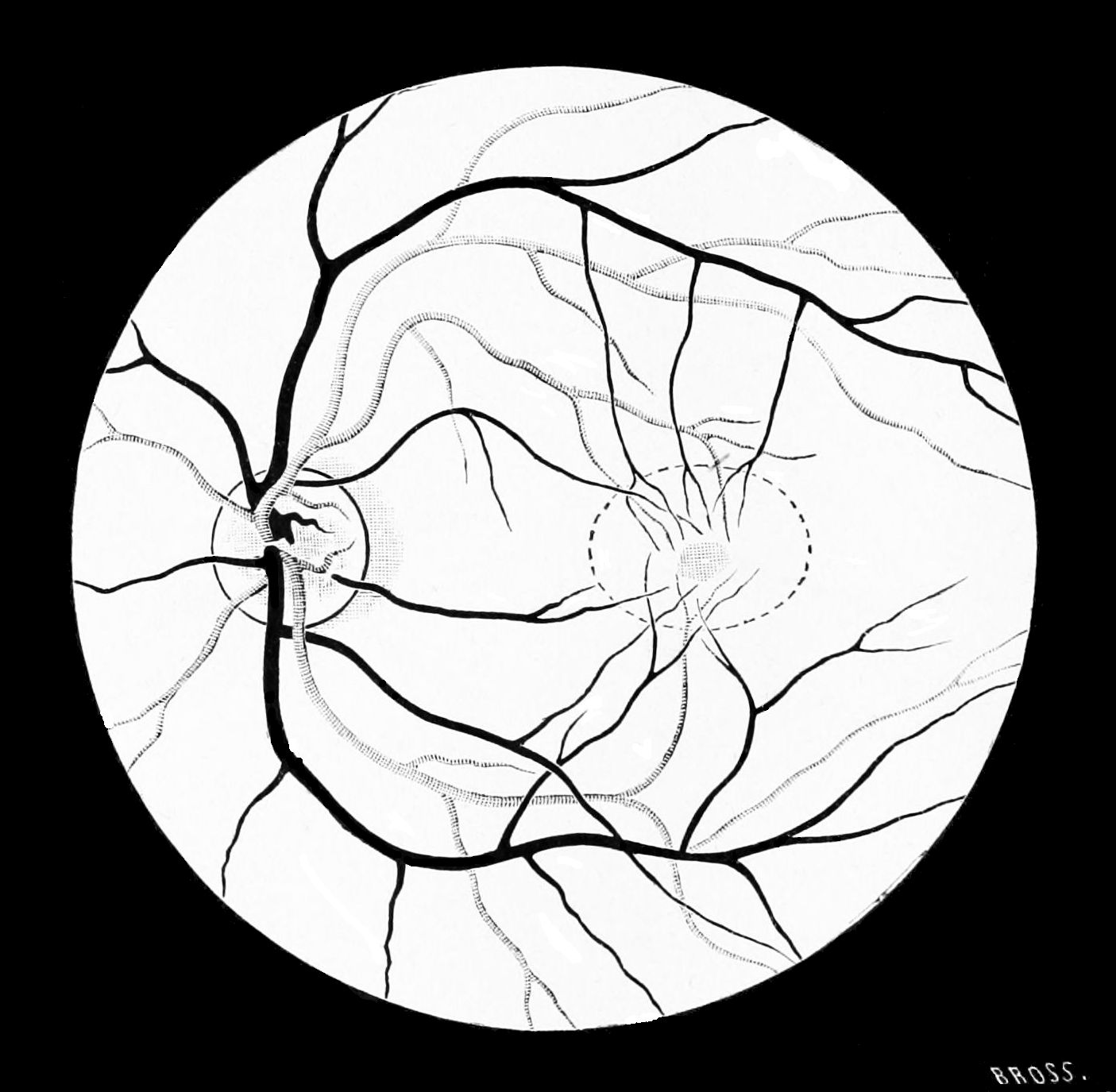
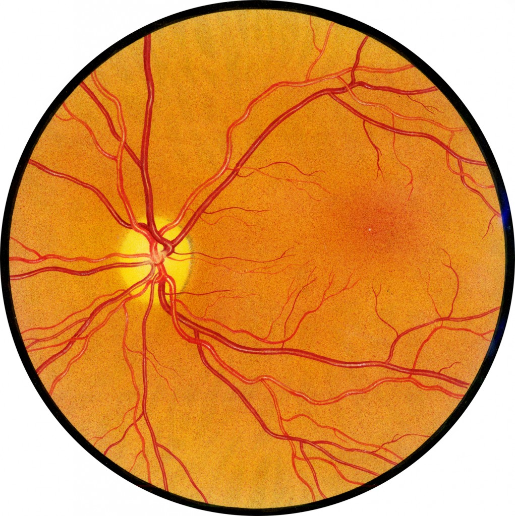
8.2k views 4 years ago. Web a true retina drawing will contain three concentric circles. Fundus drawing is universally acceptable records of the retinal disease process. It is a useful reference to monitor the clinical process and also at the time of surgery. This document provides guidance on how to draw fundus diagrams.

left eye retinal fundus drawing template Drawing templates, Templates
Not sure which apps are best for you? It lists the requisites needed which include an examination table, indirect ophthalmoscope, 20d lens, scleral depressor, and colored pencils. Aug 13, 2019 • download as pptx, pdf •. Last updated on may 11, 2023 by dee. Web if they’re documenting central findings, such as arterial occlusion or haemorrhage in the case of.
Retinal Drawing Free download on ClipArtMag
Acquired immunodeficiency syndrome (aids), cidofovir, cytomegalovirus (cmv), cmv retinitis fomivirsen, foscarnet, ganciclovir, ganciclovir implant, highly active antiretroviral therapy (hart), human immunodeficiency virus (hiv), immune recovery uveitis, valganciclovir. Are you looking to find an eye template printable to use as inspiration in your art? Pdf | on jul 1, 2011, luann dvorak and others published retinal drawing: Web retinal drawing this.
Are You Looking To Find An Eye Template Printable To Use As Inspiration In Your Art?
Pdf | on jul 1, 2011, luann dvorak and others published retinal drawing: How to draw an iris. 24 likes • 3,501 views. Ophthalmologists needs for a better practice and skills, as well as for documentation in electronic.
Web If They’re Documenting Central Findings, Such As Arterial Occlusion Or Haemorrhage In The Case Of Retinal Vein Thrombosis, Beginners Should Remember To Simply Turn The Drawing Template Upside Down, Then They’ll Be Viewing And Copying The Fundus In.
Learn how to draw eyes with these eye drawing ideas and tutorials. Web how to draw eyes in a few simple steps. The correct code for this eo would be 92225 ophthalmoscopy, extended, initial. 8.2k views 4 years ago.
There Are Some Stencils Available Commercially Which Aid In The Quick Reproduction Of Cornea And Retina Diagrams, Front Of Eye, And Slit Shape Outlines.
How to shade a realistic eyeball. It is a useful reference to monitor the clinical process and also at the time of surgery. Existing color coding addresses most of the common retinal pathologies including preretinal, intraretinal, and subretinal lesions. Last updated on may 11, 2023 by dee.
There Should Also Be 12 Tick Marks Indicating Each Clock Hour Of The Retina.
Look no further—in this blog post, we have seven different free printables that are sure to provide clarity and help take your artwork up a notch! Web skip to one of these steps: It lists the requisites needed which include an examination table, indirect ophthalmoscope, 20d lens, scleral depressor, and colored pencils. In this video, i discussed about how to draw fundus picture and colour coding in normal and various retinal pathological conditions….more.