Simple Columnar Drawing
Simple Columnar Drawing - Web simple epithelium is one of the types of epithelium that is divided into simple columnar epithelium, simple squamous epithelium, and simple cuboidal epithelium. Ciliated columnar epithelium is composed of simple columnar epithelial cells with cilia on their apical surfaces. Describe the structure and function of endocrine and exocrine glands. Web this simple columnar epithelium contains: This image shows the brush border of intestinal epithelium, which features simple columnar epithelial cells with basal nuclear polarity and an apical surface with microvilli. A squamous epithelial cell looks flat under a microscope. Note the size of the microvilli relative to the cells that they cover. The cells of this epithelium are arranged in a neat row with the nuclei at the same level, near the basal end. The entire layer of simple columnar epithelium is indicated by the bar. Bodytomy provides a labeled diagram to help you understand the structure and function of simple columnar epithelium.
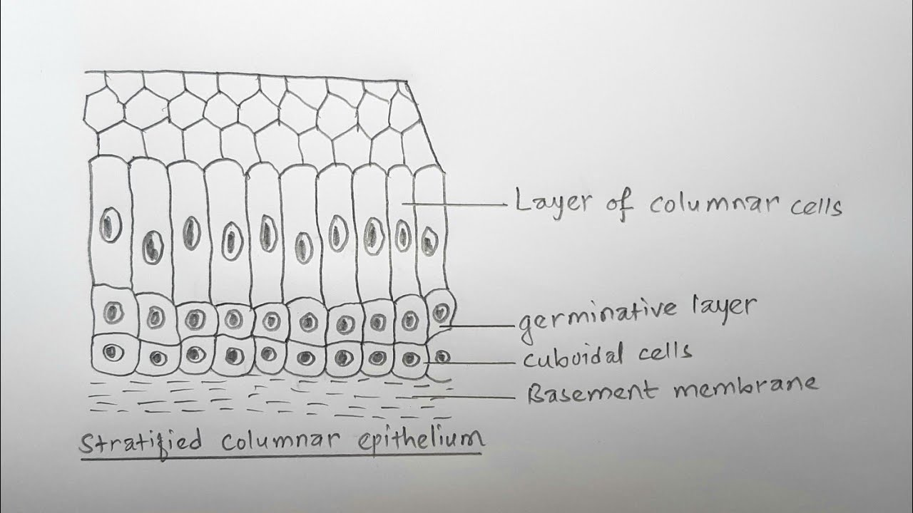
553 views 2 years ago. Ciliated columnar epithelium is composed of simple columnar epithelial cells with cilia on their apical surfaces. Web drawing histological diagram of simple columnar epithelia.useful for all medical students.drawn by using h & e pencils.explanation on epithelia while drawing. Note the size of the microvilli relative to the cells that they cover. Web this simple columnar epithelium contains: Web like the cuboidal epithelia, this epithelium is active in the absorption and secretion of molecules. This image shows the brush border of intestinal epithelium, which features simple columnar epithelial cells with basal nuclear polarity and an apical surface with microvilli. Describe the structure and function of endocrine and exocrine glands. Web simple epithelium is one of the types of epithelium that is divided into simple columnar epithelium, simple squamous epithelium, and simple cuboidal epithelium. Please include total magnification in the image key.
Use the image slider below to study numerous examples of simple columnar epithelium (all images are magnified 400x). Web there are three basic shapes used to classify epithelial cells. This is known as a brush border. Ciliated columnar epithelium is composed of simple columnar epithelial cells with cilia on their apical surfaces. Like cuboidal epithelium, the cells in the columnar epithelium are also modified to suit the function and structure of the organ better. Web simple columnar location: Describe the structure and function of endocrine and exocrine glands. Read more about modified simple columnar epithelium, dog jejenum, 20x; A squamous epithelial cell looks flat under a microscope. Web simple epithelium is one of the types of epithelium that is divided into simple columnar epithelium, simple squamous epithelium, and simple cuboidal epithelium.
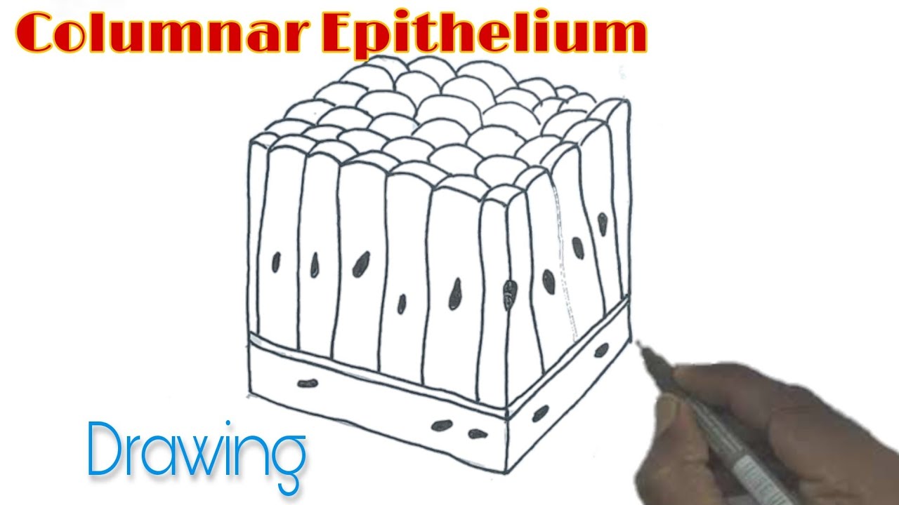
Columnar Epithelium Drawing/ How to draw Columnar Epithelium YouTube
Web simple columnar with microvilli. Web simple columnar location: Web distinguish between simple epithelia and stratified epithelia, as well as between squamous, cuboidal, and columnar epithelia. Trachea and most of the upper respiratory tract (ciliated cells) A columnar epithelial cell looks like a column or a tall rectangle.
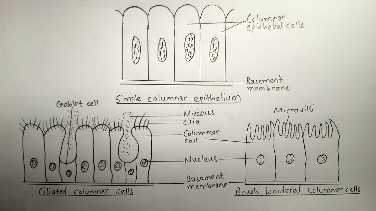
How to draw simple columnar epithelium types of comlumnar epithelium
Please include total magnification in the image key. Web there are three basic shapes used to classify epithelial cells. Like cuboidal epithelium, the cells in the columnar epithelium are also modified to suit the function and structure of the organ better. Web drawing histological diagram of simple columnar epithelia.useful for all medical students.drawn by using h & e pencils.explanation on.

Simple columnar Simple columnar epithelium, Tissue types, Types of
Ciliated columnar epithelium is composed of simple columnar epithelial cells with cilia on their apical surfaces. Describe the structure and function of endocrine and exocrine glands. The arrows are pointing to goblet cells that produce and release mucus. In humans, simple columnar epithelium lines most organs of the digestive tract including the stomach, and intestines. Use the image slider below.
simple columnar Medical Study Zone
Allows absorbtion, secretes mucous and enzymes pseudostratified columnar location: This image shows the brush border of intestinal epithelium, which features simple columnar epithelial cells with basal nuclear polarity and an apical surface with microvilli. What is the function of the microvilli? Web simple columnar epithelium is a single layer of tall, narrow cells with oval nuclei that are close to.

How to draw Pseudostratified columnar epithelium most easy way
Web this simple columnar epithelium contains: A cuboidal epithelial cell looks close to a square. What is the function of the microvilli? Web distinguish between simple epithelia and stratified epithelia, as well as between squamous, cuboidal, and columnar epithelia. Web simple columnar epithelium (400x) primate small intestine.
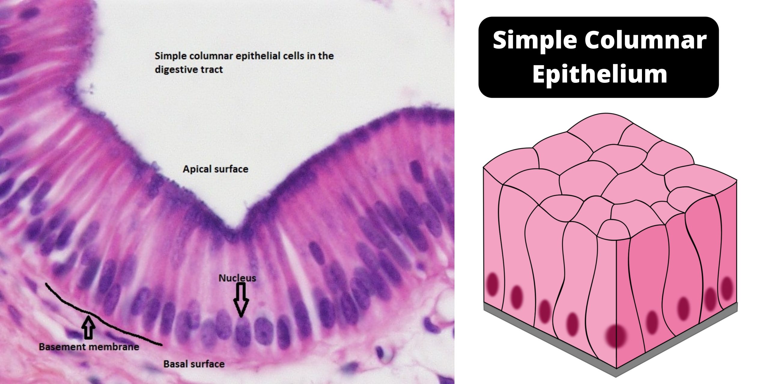
Simple columnar epithelium definition, structure, functions, examples
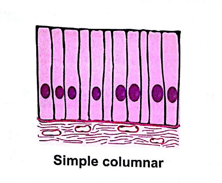
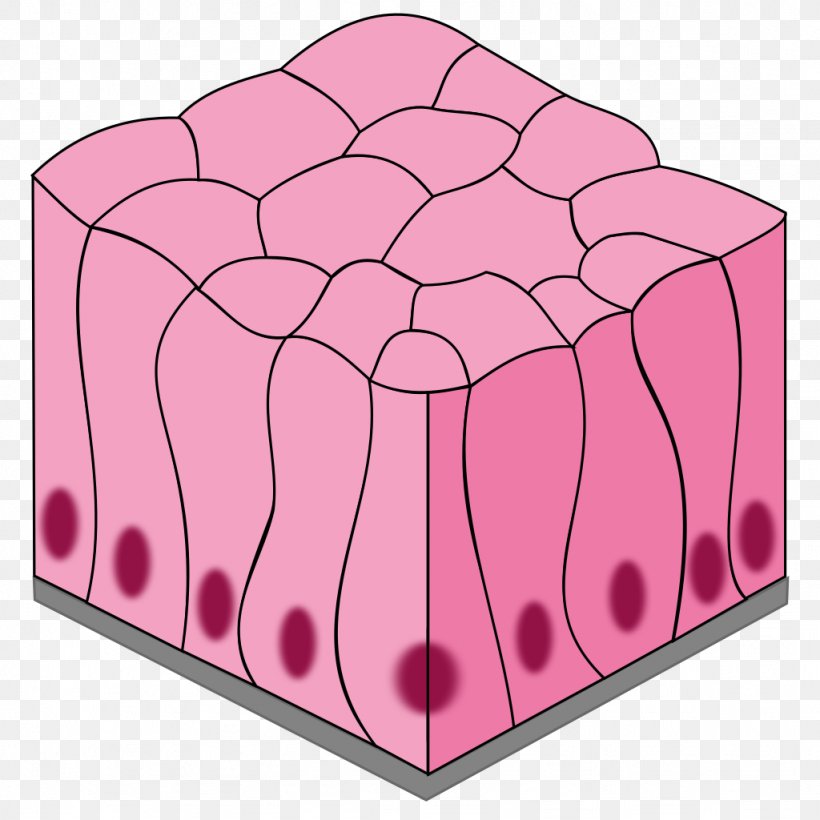
553 views 2 years ago. A squamous epithelial cell looks flat under a microscope. These cells are placed side by s. The cells of this epithelium are arranged in a neat row with the nuclei at the same level, near the basal end. Web the simple columnar epithelium under the microscope shows a single layer of cells resting on the.

Simple Columnar Tutorial Histology Atlas for Anatomy and Physiology
A squamous epithelial cell looks flat under a microscope. Web simple columnar with microvilli. Web this simple columnar epithelium contains: Web like the cuboidal epithelia, this epithelium is active in the absorption and secretion of molecules. Note the size of the microvilli relative to the cells that they cover.
Simple Columnar Epithelium Simple Squamous Epithelium Stratified
Web simple epithelium is one of the types of epithelium that is divided into simple columnar epithelium, simple squamous epithelium, and simple cuboidal epithelium. This is known as a brush border. Trachea and most of the upper respiratory tract (ciliated cells) Please include total magnification in the image key. Web simple columnar epithelia are tissues made of a single layer.

Simple Columnar Tutorial Histology Atlas for Anatomy and Physiology
Web there are three basic shapes used to classify epithelial cells. Web distinguish between simple epithelia and stratified epithelia, as well as between squamous, cuboidal, and columnar epithelia. Web simple columnar with microvilli. Web simple epithelium is one of the types of epithelium that is divided into simple columnar epithelium, simple squamous epithelium, and simple cuboidal epithelium. These cells are.
How to draw stratified columnar epithelium easy way YouTube
Read more about modified simple columnar epithelium, dog jejenum, 20x; Web drawing histological diagram of simple columnar epithelia.useful for all medical students.drawn by using h & e pencils.explanation on epithelia while drawing. These cells are placed side by s. This is known as a brush border. Web simple columnar location:
The Cells Of This Epithelium Are Arranged In A Neat Row With The Nuclei At The Same Level, Near The Basal End.
Note the size of the microvilli relative to the cells that they cover. These columnar epithelium consist of tall columnar cells. Web the simple columnar epithelium under the microscope shows a single layer of cells resting on the basement membrane. Web this simple columnar epithelium contains:
Simple Columnar Epithelium Forms The Lining Of Some Sections Of The Digestive System And Parts Of The Female Reproductive Tract.
What is the function of the microvilli? Describe the structure and function of endocrine and exocrine glands. Use the image slider below to study numerous examples of simple columnar epithelium (all images are magnified 400x). Web simple columnar epithelium (400x) primate small intestine.
Web Drawing Histological Diagram Of Simple Columnar Epithelia.useful For All Medical Students.drawn By Using H & E Pencils.explanation On Epithelia While Drawing.
Like cuboidal epithelium, the cells in the columnar epithelium are also modified to suit the function and structure of the organ better. Web simple columnar epithelia are tissues made of a single layer of long epithelial cells that are often seen in regions where absorption and secretion are important features. These cells are placed side by s. They look clear because the molecules in the mucus do not absorb a lot of stain.
Web Simple Columnar With Microvilli.
Learn its anatomy on kenhub! The arrows are pointing to goblet cells that produce and release mucus. A columnar epithelial cell looks like a column or a tall rectangle. Web the simple columnar epithelium is a type of epithelium that is formed of a single layer of long, elongated cells mostly in areas where absorption and secretion are the main functions.