Simple Cuboidal Epithelium Drawing
Simple Cuboidal Epithelium Drawing - It is sometimes referred to as the “basal lamina”. Epithelial tissue is often classified according to numbers of layers of cells present, and by the shape of the cells. Every cell attaches to the basement membrane. Web simple cuboidal epithelium: Web a simple epithelium is an epithelial tissue that is composed of a single layer of epithelial cells. Read more about simple cuboidal thyroid gland 40x; Cuboidal cells are about as tall as they are wide. The basement membrane is a thin but strong, acellular layer which lies between the epithelium and the adjacent connective tissue. While both figures show simple cuboidal epithelium of the renal tubules in the kidney, figure 4 shows a cross section. Forming sheets that cover the internal and external body surfaces (surface epithelium) and secreting organs (glandular epithelium).
The basement membrane is a thin but strong, acellular layer which lies between the epithelium and the adjacent connective tissue. Like the cuboidal epithelia, this epithelium is active in the absorption and secretion of molecules using. Simple cuboidal cells are also characterized by a single, large, round (spherical) nucleus located near the center of each cell. Web this type of epithelium serves as a lining or covering and can be specialized for secretion and sometimes for absorption (e.g. The cells in this tissue are tightly packed within a thin ecm. This ensures that the amount of substances in the lens, and its size, are maintained. Every cell attaches to the basement membrane. Web simple cuboidal epithelia occur widely in the body in many glands and glandular ducts, such as the salivary ducts, pancreatic duct, bile duct, and kidney tubules.these epithelia are composed of cells that are short prisms with a top, bottom, and six sides. While both figures show simple cuboidal epithelium of the renal tubules in the kidney, figure 4 shows a cross section. These cells are in direct contact with the basement membrane.
Thyroid gland, 40x objective 400x total magnification, simple cuboidal. Web links:simple squamous epithelium: Web simple cuboidal epithelium also covers the lens of the eye, where it controls the movement of nutrients and water, into and out of the lens from the surrounding eye fluid. Like the cuboidal epithelia, this epithelium is active in the absorption and secretion of molecules. The side of the cell that faces the free surface is called the apex, and the side that. The cells in this tissue are tightly packed within a thin ecm. On these surfaces, the cells. Web a simple epithelium is an epithelial tissue that is composed of a single layer of epithelial cells. While both figures show simple cuboidal epithelium of the renal tubules in the kidney, figure 4 shows a cross section. Simple cuboidal epithelium is found on the surface of ovaries, the lining of nephrons, the walls of the renal tubules, parts of the eye and thyroid, and in salivary glands.
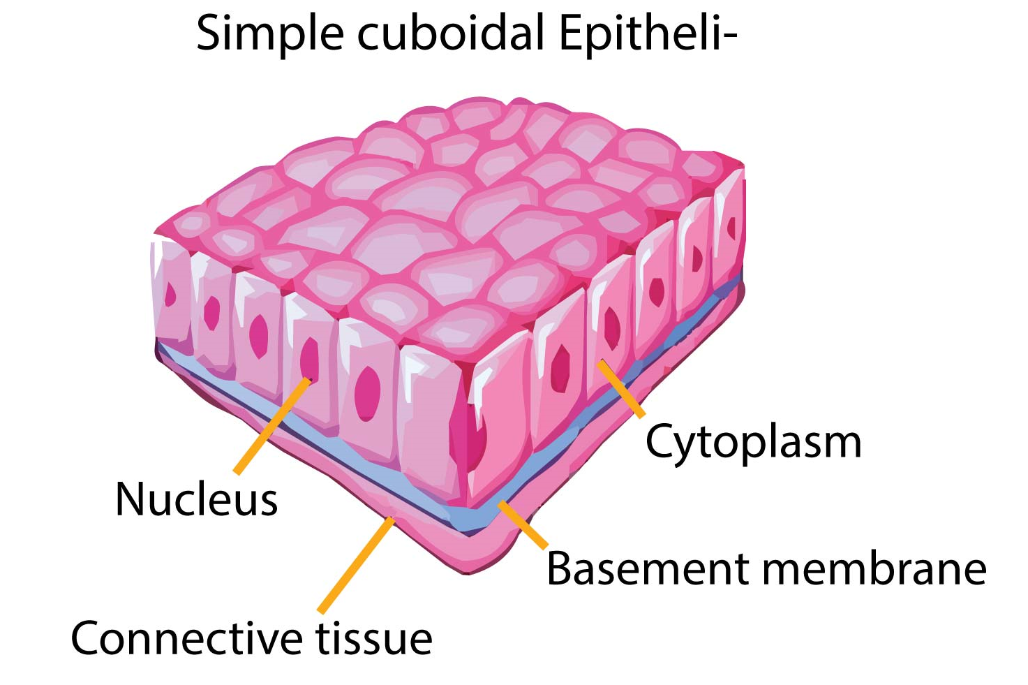
Simple Cuboidal Epithelium Diagram
The side of the cell that faces the free surface is called the apex, and the side that. Web simple cuboidal epithelia are observed in the lining of the kidney tubules and in the ducts of glands. With large, rounded, centrally located nuclei, all the cells of this epithelium are directly attached to the basement membrane. On these surfaces, the.
Epithelial Tissues Simple Tissue Biology Tissue Types Anatomy And
Web simple cuboidal epithelia are observed in the lining of the kidney tubules and in the ducts of glands. Web simple cuboidal epithelia are observed in the lining of the kidney tubules and in the ducts of glands. This ensures that the amount of substances in the lens, and its size, are maintained. Thyroid gland, 40x objective 400x total magnification,.

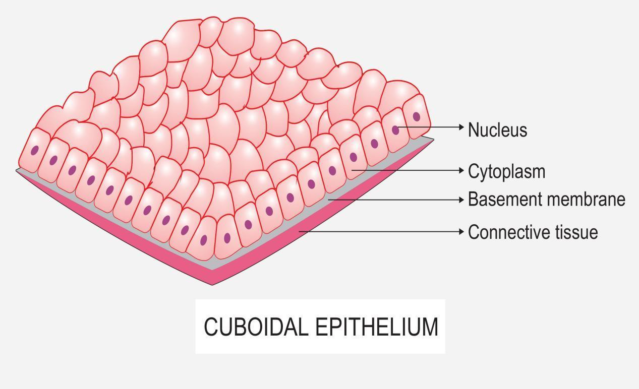
Simple Cuboidal Epithelium Labeled Basement Membrane
The basement membrane is a thin but strong, acellular layer which lies between the epithelium and the adjacent connective tissue. Like the cuboidal epithelia, this epithelium is active in the absorption and secretion of molecules. Web simple cuboidal epithelia are observed in the lining of the kidney tubules and in the ducts of glands. Like the cuboidal epithelia, this epithelium.

How To Draw Cuboidal Epithelial Tissue (step by step) how_to_draw
Every cell attaches to the basement membrane. Web simple cuboidal epithelia are observed in the lining of the kidney tubules and in the ducts of glands. Simple cuboidal cells are also characterized by a single, large, round (spherical) nucleus located near the center of each cell. Each cell have centrally located round nucleus. They function as a covering for several.

Simple cuboidal epithelium Diagram Quizlet
Types of simple cuboidal epithelia. Like the cuboidal epithelia, this epithelium is active in the absorption and secretion of molecules. Read more about simple cuboidal thyroid gland 40x; It is sometimes referred to as the “basal lamina”. Allows absorbtion, secretes mucous and enzymes.

Simple Cuboidal 400x Labeled
Each cell have centrally located round nucleus. Web sweat glands, salivary glands, mammary glands, adrenal glands, and pituitary glands are examples of glands made of epithelial tissue. Like the cuboidal epithelia, this epithelium is active in the absorption and secretion of molecules. It is sometimes referred to as the “basal lamina”. To help you understand how to identify simple squamous.
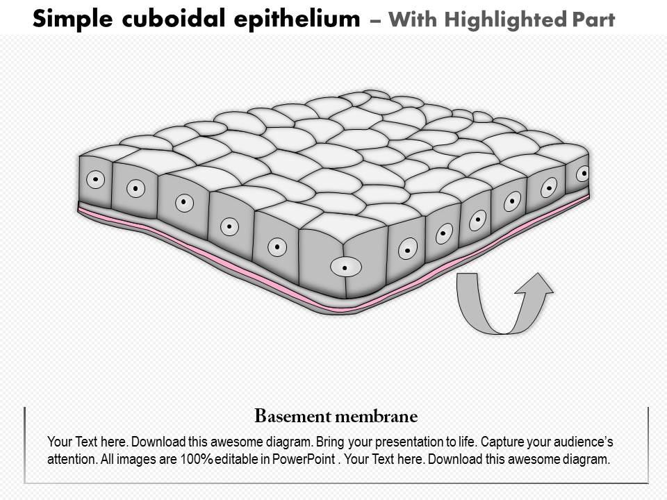
0614 Simple Cuboidal Epithelium Medical Images For Powerpoint
Simple epithelial tissues have only one layer of cells. Like the cuboidal epithelia, this epithelium is active in the absorption and secretion of molecules. Simple cuboidal cells are also characterized by a single, large, round (spherical) nucleus located near the center of each cell. Each cell have centrally located round nucleus. The cuboidal epithelium in the ovaries forms the ovarian.

Simple cuboidal epithelium Diagram Quizlet
Web simple cuboidal epithelia are observed in the lining of the kidney tubules and in the ducts of glands. Like the cuboidal epithelia, this epithelium is active in the absorption and secretion of molecules. Like the cuboidal epithelia, this epithelium is active in the absorption and secretion of molecules using. Web simple cuboidal epithelium can look a little different depending.

How to draw stratified cuboidal epithelium easy way YouTube
Like the cuboidal epithelia, this epithelium is active in the absorption and secretion of molecules using. The proximal convoluted tubules of the kidney). Web simple cuboidal epithelia are observed in the lining of the kidney tubules and in the ducts of glands. Web simple cuboidal epithelium: Every cell attaches to the basement membrane.
6 Epithelial tissue. A) Representative model of simple cuboidal
Types of simple cuboidal epithelia. Thyroid gland, 40x objective 400x total magnification, simple cuboidal. This tissue consists of cubical cells. These cells are in direct contact with the basement membrane. Web epithelium is one of only 4 types of human body tissues.like all types, it is formed by cells within an extracellular matrix (ecm).
Web Simple Cuboidal Epithelium Can Look A Little Different Depending On The Direction The Tissue Has Been Sectioned.
Knowing about the shape and position of the nucleus in the cells will allow you to recognize tissues even when you can't. Web sweat glands, salivary glands, mammary glands, adrenal glands, and pituitary glands are examples of glands made of epithelial tissue. Forming sheets that cover the internal and external body surfaces (surface epithelium) and secreting organs (glandular epithelium). Web histology diagram for simple cuboidal epithelium histology diagram.
This Ensures That The Amount Of Substances In The Lens, And Its Size, Are Maintained.
Each cell have centrally located round nucleus. Web welcome to diya's art tutorial youtube channel today in this video i'm showing how to draw cuboidal. They function as a covering for several organs providing protection against damage and other chemicals. Web simple cuboidal epithelia are observed in the lining of the kidney tubules and in the ducts of glands.
On These Surfaces, The Cells.
Although the height and shape of the cells may vary some, simple cuboidal epithelial cells are generally about as tall as they are wide, with a centrally located nucleus. Simple cuboidal epithelium is a type of epithelium that consists of a single layer of cub. Web owing to the shape of the cells, the primary functions of the simple cuboidal epithelium are secretion, absorption, and covering. This tissue consists of cubical cells.
Secretes Mucus Which Is Moved With.
Web drawing histological diagram of simple cuboidal epithelia.useful for all medical students.drawn by using h & e pencils.explanation on epithelia while drawing. Like the cuboidal epithelia, this epithelium is active in the absorption and secretion of molecules using. This tissue may be classified histologically according to the shape of the cells it is made up of. To help you understand how to identify simple squamous epithelium, we have included two examples of this tissue.