Simple Squamous Epithelium Drawing
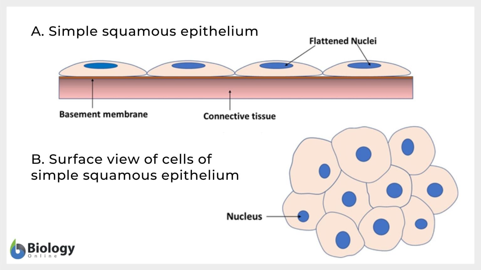
Simple Squamous Epithelium Drawing - Web peritoneum in the intestine is an example of the mesothelium that forms the epithelia of the serous intestinal membrane. Epithelial tissue is often classified according to numbers of layers of cells present, and by the shape of the cells. Web simple squamous simple epithelium can be divided into 4 major classes, depending on the shapes of constituent cells. Web compare this image to the drawing of simple squamous epithelium in your textbook, which shows what an intact layer of cells should look like. The simple squamous epithelium also lines the bowman’s capsule of the nephrons in the kidney. But when we draw a sketch of. Introduction to simple squamous epithelium. Squamous cells are large, thin, and flat and contain a rounded nucleus. Web simple squamous epithelium: First, you should draw the basement membrane of the simple squamous epithelium.
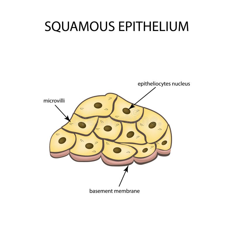
Notice that the location of the. The cells found in this epithelium type are flat and thin, making simple squamous epithelium ideal for lining areas where passive diffusion of gases occur.areas where it can be found include: Web browse 140+ simple squamous epithelium stock illustrations and vector graphics available royalty. This epithelium is shown better in em 080 bowman's capsule by scanning electron microscopy. Web now, let’s learn how to draw the simple squamous epithelium that finds under a microscope. And so this is also going to allow for the exchange of nutrients, allowing nutrients such as glucose to diffuse out of the capillaries and into the neighboring tissues. The endothelium is the epithelial tissue that lines vessels of the lymphatic and cardiovascular system, and it is made up of a single layer of squamous cells. Web links:simple squamous epithelium: Web the cells in simple squamous epithelium have the appearance of thin scales. The epithelial tissue is in direct contact with the basement membrane that.
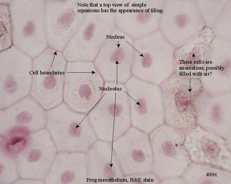
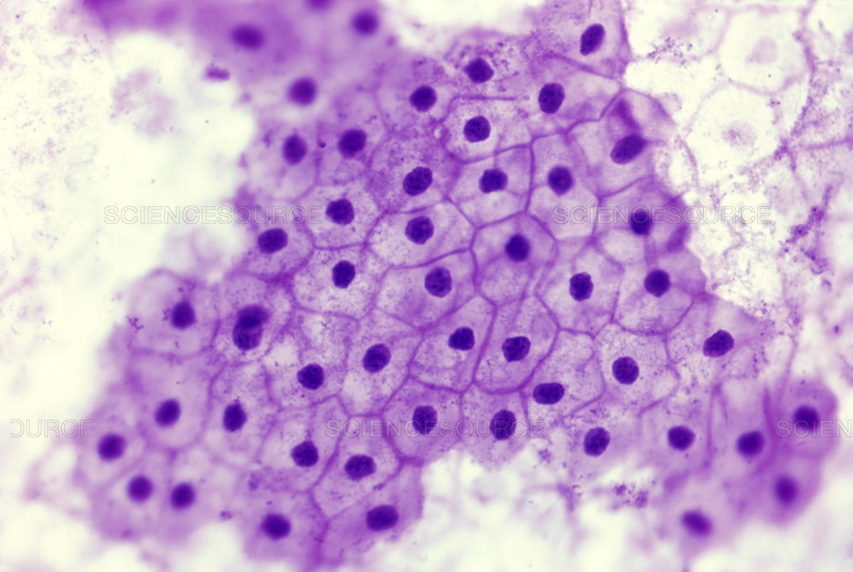
Web links:simple squamous epithelium: Web drawing histological diagram of simple squamous epithelia.useful for all medical students.drawn by using h & e pencils Web simple squamous epithelium is a type of tissue found in the alveoli of the lungs and interior walls of blood vessels and lymphatic vessels. Simple squamous epithelium, isolated (400x) buccal mucosal in the center of this image are two simple squamous epithelial cells that are still attached to each other. Web histology diagram of simple squamous epithelium histology diagram. Simple squamous epithelium is composed of a single layer of thin, flat, somewhat roundish cells (shaped like irregular pancakes) that all remain in contact with the basement membrane (simple. The epithelial tissue is in direct contact with the basement membrane that. Histology of the simple squamous epithelium that lines the renal corpuscles in the kidney. Like other epithelial cells, they have polarity and contain a distinct apical surface with specialized membrane proteins. Web simple squamous simple epithelium can be divided into 4 major classes, depending on the shapes of constituent cells.

Simple Squamous Epithelium Diagram Quizlet
Tall columnar epithelium lines the ducts of many exocrine glands. The only part of these cells that can be seen are their nuclei bulging into the interior. That rests on a thick basement membrane. Histology of the simple squamous epithelium that lines the renal corpuscles in the kidney. Web simple squamous simple epithelium can be divided into 4 major classes,.
Epithelial Tissue Anatomy & Physiology
[1] this type of epithelium is often permeable and occurs where small molecules need to pass quickly. I will show you so simple method to draw this simple squamous epithelium. But when we draw a sketch of. Web peritoneum in the intestine is an example of the mesothelium that forms the epithelia of the serous intestinal membrane. Web compare this.

simple squamous epithelium Google Search Histology slides, Squamous
The cells in a simple squamous epithelium have the appearance of thin scales. Web now, let’s learn how to draw the simple squamous epithelium that finds under a microscope. Both surface and side view has been demonstrated in this video. Squamous cells are large, thin, and flat and contain a rounded nucleus. The simple squamous epithelium also lines the bowman’s.
Simple Squamous Epithelium Inrtroducrion , Anatomy & Function
The only part of these cells that can be seen are their nuclei bulging into the interior. Web simple squamous epithelium is a type of tissue found in the alveoli of the lungs and interior walls of blood vessels and lymphatic vessels. And so this is also going to allow for the exchange of nutrients, allowing nutrients such as glucose.

Stratified Squamous Epithelium Labeled Diagram
Web simple squamous simple epithelium can be divided into 4 major classes, depending on the shapes of constituent cells. This epithelium is shown better in em 080 bowman's capsule by scanning electron microscopy. At the same time, the. First, you should draw the basement membrane of the simple squamous epithelium. Simple squamous epithelium, because of the thinness of the cells,.
Simple squamous epithelium Definition and Examples Biology Online
This is made up of thin, flat and hexagonal cells. Web a simple squamous epithelium, also known as pavement epithelium and tessellated epithelium, is a single layer of flattened, polygonal cells in contact with the basal lamina (one of the two layers of the basement membrane) of the epithelium. Web simple squamous epithelium is a type of tissue found in.
Squamous Epithelium Diagram
But when we draw a sketch of. The simple squamous epithelium also lines the bowman’s capsule of the nephrons in the kidney. [1] this type of epithelium is often permeable and occurs where small molecules need to pass quickly. The cells in a simple squamous epithelium have the appearance of thin scales. Introduction to simple squamous epithelium.

Draw A Labelled Diagram Of Squamous Epithelial Tissue vrogue.co
But when we draw a sketch of. Web simple squamous epithelium, also known as simple squamous epithelial tissue or pavement epithelium, is an epithelial tissue that is composed of a single layer of epithelial cells. A large central rounded nucleus contai. Simple squamous epithelium, isolated (400x) buccal mucosal in the center of this image are two simple squamous epithelial cells.

34+ Simple Squamous Epithelium Drawing NeeraNatania
Web sweat glands, salivary glands, mammary glands, adrenal glands, and pituitary glands are examples of glands made of epithelial tissue. [1] this type of epithelium is often permeable and occurs where small molecules need to pass quickly. Simple squamous epithelium stock illustrations. The outer wall of the bowman’s capsule is bordered by a single layer of squamous cells. Like other.
Epithelial Tissues Simple Tissue Biology Tissue Types Anatomy And
This type of epithelium is adapted for secretion and/or absorption, and can also be protective. The nuclei of squamous cells tend to appear flat, horizontal, and elliptical, mirroring the form of the cell. Web sweat glands, salivary glands, mammary glands, adrenal glands, and pituitary glands are examples of glands made of epithelial tissue. Squamous cell nuclei tend to be flat,.
First, You Should Draw The Basement Membrane Of The Simple Squamous Epithelium.
Functions of simple squamous epithelium. Web browse 140+ simple squamous epithelium stock illustrations and vector graphics available royalty. Web simple squamous epithelium, also known as simple squamous epithelial tissue or pavement epithelium, is an epithelial tissue that is composed of a single layer of epithelial cells. Web this is a single layer of cells, and the cells are all tall columnar.
The Endothelium Is The Epithelial Tissue That Lines Vessels Of The Lymphatic And Cardiovascular System, And It Is Made Up Of A Single Layer Of Squamous Cells.
Notice that the location of the. Squamous cells are large, thin, and flat and contain a rounded nucleus. The epithelial tissue is in direct contact with the basement membrane that. At the same time, the.
Web Drawing Histological Diagram Of Simple Squamous Epithelia.useful For All Medical Students.drawn By Using H & E Pencils
Histology of the simple squamous epithelium that lines the renal corpuscles in the kidney. This is made up of thin, flat and hexagonal cells. But when we draw a sketch of. The simple squamous epithelium also lines the bowman’s capsule of the nephrons in the kidney.
Simple Secretory Columnar Epithelium Lines The Stomach And Uterine Cervix.the Simple Columnar Epithelium That.
Squamous cell nuclei tend to be flat, horizontal, and elliptical, mirroring the form of the cell. Tall columnar epithelium lines the ducts of many exocrine glands. A large central rounded nucleus contai. The outer wall of the bowman’s capsule is bordered by a single layer of squamous cells.