Skeletal Muscle Drawing
Skeletal Muscle Drawing - Labelled diagram of skeletal muscle. Web this article describes the histology of skeletal muscle, focusing on structure, types, contraction and clinical points. Bones are the foundation of the body. Web 90 skeletal muscle stock illustrations and clipart. Web how to draw muscles. Get free printable coloring page of this drawing. Web learn about the three types of muscle as you use our 3d models to explore the anatomical structure and physiology of human muscles. Turn your eyes—a tiny movement, considering the conspicuously large and strong external eye muscles that control eyeball movements. New 3d rotate and zoom. Describe the connective tissue layers surrounding skeletal muscle.
Web how to draw human muscular system. The musculoskeletal system (locomotor system) is a human body system that provides our body with movement, stability, shape, and support. Mastering the human skeleton will mean you get figure drawing right every time. Web we’ve created muscle anatomy charts for every muscle containing region of the body: This type of muscle creates movement in the body. Describe the structure and function of skeletal muscle fibers. Define a muscle fiber, myofibril, and sarcomere. Explain how muscles work with tendons to move the body. December 31, 2022 | published on: See skeletal muscle diagram stock video clips.
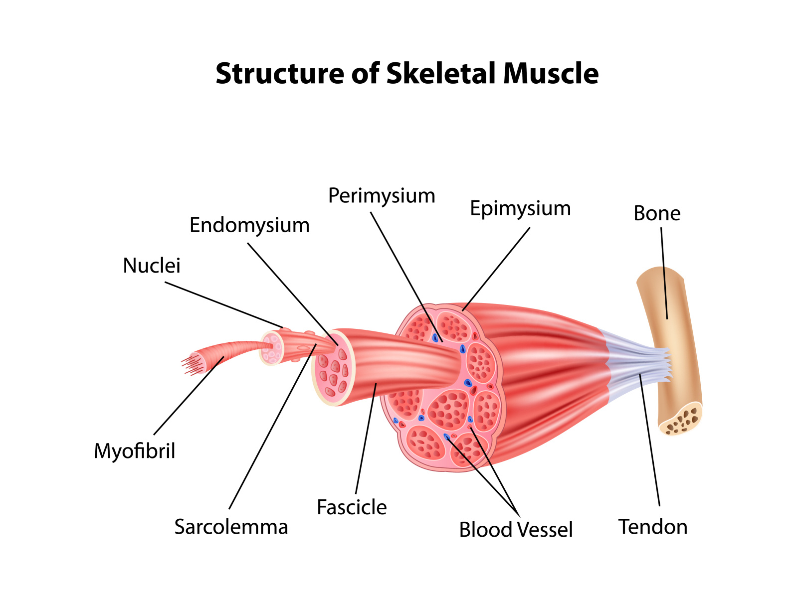
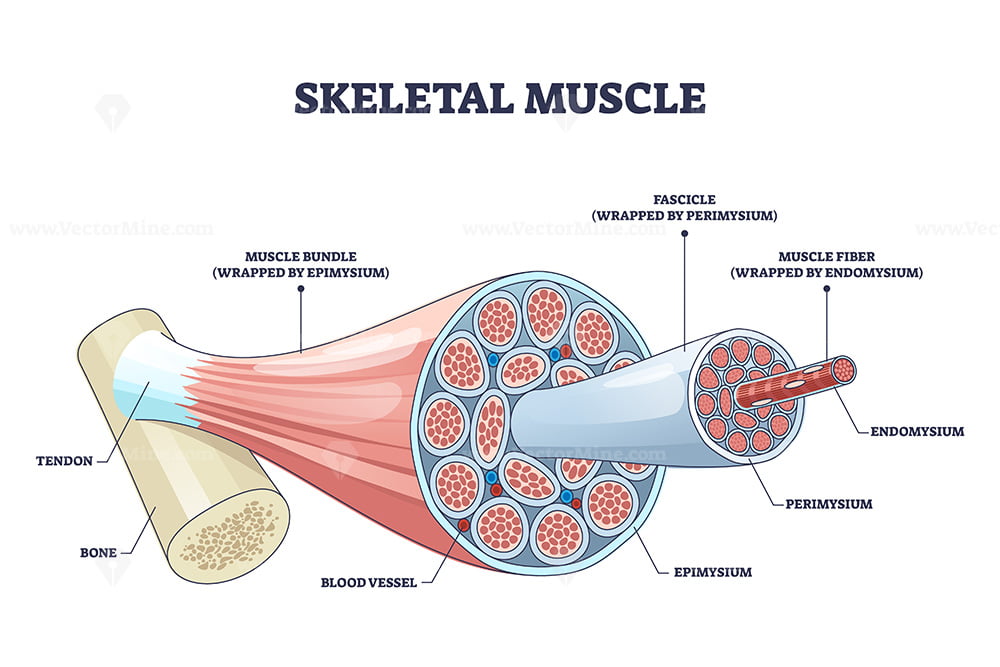
Smooth muscles are found in organs and blood vessels and are involuntary. There are more than 600 skeletal muscles, and they makes up about 40 percent of a person’s body weight. Within muscles, there are layers of connective tissue called the epimysium, perimysium, and endomysium. Web 99k views 3 years ago science diagrams | explained and labelled science diagrams. Learn step by step drawing tutorial. Web skeletal muscles are the ones we can control voluntarily and are responsible for movement. Once you’re feeling confident, it’s time to test yourself. By the end of this section, you will be able to: Learn this topic now at kenhub! These layers cover muscle subunits, individual muscle cells, and myofibrils respectively.
Structure Skeletal Muscle Anatomy by Tigatelu on Dribbble
Learn step by step drawing tutorial. Turn your eyes—a tiny movement, considering the conspicuously large and strong external eye muscles that control eyeball movements. Bones are the foundation of the body. New 3d rotate and zoom. Web different type of muscle cells have different unique characteristics.

Skeletal Muscle Cell Structure
Learn about the bones, joints, and skeletal anatomy of the human body. Web this article describes the histology of skeletal muscle, focusing on structure, types, contraction and clinical points. Web learn about the three types of muscle as you use our 3d models to explore the anatomical structure and physiology of human muscles. Web different type of muscle cells have.
Skeletal Muscle Drawing at Explore collection of
Use the location, shape and surrounding structures to help you memorize each muscle. Turn your eyes—a tiny movement, considering the conspicuously large and strong external eye muscles that control eyeball movements. Get free printable coloring page of this drawing. Web different type of muscle cells have different unique characteristics. Overview of the anatomy, function and main structures of the muscular.

(A) Illustration of skeletal muscle structure copied with permission
Get free printable coloring page of this drawing. These layers cover muscle subunits, individual muscle cells, and myofibrils respectively. View the muscles of the upper and lower extremity in the diagrams below. Smooth muscles are found in organs and blood vessels and are involuntary. Describe the connective tissue layers surrounding skeletal muscle.
Skeletal muscle structure with anatomical inner layers outline diagram
39k views 2 years ago. Get free printable coloring page of this drawing. Explain how muscles work with tendons to move the body. Within muscles, there are layers of connective tissue called the epimysium, perimysium, and endomysium. The skeletal muscle fibres are multinucleated.

How To Draw Skeletal, Smooth and Cardiac Muscle Diagram Types Of
The muscle tissue is composed of a large number of myocytes or muscle cells. Web explore the skeletal system with our interactive 3d anatomy models. See skeletal muscle diagram stock video clips. Use the location, shape and surrounding structures to help you memorize each muscle. Describe the layers of connective tissues packaging skeletal muscle.

Skeletal Muscle Drawing HighRes Vector Graphic Getty Images
Identify areas of the skeletal muscle fibers. Muscle and fat, in contrast, can vary wildly from person to person and even throughout a lifetime. View the muscles of the upper and lower extremity in the diagrams below. Web 23 september 2023 by proactive creative. List the major sarcomeric proteins involved with contraction.

How to draw " Skeletal ( voluntary Muscles )" step by step in a easy
Bones are the foundation of the body. Mastering the human skeleton will mean you get figure drawing right every time. For example, the skeletal muscle is the only type of muscle cell that is always multinucleated (for more info see the latter half of sal's video). Web 99k views 3 years ago science diagrams | explained and labelled science diagrams..

Shapes of skeletal muscles with various muscular types outline diagram
Use the location, shape and surrounding structures to help you memorize each muscle. Learn about the bones, joints, and skeletal anatomy of the human body. It’s a complex puzzle of bones and muscles that gives life and energy to every pose we draw. Each chart groups the muscles of that region into its component groups, making your revision a million.

How To Draw Structure Of Skeletal Muscle YouTube
Describe the layers of connective tissues packaging skeletal muscle. Web skeletal muscles are the ones we can control voluntarily and are responsible for movement. New 3d rotate and zoom. Labelled diagram of skeletal muscle. Web this article describes the histology of skeletal muscle, focusing on structure, types, contraction and clinical points.
Within Muscles, There Are Layers Of Connective Tissue Called The Epimysium, Perimysium, And Endomysium.
Identify areas of the skeletal muscle fibers. List the major sarcomeric proteins involved with contraction. Mastering the human skeleton will mean you get figure drawing right every time. In this video i have shown the simplest way of drawing muscle drawing.
For Example, Upper Limb Muscles Are Grouped By Shoulder And Arm, Forearm.
The skeletal muscle fibres are multinucleated. Bones are the foundation of the body. There are more than 600 skeletal muscles, and they makes up about 40 percent of a person’s body weight. Web 90 skeletal muscle stock illustrations and clipart.
Turn Your Eyes—A Tiny Movement, Considering The Conspicuously Large And Strong External Eye Muscles That Control Eyeball Movements.
As an artist, i’ve always been fascinated by the human form. Explain how muscles work with tendons to move the body. Web how to draw muscles. Web each skeletal muscle has three layers of connective tissue (called “mysia”) that enclose it and provide structure to the muscle as a whole, and also compartmentalize the muscle fibers within the muscle (figure 10.3).
Web 23 September 2023 By Proactive Creative.
New 3d rotate and zoom. Cardiac muscles are found in the heart and are also involuntary. It is the pen diagram of skeletal,. Each chart groups the muscles of that region into its component groups, making your revision a million times easier.