Skeletal Muscle Tissue Drawing
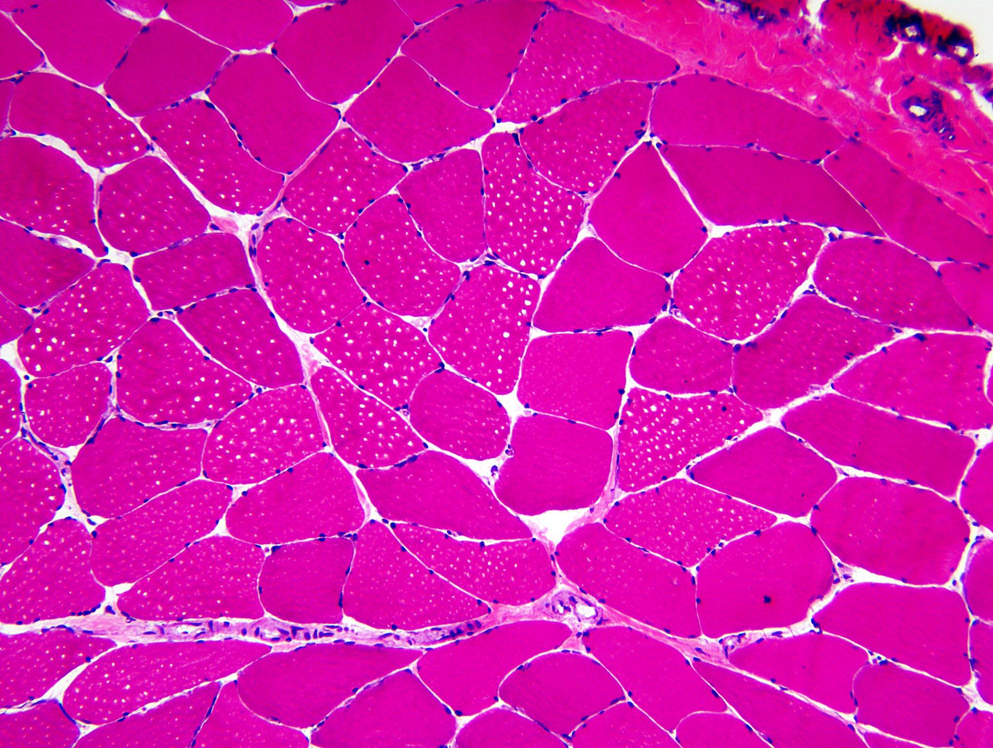
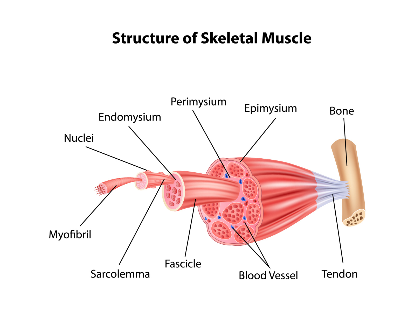
Skeletal Muscle Tissue Drawing - Identifying features are cylindrical cells and multiple peripheral nuclei. Skeletal muscles vary considerably in size, shape, and arrangement of fibers. Web skeletal muscle fibers are organized into groups called fascicles. Some skeletal muscle cells can be followed for over 2.5 µm on the left side of the specimen. Contractile tissue is able to generate tension of force. Web also, the epimysium anchors the muscles to tendons. Skeletal muscles maintain posture, stabilize bones and joints, control internal movement, and generate heat. Your bones move when skeletal muscles contract and pull on the tendons. These layers cover muscle subunits, individual muscle cells, and myofibrils respectively. Muscular dystrophy is a type of genetic disorder described by weakening and atrophy of muscular tissue.
Web using skeletal muscle tissue engineering, this transformative technology can potentially revolutionise the food industry, offering solutions to environmental and ethical challenges 1,2,3,4,20,32,33. These muscle cells are long and multinucleated. Expanding and contracting your chest cavity so you can inhale and exhale at will. Web skeletal muscle tissue engineering (smte) seeks to meet this clinical demand. The tension created by contraction of the muscle. Each organ or muscle consists of skeletal muscle tissue, connective tissue, nerve tissue, and blood or vascular tissue. Web skeletal muscle is the most common type of muscle tissue found in the body and consists of highly elongated, multinucleate, non branching cells which are arranged in a parallel manner. It is classified as a striated muscle tissue, which functions to contract and permit movements under voluntary control. At the ends of each skeletal muscle a tendon connects the muscle to. They range from extremely tiny strands such as the stapedium muscle of the.
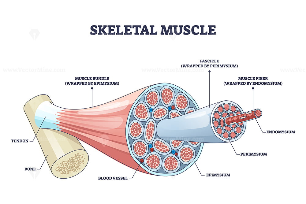
All three muscle tissues have some properties in common; Some skeletal muscle cells can be followed for over 2.5 µm on the left side of the specimen. Web skeletal muscle fibers are organized into groups called fascicles. Muscular dystrophy is a type of genetic disorder described by weakening and atrophy of muscular tissue. Each cell is from 50 to 150 µm in diameter. Skeletal myocytes often measure several centimeters, or tens of centimeters in length, with the number of nuclei contained within being. Expanding and contracting your chest cavity so you can inhale and exhale at will. Bundles of muscle fibers make up a muscle fascicle, and fascicles' bundles make up a skeletal muscle. Web inside each skeletal muscle, muscle fibers are organized into individual bundles, each called a fascicle, by a middle layer of connective tissue called the perimysium.this fascicular organization is common in muscles of the limbs; Muscle cell / fibroblast nuclei.

Skeletal muscle tissue. Skeletal muscle consists of muscle fibers that
Identifying features are cylindrical cells and multiple peripheral nuclei. Web of skeletal muscle cells is seen on the left of the specimen. Web skeletal muscle is one of the three types of muscle tissue, alongside cardiac and smooth muscle. Skeletal myocytes often measure several centimeters, or tens of centimeters in length, with the number of nuclei contained within being. Some.

Illustrations Skeletal Muscle General Histology
Under the microscope, this appears as disorganized and reduced. Tendons are flexible but tough cords of tissue. All the components of the skeletal muscle contribute. Your bones move when skeletal muscles contract and pull on the tendons. Web skeletal muscle is one of the three types of muscle tissue, alongside cardiac and smooth muscle.

How to draw " Skeletal ( voluntary Muscles )" step by step in a easy
It attaches to bones and the orbits through tendons. (c) cardiac muscle cells appear striated and have a single nucleus. They range from extremely tiny strands such as the stapedium muscle of the. Web skeletal musculature structure of the skeletal muscle. Excitable tissue responds to stimuli through electrical signals.
skeletal muscle tissue drawing
These muscle cells are long and multinucleated. Muscle tissue has a unique histological appearance which enables it to carry out its function. Muscles work on a macro level, starting with tendons that attach muscles to bones. Web muscle is one of the four primary tissue types of the body, and the body contains three types of muscle tissue: Web so.
Skeletal muscle structure with anatomical inner layers outline diagram
Skeletal muscles are voluntary and striated in nature. Web there are six actin molecules around a single myosin molecules and there are more than 100,000 sarcomeres (one myosin and six actin make 1 sarcomere) in a single bicep muscle fibre (a single cell) and 253000 such fibres in a young man's bicep. These layers cover muscle subunits, individual muscle cells,.

Skeletal Muscle Tissue Diagram Quizlet
Each skeletal muscle has a structure of bundles within bundles. The musculoskeletal system (locomotor system) is a human body system that provides our body with movement, stability, shape, and support. Skeletal muscles are voluntary and striated in nature. (a) skeletal muscle cells have prominent striation and nuclei on their periphery. Bones are also connected to other bones by ligaments.
Structure Skeletal Muscle Anatomy by Tigatelu on Dribbble
Under the microscope, this appears as disorganized and reduced. Tendons are flexible but tough cords of tissue. They range from extremely tiny strands such as the stapedium muscle of the. The solid components include proteins and other organic and inorganic substances. Web skeletal musculature structure of the skeletal muscle.

What Is Skeletal System Anatomy Design Talk
Web skeletal muscle tissue engineering (smte) seeks to meet this clinical demand. Identifying features are cylindrical cells and multiple peripheral nuclei. Bones are also connected to other bones by ligaments. (a) skeletal muscle cells have prominent striation and nuclei on their periphery. Web the skeletal muscles are a vital part of your musculoskeletal system.

How To Draw Structure Of Skeletal Muscle YouTube
Skeletal muscle is formed by 75% of water and 25% of solids. It allows the nervous system to trigger a specific movement of a muscle by activating a subset of muscle fibers within a. Tendons are flexible but tough cords of tissue. At the ends of each skeletal muscle a tendon connects the muscle to. Web of skeletal muscle cells.

(A) Illustration of skeletal muscle structure copied with permission
Skeletal muscle, cardiac muscle, and smooth muscle ( figure 10.2 ). Web skeletal muscle is the most common type of muscle tissue found in the body and consists of highly elongated, multinucleate, non branching cells which are arranged in a parallel manner. Bones are also connected to other bones by ligaments. Skeletal muscle is formed by 75% of water and.
Each Organ Or Muscle Consists Of Skeletal Muscle Tissue, Connective Tissue, Nerve Tissue, And Blood Or Vascular Tissue.
They range from extremely tiny strands such as the stapedium muscle of the. Web inside each skeletal muscle, muscle fibers are organized into individual bundles, each called a fascicle, by a middle layer of connective tissue called the perimysium.this fascicular organization is common in muscles of the limbs; The musculoskeletal system (locomotor system) is a human body system that provides our body with movement, stability, shape, and support. They serve a variety of functions, including:
Each Cell Is From 50 To 150 Μm In Diameter.
Web skeletal muscle tissue engineering (smte) seeks to meet this clinical demand. Muscle tissue has a unique histological appearance which enables it to carry out its function. At each level of bundling, a connective tissue membrane surrounds the bundle. Skeletal muscle is comprised of a series of muscle fibers made of muscle cells.
Muscle Cell / Fibroblast Nuclei.
(b) smooth muscle cells have a single nucleus and no visible striations. Web the skeletal muscles are a vital part of your musculoskeletal system. They are responsible for the movement of appendages and locomotion. Skeletal muscles are voluntary and striated in nature.
It Is Subdivided Into Two Broad Systems:
Bones are also connected to other bones by ligaments. Skeletal muscles maintain posture, stabilize bones and joints, control internal movement, and generate heat. The tension created by contraction of the muscle. Web muscle is one of the four primary tissue types of the body, and the body contains three types of muscle tissue: