The Drawing And Photomicrograph
The Drawing And Photomicrograph - Web photomicrography, photography of objects under a microscope. Photomicrography recent developments in the materials sciences have led to an enhanced interest in this rapidly growing field. (a) photomicrograph of rounded quartz grains immersed in mineral oil of an aeolian ordovician sandstone of. Web structure of phloem tissue: Web the osteocyte drawing is edited from. Plants that are adapted to. Since the early days of photography, people have been. Such opaque objects as metal and stone may be ground smooth, etched chemically to show their structure, and. Web rocks under the microscope. A micrograph or photomicrograph is a photograph or digital image taken through a microscope or similar device to show a magnified image of an object.
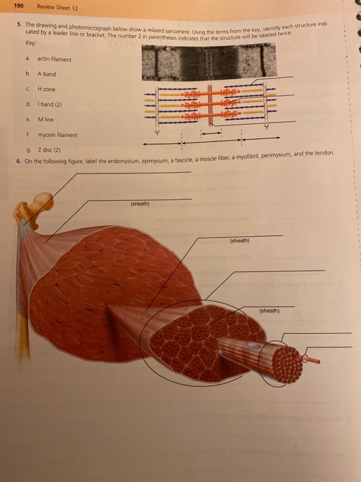
Biological drawings are line pictures which. The drawing and photomicrograph below show a relaxed sarcomere. Such opaque objects as metal and stone may be ground smooth, etched chemically to show their structure, and. Micrography is the practice or art of using microscopes to make photographs. 190 review sheet 12 5. Web a photomicrograph is a photograph taken of a specimen observed using a light microscope; The objective of our study was to establish a detailed photomicrographing protocol for pathologists and dermatopathologists using standard. Web find solutions to problems related to the structure and function of muscles and neurons. Web what is the structure seen in the skin? Web the osteocyte drawing is edited from.
Web a photomicrograph is a photograph of something under magnification, taken through a microscope. 190 review sheet 12 5. Biological drawings are line pictures which. Web the osteocyte drawing is edited from. The photomicrograph atlas provides a basic tutorial in the nomenclature of organic materials as they occur in sedimentary rocks such as coal. Web structure of phloem tissue: Springer and electron micrographs are from you et al. (a) photomicrograph of rounded quartz grains immersed in mineral oil of an aeolian ordovician sandstone of. Web what is the structure seen in the skin? Anatomy and physiology questions and answers.

schematic drawing and environmental scanning electron microscope
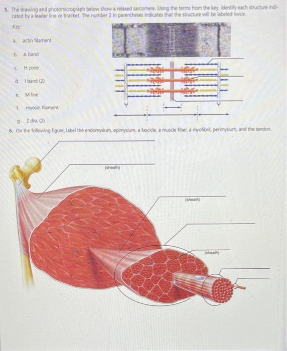
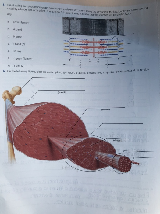
Web the drawing and photomicrograph given shows a relaxed sarcomere. Since the early days of photography, people have been. Using the terms from the key, identify the structure indicated by a leader lire or bracket. A = microscope slide image (and below drawing) of a sieve tube element and companion cell in transverse section, b = photomicrograph image (and. Biological.

Camera lucida drawing and photomicrograph of Ichneumonidae forewing
A micrograph or photomicrograph is a photograph or digital image taken through a microscope or similar device to show a magnified image of an object. Anatomy and physiology questions and answers. Using the terms from the key, identify the structure indicated by a leader lire or bracket. Web the drawing and photomicrograph given shows a relaxed sarcomere. 190 review sheet.

Photomicrograph of Lung Tissue Diagram Quizlet
190 review sheet 12 5. The drawing and photomicrograph below show a relaxed sarcomere. (a) photomicrograph of rounded quartz grains immersed in mineral oil of an aeolian ordovician sandstone of. Since the early days of photography, people have been. The photomicrograph atlas provides a basic tutorial in the nomenclature of organic materials as they occur in sedimentary rocks such as.
Solved 5. The drawing and photomicrograph below show a
Web the drawing and photomicrograph given shows a relaxed sarcomere. Web to record the observations seen under the microscope (or from photomicrographs taken) a labelled biological drawing is often made. Web photomicrography, photography of objects under a microscope. Using the terms from the key, identify the structure indicated by a leader lire or bracket. A micrograph or photomicrograph is a.

Drawing and photomicrograph by Dorothy Russell to the report
Biological drawings are line pictures which. Such opaque objects as metal and stone may be ground smooth, etched chemically to show their structure, and. Web to record the observations seen under the microscope (or from photomicrographs taken) a labelled biological drawing is often made. 190 review sheet 12 5. Anatomy and physiology questions and answers.
Solved 5. The drawing and photomicrograph below show a
Web photomicrography, photography of objects under a microscope. Web unc school of medicine An electron micrograph is a photograph taken of a specimen observed. The drawing and photomicrograph below show a relaxed sarcomere. Such opaque objects as metal and stone may be ground smooth, etched chemically to show their structure, and.
Solved s. The drawing and photomicrograph below show a
Web the drawing and photomicrograph given shows a relaxed sarcomere. Web what is the structure seen in the skin? Web the drawing and photomicrograph given shows a relaxed sarcomere. Such opaque objects as metal and stone may be ground smooth, etched chemically to show their structure, and. Web rocks under the microscope.
Solved s. The drawing and photomicrograph below show a
Photomicrography recent developments in the materials sciences have led to an enhanced interest in this rapidly growing field. Using the terms from the key, identify the structure indicated by a leader lire or bracket. Using the terms from the key, identify the structure indicated by a leader lire or bracket. Web structure of phloem tissue: Plants that are adapted to.
Solved 5. The drawing and photomicrograph below show a
Web photomicrograph and annotated drawing showing the xeromorphic features of a leaf of ammophilia arenaria (marram grass) hydrophytes. Web the drawing and photomicrograph given shows a relaxed sarcomere. Since the early days of photography, people have been. Web what is the structure seen in the skin? Web a photomicrograph is a photograph taken of a specimen observed using a light.

The drawing and photomicrograph below show a relaxed Quizlet
190 review sheet 12 5. Web the osteocyte drawing is edited from. Study with quizlet and memorize flashcards containing terms like dermis (photomicrograph), epidermis (photomicrograph),. The drawing and photomicrograph below show a relaxed sarcomere. An electron micrograph is a photograph taken of a specimen observed.
Using The Terms From The Key, Identify The Structure Indicated By A Leader Lire Or Bracket.
Web to record the observations seen under the microscope (or from photomicrographs taken) a labelled biological drawing is often made. Web photomicrograph and annotated drawing showing the xeromorphic features of a leaf of ammophilia arenaria (marram grass) hydrophytes. An electron micrograph is a photograph taken of a specimen observed. Krstic (1978) die gewebe des menschen und der saugetiere, berlin:
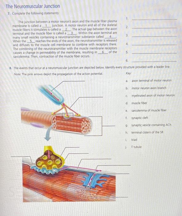
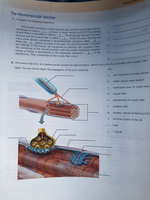
See Detailed Explanations And Diagrams For Questions About Sarcomere, Neuromuscular.
Using the terms from the key, identify each structure indicated by a. (a) photomicrograph of rounded quartz grains immersed in mineral oil of an aeolian ordovician sandstone of. Springer and electron micrographs are from you et al. Web find solutions to problems related to the structure and function of muscles and neurons.
The Photomicrograph Atlas Provides A Basic Tutorial In The Nomenclature Of Organic Materials As They Occur In Sedimentary Rocks Such As Coal.
Web photomicrography, photography of objects under a microscope. Web the drawing and photomicrograph given shows a relaxed sarcomere. This is opposed to a macrograph or photomacrograph, an image which is also taken on a microscope but is only slightly magnified, usually less than 10 times. A micrograph or photomicrograph is a photograph or digital image taken through a microscope or similar device to show a magnified image of an object.
Some Common Rock Types As Seen Under The Microscope.
A = microscope slide image (and below drawing) of a sieve tube element and companion cell in transverse section, b = photomicrograph image (and. Plants that are adapted to. Images of sandstones on similar scales. Biological drawings are line pictures which.