Thoracic Drawing
Thoracic Drawing - Anatomy of the thoracic wall and the breast (illustrations) : Also shown are the spinal cord, vertebra (back bone), conus medullaris (the end of the spinal cord), cauda equina (the. Web subscribe to my channel to get more drawing videos Vertebrae are the 33 individual, interlocking bones that form your spinal column. Web in this episode, i’ll show you how to draw the forms of the rib cage step by step.giveaway! Web an understanding of thoracic imaging requires knowledge of the anatomy being imaged, as described in this chapter, as well as the imaging techniques applied to the thorax, covered in chapter 2. Web the thoracic cage, also known as the rib cage, is the osteocartilaginous structure that encloses the thorax.it is formed by the 12 thoracic vertebrae, 12 pairs of ribs and associated costal cartilages and the sternum. It starts at the base of your neck and ends at the bottom of your ribs. Web thoracic spine anatomy and upper back pain. Web what is thoracic spine.
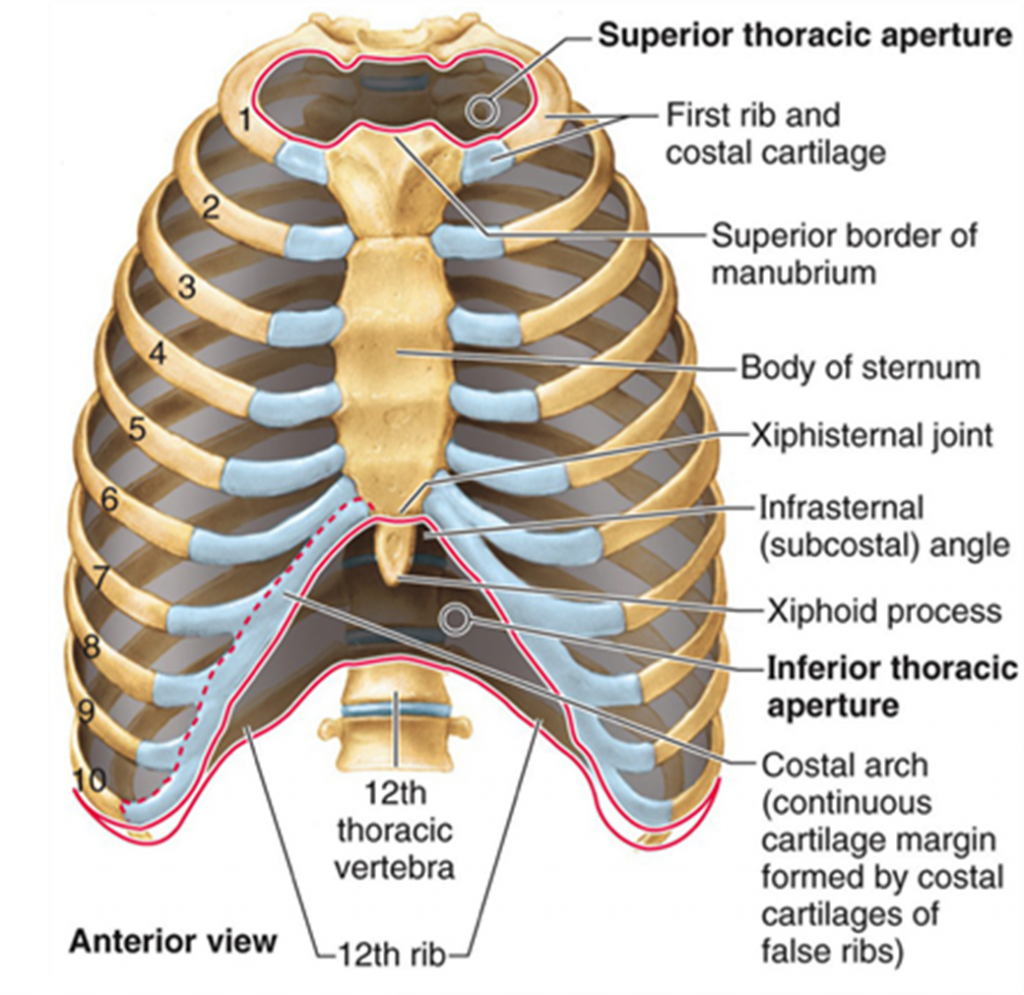
The thoracic spine is the longest region of the spine, and by some measures it is also the most complex. Anatomy of the thoracic wall and the breast (illustrations) : The thorax is bound by bony structures including the 12 pairs of ribs and thoracic vertebrae, whilst also being supported by many ligaments and muscles. It contains the lungs, the middle and lower airways—the. Web thoracic spine anatomy and upper back pain. Web thoracic cavity, the second largest hollow space of the body. The superior thoracic aperture found. Web the thoracic duct is the largest lymphatic vessel in the human body. Separates the thoracic cavity from the abdominal cavity (the word diaphragm is derived from the greek ‘diáphragma’, meaning partition).; Web an understanding of thoracic imaging requires knowledge of the anatomy being imaged, as described in this chapter, as well as the imaging techniques applied to the thorax, covered in chapter 2.
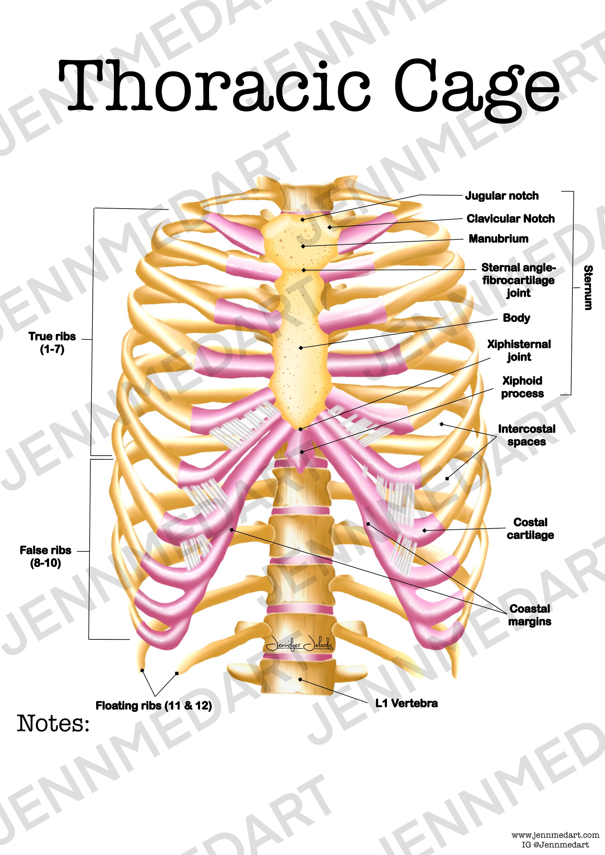
Web thoracic cavity, the second largest hollow space of the body. Web in this episode, i’ll show you how to draw the forms of the rib cage step by step.giveaway! Web thoracic spine anatomy and upper back pain. The thoracic cage (rib cage) forms the thorax (chest) portion of the body. It is the only spinal region attached to the rib cage. The thoracic cage takes the form of a domed bird cage with the horizontal bars formed by ribs and costal cartilages. Web your thoracic spine is the middle section of your spine. Around 75% of the lymph from the entire body (aside from the right upper limb, right breast, right lung and right side of the head and neck) passes through the thoracic duct. Their primary role is to form the thoracic cage that protects the heart, lungs, and esophagus. Collectively, these three sections make a tower of 24 bones that gives the body structure and.

Gross Anatomy Glossary Thoracic Cage ditki medical & biological sciences
It consists of the 12 pairs of ribs with their costal cartilages and the sternum ( figure 7.5.1 ). It serves two main functions: The thorax itself can be split up into various areas that contain important structures. Web the thoracic cage, also known as the rib cage, is the osteocartilaginous structure that encloses the thorax.it is formed by the.
Thoracic Cage Anatomy Worksheet Single FILLED Digital Download Human
Web anatomical position and relations. It starts at the base of your neck and ends at the bottom of your ribs. The cells of the immune system circulate through the lymphatic system.also, large molecular. The thoracic cage protects the heart and lungs. Connecting with the cervical spine above and the lumbar spine below, the thoracic spine runs from the base.
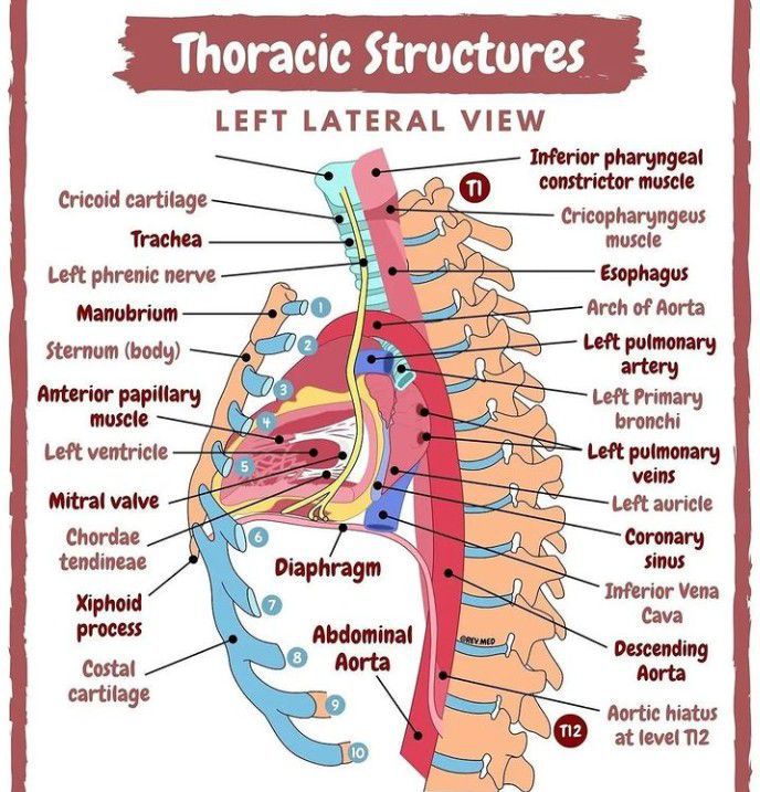
Thoracic Structures from Left Lateral View MEDizzy
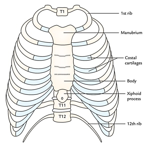
Patterns of bony anatomy of the thoracic cavity and rib cage in anterior and posterior view. Anatomy of the thoracic wall and the breast (illustrations) : It is enclosed by the ribs, the vertebral column, and the sternum, or breastbone, and is separated from the abdominal cavity (the body’s largest hollow space) by a muscular and membranous partition, the diaphragm..

1 Schematic illustration of the anatomy of the thoracic cage. 1R fi rst
Web the thoracic cage, also known as the rib cage, is the osteocartilaginous structure that encloses the thorax.it is formed by the 12 thoracic vertebrae, 12 pairs of ribs and associated costal cartilages and the sternum. The thoracic cage (rib cage) forms the thorax (chest) portion of the body. Collectively, these three sections make a tower of 24 bones that.

Human Anatomy Chest Cavity Anatomy Of Chest Bones Human Anatomy Diagram
The thoracic cage protects the heart and lungs. Around 75% of the lymph from the entire body (aside from the right upper limb, right breast, right lung and right side of the head and neck) passes through the thoracic duct. These 12 bones are separated from each other by intervertebral discs. *completed*if you’d like to win a free membership to.

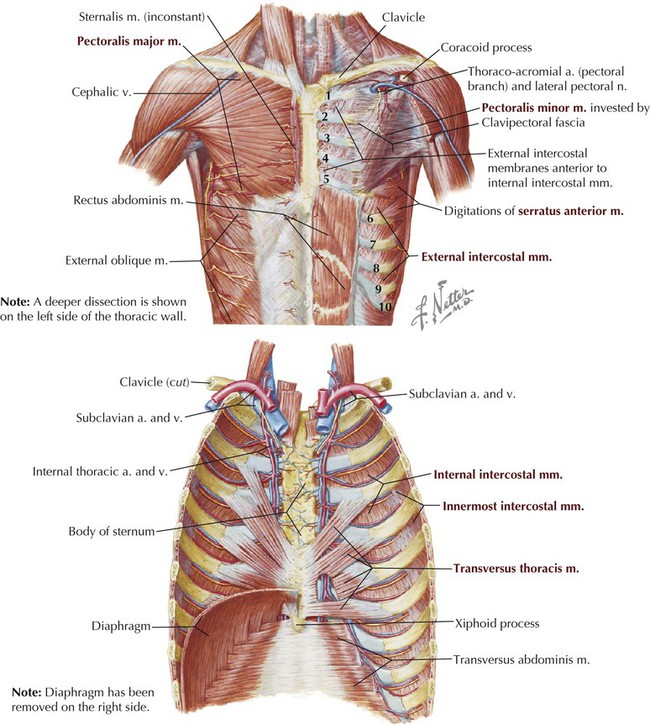
Vasculature of the Anterior Thoracic Wall. Art as Applied to Medicine
Connecting with the cervical spine above and the lumbar spine below, the thoracic spine runs from the base of the neck down to the abdomen. Anatomy of the thoracic wall and the breast (illustrations) : The anatomic illustrations are presented as either line drawings or. Their primary role is to form the thoracic cage that protects the heart, lungs, and.
Thorax Basicmedical Key
Web in this episode, i’ll show you how to draw the forms of the rib cage step by step.giveaway! The anatomic illustrations are presented as either line drawings or. Web thoracic cavity, the second largest hollow space of the body. It starts at the base of your neck and ends at the bottom of your ribs. The lungs lie either.
Thoracic, Chest & Rib Pain Aligned for Life
Their primary role is to form the thoracic cage that protects the heart, lungs, and esophagus. The thoracic cage protects the heart and lungs. Web the thoracic duct is the largest lymphatic vessel in the human body. Also shown are the spinal cord, vertebra (back bone), conus medullaris (the end of the spinal cord), cauda equina (the. Anatomy of the.
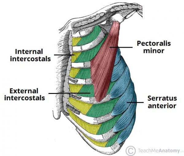
Thoracic Muscles Attachments Actions TeachMeAnatomy
Table 1.1 lists the major anatomic structures within the thorax that are discussed. Web the thoracic spine sits between the cervical spine in the neck and the lumbar spine in the lower back. It contains the lungs, the middle and lower airways—the. Web subscribe to my channel to get more drawing videos Also shown are the spinal cord, vertebra (back.
Thoracic Cage Intrinsic Muscles, Formation and Shape Earth's Lab
Web subscribe to my channel to get more drawing videos It is enclosed by the ribs, the vertebral column, and the sternum, or breastbone, and is separated from the abdominal cavity (the body’s largest hollow space) by a muscular and membranous partition, the diaphragm. It’s the longest section of your spine. Anatomy of the thoracic wall and the breast (illustrations).
Collectively, These Three Sections Make A Tower Of 24 Bones That Gives The Body Structure And.
Undergoes contraction and relaxation, altering the. Patterns of bony anatomy of the thoracic cavity and rib cage in anterior and posterior view. Anatomy of the thoracic wall and the breast (illustrations) : Web thoracic wall the first step in understanding thorax anatomy is to find out its boundaries.
The Thoracic Cage (Rib Cage) Forms The Thorax (Chest) Portion Of The Body.
Around 75% of the lymph from the entire body (aside from the right upper limb, right breast, right lung and right side of the head and neck) passes through the thoracic duct. Web anatomical position and relations. Web the thoracic spine sits between the cervical spine in the neck and the lumbar spine in the lower back. Web the thoracic duct is the largest lymphatic vessel in the human body.
Also Shown Are The Spinal Cord, Vertebra (Back Bone), Conus Medullaris (The End Of The Spinal Cord), Cauda Equina (The.
Web an understanding of thoracic imaging requires knowledge of the anatomy being imaged, as described in this chapter, as well as the imaging techniques applied to the thorax, covered in chapter 2. The thoracic cage protects the heart and lungs. Separates the thoracic cavity from the abdominal cavity (the word diaphragm is derived from the greek ‘diáphragma’, meaning partition).; *completed*if you’d like to win a free membership to the premium.
It Contains The Lungs, The Middle And Lower Airways—The.
It serves two main functions: Vertebrae are the 33 individual, interlocking bones that form your spinal column. Web what is thoracic spine. Web don't waste your time reading this description.