Thymus Drawing
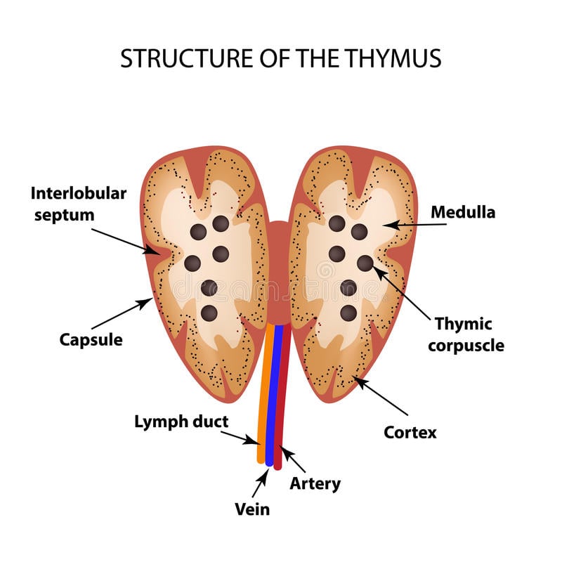
Thymus Drawing - It is a pinkish gray gland in color with a lobulated surface (fig. Their respective parts are labeled, vintage line drawing or engraving illustration. It makes white blood cells, called lymphocytes, which protect the body against infections. Partially differentiated stem cells, which are to become lymphocytes, originate in the bone marrow and migrate to the the thymus gland. The thymus gland is a pink, lobulated lymphoid organ, located in the thoracic cavity and neck. In adults, it is commonly removed 'cause the patient has myasthenia gravis. 27.9 mb (427.8 kb compressed) 2690 x 3630 pixels. If you need more update thymus slide pictures then you may follow anatomy learner at here in social media. Within the thymus, thymus cell lymphocytes or t cells mature. Web the thymus is an encapsulated primary lymphoid organ.histologically, it is divided into subcapsular cortical, cortical and medullary regions within each lobule, created by the intervening connective tissue septae extending from the capsule.
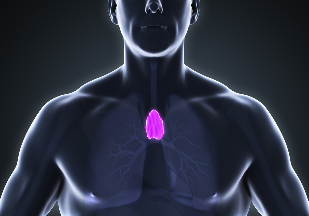
Histologically, the thymus is divided into lobules, each one consisting of a central medulla and a peripheral cortex. Web rejuvenating them during the generation process would make production quicker and more robust, says sebastiano. Web the thymus is a flat encapsulated lymphoid organ located in the anterior superior mediastinum, right behind the sternum. Web the thymus gland is an important part of your immune system. Web the thymus ( pl.: Therefore, adults don’t really need a thymus. Thymus is a little organ that is in the mediastinum. Embryologically, the thymus gland is derived from the third pharyngeal pouch. The thymus gland is a pink, lobulated lymphoid organ, located in the thoracic cavity and neck. The thymus gland, despite containing glandular tissue and producing several hormones, is much more closely associated with the immune system than with the endocrine system.
Embryologically, the thymus gland is derived from the third pharyngeal pouch. Representation of the thymus, endocrine gland located ahead of the heart playing a role in the immune system and atrophies progressively after puberty. The thymus gland, despite containing glandular tissue and producing several hormones, is much more closely associated with the immune system than with the endocrine system. Web anatomy of the thymus gland; T cells are critical to the adaptive immune system, where the body adapts to specific foreign invaders. Web thymus (anterior view) the thymus is a primary lymphoid organ located in the mediastinum. Thymus is a little organ that is in the mediastinum. Although the thymus is sometimes called a gland, it is. Web the thymus gland. Web thymus gland drawing stock photos and images.
Thymus Structure and Functions
The organ is called thymus because its shape resembles that of a thyme leaf. Representation of the thymus, endocrine gland located ahead of the heart playing a role in the immune system and atrophies progressively after puberty. After puberty, it decreases in size and is slowly replaced by fat. Thymuses or thymi) is a specialized primary lymphoid organ of the.
Thymus Facts, Function & Diseases Live Science
Within the thymus, thymus cell lymphocytes or t cells mature. Gross cross sectional dissection of the thymus reveals a darker cortical region that is more. Web the thymus ( pl.: Web thymus (anterior view) the thymus is a primary lymphoid organ located in the mediastinum. Web thymus histology drawing.

Anatomy, Location, and Functions of the Thymus Facty Health
Micrograph of a thymic corpusle (hassall's corpusle). After puberty, it decreases in size and is slowly replaced by fat. How to draw diagram of thymus gland step by step for beginnershello friends in this video i tell you about how to draw thymus gland. Its functions include producing white blood cells known as t cells, which aid in immunity. In.

how to draw diagram of thymus gland step by step for beginners YouTube
Web anatomy of the thymus gland; If you need more update thymus slide pictures then you may follow anatomy learner at here in social media. The thymus is located in the upper front part of the chest, in the anterior. T cells are critical to the adaptive immune system, where the body adapts to specific foreign invaders. The capsule and.

how to draw thymus gland how to draw diagram of thymus gland YouTube
Embryologically, the thymus gland is derived from the third pharyngeal pouch. Its functions include producing white blood cells known as t cells, which aid in immunity. It helps train the white blood cells that protect your immune system. Web anatomy of the thymus gland; Histologically, the thymus is divided into lobules, each one consisting of a central medulla and a.

how to draw diagram of thymus gland step by step in simple way YouTube
Web rejuvenating them during the generation process would make production quicker and more robust, says sebastiano. After puberty, it decreases in size and is slowly replaced by fat. Web thymus (anterior view) the thymus is a primary lymphoid organ located in the mediastinum. Embryologically, the thymus gland is derived from the third pharyngeal pouch. In the adolescent, it is involved.
:max_bytes(150000):strip_icc()/human-thymus-anatomy-513000737-d60061aa3a334e67bc64f6177465698c.jpg)
Thymus Anatomy, Function, and Treatment
Web the thymus ( pl.: Web the thymus is an encapsulated primary lymphoid organ.histologically, it is divided into subcapsular cortical, cortical and medullary regions within each lobule, created by the intervening connective tissue septae extending from the capsule. Florian’s approach, too, aims to produce healthier immune cells — inside the. Representation of the thymus, endocrine gland located ahead of the.

how to draw thymus gland how to draw labelled diagram of thymus gland
Although the thymus is sometimes called a gland, it is. These cells find and destroy pathogens like bacteria circulating in the bloodstream. How to draw diagram of thymus gland step by step for beginnershello friends in this video i tell you about how to draw thymus gland. Gross cross sectional dissection of the thymus reveals a darker cortical region that.

how to draw diagram of thymus gland how to draw thymus gland easily
It is a pinkish gray gland in color with a lobulated surface (fig. Its functions include producing white blood cells known as t cells, which aid in immunity. The capsule and septa contain blood. Gross cross sectional dissection of the thymus reveals a darker cortical region that is more. Web anatomy of the thymus gland;

THYMUS GLAND, DRAWING Stock Photo Alamy
Web anatomy of the thymus gland; It helps train the white blood cells that protect your immune system. Also shown are the ribs, lungs, and heart. T cells are critical to the adaptive immune system, where the body adapts to specific foreign invaders. Anatomy of the thymus gland.
Thymuses Or Thymi) Is A Specialized Primary Lymphoid Organ Of The Immune System.
Web anatomically, the earliest preserved drawings of the gland were the line drawings of vesalius. Web the thymus gland is located in the chest behind the breastbone. In the adolescent, it is involved the development of the immune system. Web rejuvenating them during the generation process would make production quicker and more robust, says sebastiano.
Although The Thymus Is Sometimes Called A Gland, It Is.
Drawing shows the thymus gland in the upper chest under the breastbone. Therefore, adults don’t really need a thymus. The thymus is located in the upper front part of the chest, in the anterior. It makes white blood cells, called lymphocytes, which protect the body against infections.
The Organ Is Called Thymus Because Its Shape Resembles That Of A Thyme Leaf.
I am so excited to share thymus histology drawing pictures with you so that you might understand the every single part of thymus easily. Also shown are the ribs, lungs, and heart. Their respective parts are labeled, vintage line drawing or engraving illustration. Embryologically, the thymus gland is derived from the third pharyngeal pouch.
Within The Thymus, Thymus Cell Lymphocytes Or T Cells Mature.
After puberty, it decreases in size and is slowly replaced by fat. If you need more update thymus slide pictures then you may follow anatomy learner at here in social media. Web thymus histology drawing. Gross cross sectional dissection of the thymus reveals a darker cortical region that is more.